. 2013 Apr 9;3(4):585-595.
doi: 10.1534/g3.112.005165.

Assembly of the Synaptonemal Complex Is a Highly Temperature-Sensitive Process That Is Supported by PGL-1 During Caenorhabditis elegans Meiosis

Affiliations

Assembly of the Synaptonemal Complex Is a Highly Temperature-Sensitive Process That Is Supported by PGL-1 During Caenorhabditis elegans Meiosis

Ceyda Bilgir et al. G3 (Bethesda). .

Abstract

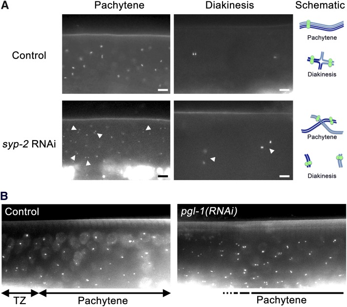
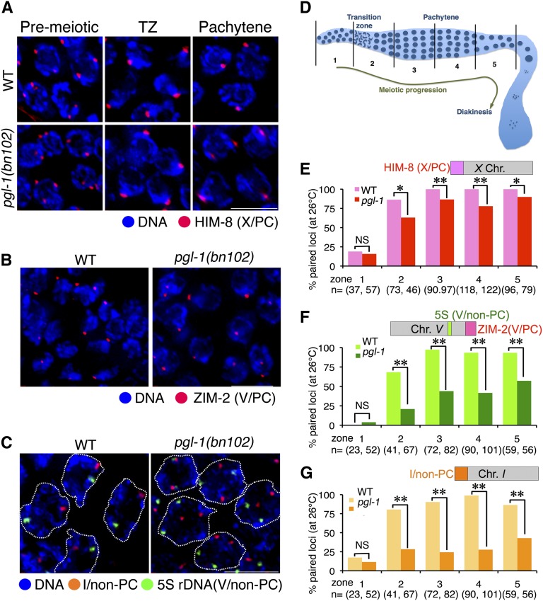
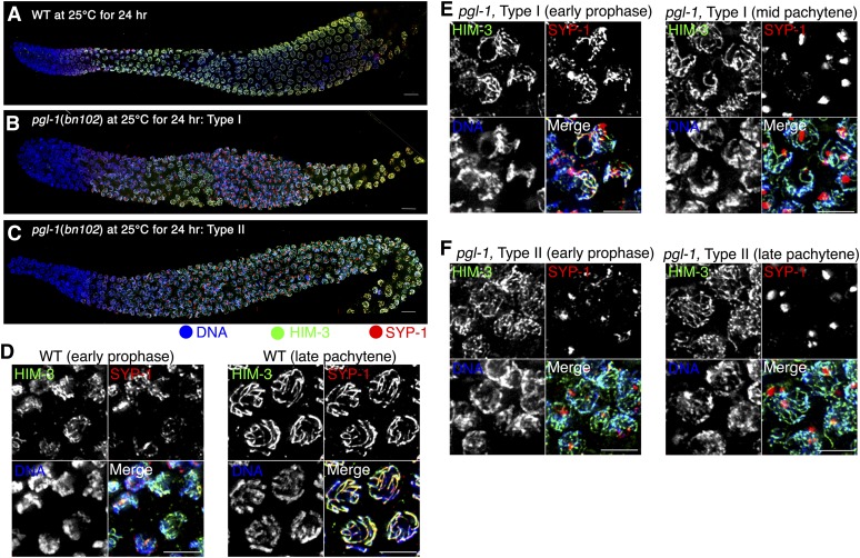
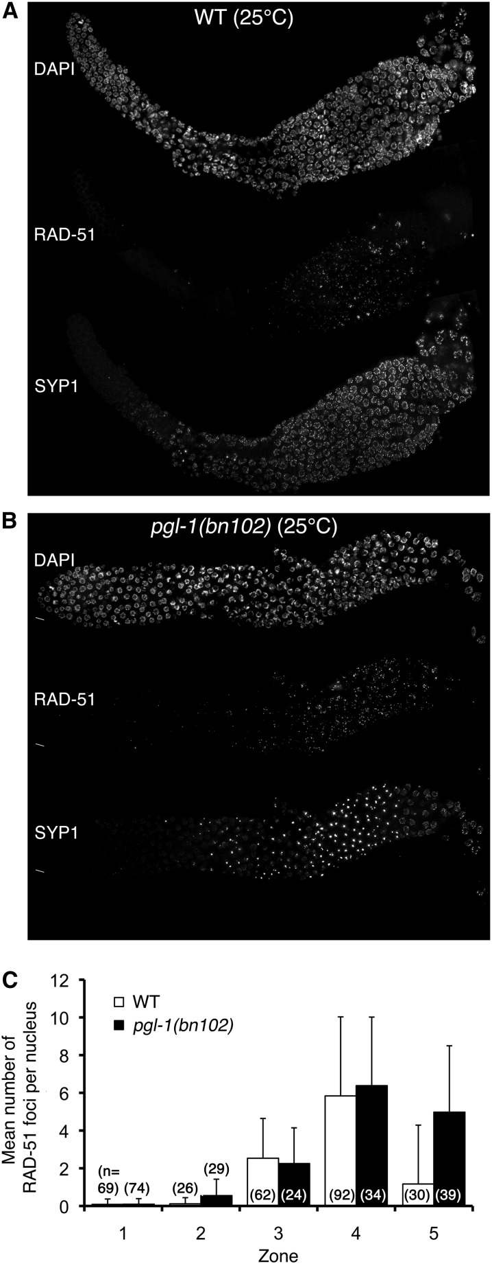
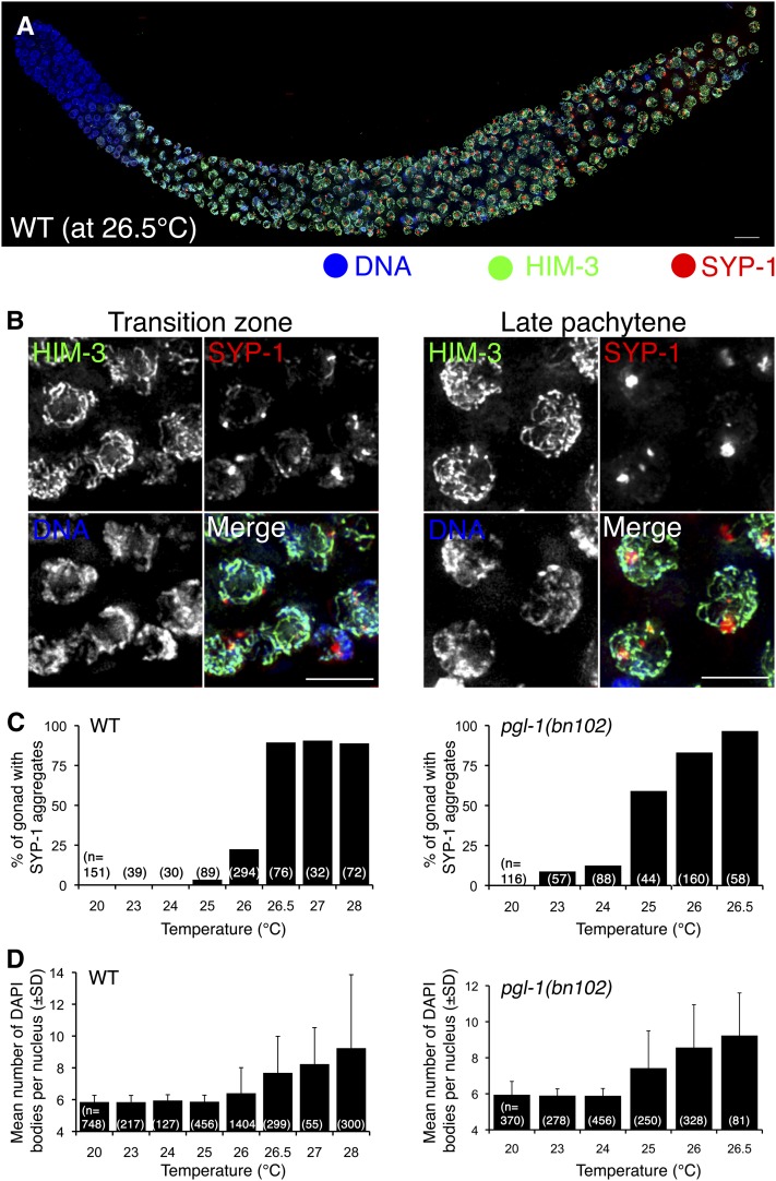
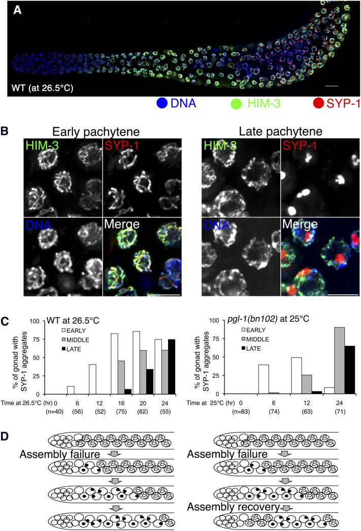
Successful chromosome segregation during meiosis depends on the synaptonemal complex (SC), a structure that stabilizes pairing between aligned homologous chromosomes. Here we show that SC assembly is a temperature-sensitive process during Caenorhabditis elegans meiosis. Temperature sensitivity of SC assembly initially was revealed through identification of the germline-specific P-granule component PGL-1 as a factor promoting stable homolog pairing. Using an assay system that monitors homolog pairing in vivo, we showed that depletion of PGL-1 at 25° disrupts homolog pairing. Analysis of homolog pairing at other chromosomal loci in a pgl-1-null mutant revealed a pairing defect similar to that observed in mutants lacking SC central region components. Furthermore, loss of pgl-1 function at temperatures ≥25° results in severe impairment in loading of SC central region component SYP-1 onto chromosomes, resulting in formation of SYP-1 aggregates. SC assembly is also temperature sensitive in wild-type worms, which exhibit similar SYP-1 loading defects and formation of SYP-1 aggregates at temperatures ≥26.5°. Temperature shift analyses suggest that assembly of the SC is temperature sensitive, but maintenance of the SC is not. We suggest that the temperature sensitive (ts) nature of SC assembly may contribute to fitness and adaptation capacity in C. elegans by enabling meiotic disruption in response to environmental change, thereby increasing the production of male progeny available for outcrossing.

Keywords: P-granule; aneuploid gametes; pachytene; polycomplex; synapsis.


Figures

Figure 1
Figure 1
Fluorescent signals in live animals carrying a GFP-LacI/lacO system report the status of meiotic homologous pairing. (A) Shown are the pachytene (left) and diakinesis (right) regions of a gonad in live AV221 worms without any treatment (top) and after syp-2(RNAi) by injection of double-stranded RNA targeted to the syp-2 gene into their parental worms (bottom). The locations of nuclei with unpaired signals in the syp-2(RNAi) gonad are indicated by arrowheads. In some cases, the nuclei can be identified by diffuse GFP signals. The bright diffuse fluorescent signals seen in the lower part of a panel are the autofluorescence of the gut, whereas the fluorescent dots in the upper part of a panel are the signals corresponding to LacI-GFP. Worms were cultured at 20°. Bar: 5 µm. Schematics depicting the inferred configuration of chromosomes are shown at the right. (B) The TZ and the pachytene region of a gonad in live AV221 worms fed either bacteria carrying the empty RNAi vector (left) or an RNAi plasmid expressing double-stranded RNA targeted to pgl-1 gene (right). Note that many nuclei are visualized by bright diffuse GFP signals. TZ and the pachytene regions are defined by nuclear morphology. The beginning of the pachytene zone is not clear for pgl-1(RNAi) due to the lack of a clear TZ, which is indicated by a dashed line. Worms were cultured at 20° until L4 stage, then shifted to 25° for 24 hr.
Figure 2
Figure 2
The pgl-1 mutant exhibits a decrease in meiotic homologous pairing. (A) IF staining of HIM-8 (marking the PCs, of the X chromosomes, red) with DNA counter staining (blue) in whole mount gonads from wild-type (top) and pgl-1(bn102) mutant (bottom) worms cultured at 26° for 24 hr. Bar: 5 µm. (B) IF staining of ZIM-2 (marking the PC of chromosome V and appearing in the TZ, red) in the wild type (left) and the pgl-1(bn102) mutant (right) with DNA counter staining (blue) cultured at 26°. Bar: 5 µm. (C) FISH visualizing the non-PC end of chromosome I (red) and 5S rDNA locus on the chromosome V (green) with DNA counter staining (blue) in pachytene nuclei (outlined with dashed lines) of wild-type (left) and the pgl-1(bn102) mutant (right) worms cultured at 26°. Bar: 5 µm. (D) Schematic diagram of zones (1−5) of the gonad used for scoring of pairing efficiency. The distal end is shown toward the left. (E, F, G) The pairing efficiency at the indicated locus is presented as the percentage of nuclei in each zone with paired signals. Asterisks indicate a statistically significant difference between wild type and pgl-1 in the corresponding stage (*0.001 ≤ P < 0.01, **P < 0.001). NS indicates not statistically significant.
Figure 3
Figure 3
The pgl-1 mutant exhibits SYP-1 aggregate formation. (A−C) IF staining of HIM-3 (green) and SYP-1 (red) in a wild-type (A) and a pgl-1(bn102) mutant (B and C) whole mount gonad cultured at 25° for 24 hr. In the Type I gonad (B), SYP-1 aggregates are present in the middle-late pachytene regions of the gonad but are not present in early meiotic prophase; in the Type II gonad (C), SYP-1 aggregates are detected throughout the meiotic region. The distal end is toward the left. Bar is 10 µm. (D−F) Closeup of panels (A), (B), and (C), respectively, in early meiotic prophase (left) and the middle or late pachytene region (right). Bar: 5 µm.
Figure 4
Figure 4
Accumulation of RAD-51 foci in pgl-1(bn102) worms cultured at 25°. (A and B) IF staining of RAD-51 (middle) and SYP-1 (bottom) with DNA counterstaining (top) in the wild-type (A) and the pgl-1(bn102) mutant (B) whole mount gonad cultured at 25° for 24 hr after L4 stage. (C) Average number of RAD-51 foci per nucleus in five zones of the gonad. Zones are defined as in Figure 2D. Error bar indicates SD.
Figure 5
Figure 5
The wild type exhibits SYP-1 aggregate formation similar to the pgl-1 mutant at high temperatures, which correlates with a failure in chiasma formation. (A) IF staining of HIM-3 (green) and SYP-1 (red) in the wild-type whole mount gonad cultured at 26.5° for 24 hr. This is a representative wild-type gonad, which exhibits the Type II pattern in which SYP-1 aggregates are detected both in the early and middle-late pachytene regions. The distal end is toward left. Bar: 10 µm. (B) Closeup of the (A) in the TZ (left) and the late pachytene region (right). Bar: 5 µm. (C) the frequency of the gonads exhibiting SYP-1 aggregate formation at the indicated temperature in the wild type (left) and the pgl-1(bn102) mutant (right). (D) Average number of DAPI-stained bodies in a diakinesis nucleus in the wild type and the pgl-1(bn102) mutant cultured at the indicated temperatures for 48 hr after L4 stage. Error bar indicates SD. Prior to L4 stage, worms were cultured at 20°.
Figure 6
Figure 6
SYP-1 aggregates first appear in the early pachytene stage in both the wild type and the plg-1 mutant cultured at high temperatures. (A) IF staining of HIM-3 (green) and SYP-1(red) in the wild-type whole mount gonad cultured at 26.5°. This example is a Type I gonad, with assembled SCs in the early pachytene region and aggregates in the middle-late pachytene region. (B) Closeup of (A) in the early pachytene stage (left) and the late pachytene region (right). Bar: 5 µm. (C) Graphs showing the frequencies of gonads exhibiting SYP-1 aggregate formation in the early (white), middle (gray), or late pachytene regions (black) when the worms were cultured for the indicated amount of time at elevated temperature (26.5° for the wild type; 25° for the pgl-1(bn102) mutant). (D) Schematic depicting the inferred progression through meiotic prophase of nuclei that failed in SC assembly, either without (left) or with recovery of the ability to assemble SC in nuclei entering meiotic prophase at later time points after the temperature shift.

References

    1. Brenner S., 1974. The genetics of Caenorhabditis elegans. Genetics 77: 71–94 - PMC - PubMed
    1. Colaiacovo M. P., MacQueen A. J., Martinez-Perez E., McDonald K., Adamo A., et al. , 2003. Synaptonemal complex assembly in C. elegans is dispensable for loading strand-exchange proteins but critical for proper completion of recombination. Dev. Cell 5: 463–474 - PubMed
    1. Couteau F., Nabeshima K., Villeneuve A., Zetka M., 2004. A component of C. elegans meiotic chromosome axes at the interface of homolog alignment, synapsis, nuclear reorganization, and recombination. Curr. Biol. 14: 585–592 - PubMed
    1. Dombecki C. R., Chiang A. C. Y., Kang H.-J., Bilgir C., Stefanski N. A., et al. , 2011. The Chromodomain protein MRG-1 facilitates SC-independent homologous pairing during meiosis in Caenorhabditis elegans. Dev. Cell 21: 1092–1103 - PubMed
    1. Felix M. A., Braendle C., 2010. The natural history of Caenorhabditis elegans. Curr. Biol. 20: R965–R969 - PubMed

Publication types

MeSH terms

Substances

LinkOut - more resources