. 2013 Apr 16;110(16):E1500-7.
doi: 10.1073/pnas.1300616110. Epub 2013 Apr 2.

Autoantigen can promote progression to a more aggressive TCL1 leukemia by selecting variants with enhanced B-cell receptor signaling

Affiliations

Autoantigen can promote progression to a more aggressive TCL1 leukemia by selecting variants with enhanced B-cell receptor signaling

Shih-Shih Chen et al. Proc Natl Acad Sci U S A. .

Abstract

(Auto)antigen engagement by the B-cell receptor (BCR) and possibly the sites where this occurs influence the outcome of chronic lymphocytic leukemia (CLL). To test if selection for autoreactivity leads to increased aggressiveness and if this selection plays out equally in primary and secondary tissues, we used T-cell leukemia (TCL)1 cells reactive with the autoantigen phosphatidylcholine (PtC). After repeated transfers of splenic lymphocytes from a single mouse with oligoclonal PtC-reactive cells, outgrowth of cells expressing a single IGHV-D-J rearrangement and superior PtC-binding and disease virulence occurred. In secondary tissues, increased PtC-binding correlated with enhanced BCR signaling and cell proliferation, whereas reduced signaling and division of cells from the same clone was documented in cells residing in the bone marrow, blood, and peritoneum, even though cells from the last site had highest surface membrane IgM density. Gene-expression analyses revealed reciprocal changes of genes involved in BCR-, CD40-, and PI3K-signaling between splenic and peritoneal cells. Our results suggest autoantigen-stimulated BCR signaling in secondary tissues promotes selection, expansion, and disease progression by activating pro-oncogenic signaling pathways, and that--outside secondary lymphoid tissues--clonal evolution is retarded by diminished BCR-signaling. This transferrable, antigenic-specific murine B-cell clone (TCL1-192) provides a platform to study the types and sites of antigen-BCR interactions and genetic alterations that result and may have relevance to patients.


Conflict of interest statement

The authors declare no conflict of interest.

Figures

Fig. 1.
Fig. 1.
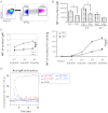
Adoptive transfer of TCL1-192 cells in SCID mice. (A) 20 × 106 nonselected splenic lymphocytes from TCL1-192 mouse were intravenously injected into two SCID mice. The recipient mice appeared moribund and were killed at 6 mo after cell engraftment; splenic lymphocytes were collected and injected into another three SCID mice (5 × 10⁶ per mouse). At the time second transferred mice appeared moribund (2 mo posttransfer), the adoptive transfer process was repeated two more times; each time recipient mice survived for only 5–6 wk. Examples of PtC binders in donor and serial adoptive transferred mice were shown. Scatter graphs indicate mean ± SD. Statistical analysis was done by one-way ANOVA and unpaired t test using GraphPad Prism software. (B) Clones identified in donor TCL1-192 splenocytes and recipient splenic and peritoneal lymphocytes after each transfer. Results shown are sequences of IGHV-D-J and IGLVκ-Jκ cDNAs from unselected splenic lymphocytes in the donor TCL1-192 mouse; and PtC+, PtC populations sorted from splenic and peritoneal lymphocytes in the transferred mice. *P < 0.05, **P < 0.01, ***P < 0.001.
Fig. 2.
Fig. 2.
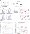
Sorted PtC+ and PtC fractions differ in smIgM expression and responses to BCR stimulation. (A) Example of gating strategy used for sorting of PtC+ (purple) and PtC (blue) fractions from B-1 or CLL cells. (B) Average smIgM expression in unselected, selected PtC+ and PtC cells collected from peritoneal cavity (PC) and spleen (SP) in fourth-transferred mice (n = 3). Data presented as mean ± SD. Statistical analysis was done by student t test using GraphPad Prism software, *P < 0.05. (C) Intensity of smIgM (Left) and PtC surface binding (Right) in serial transfer mice. Data shown are average of three mice (mean ± SD). Two-way ANOVA repeated measurement by GraphPad Prism was used to compare mean fluorescence intensity (MFI) changes between SP and PC cells during serial transfers, ***P < 0.001. (D) Ca2+ mobilization in response to anti-mouse IgM (µ-chain–specific) in splenic PtC+ (purple), PtC (blue) cells, and peritoneal PtC+ (red), PtC (light blue) cells. Data shown are representative of triplicate experiments. Results are expressed as the ratio of FL1/FL3 against time.
Fig. 3.
Fig. 3.
More aggressive CLL in mice adoptively transferred with enriched PtC+ cells. (A) In vivo analysis of proliferating cells in PtC+ and PtC fractions in fourth transferred mice (n = 5). CLL cells were collected from spleen (SP) and peritoneal cavity (PC) and then sorted for PtC+ and PtC fractions to analyze the percentage of BrdU incorporation. (B) 1 × 10⁶ PtC+ and PtC populations sorted from fourth transferred mice were injected into six mice, and mice were killed 5 wk later. Cells from peritoneal washout, spleen and blood were harvested and analyzed for the total number of CLL cells (shown in C). Bar graphs indicate mean values and SD. Statistical analysis was performed with unpaired t test using GraphPad Prism software. **P < 0.01, ***P < 0.001.
Fig. 4.
Fig. 4.
Peritoneal and splenic TCL1-192 CLL cells differ in smIgM density and response to smIgM cross-linking. (A) Intensity of smIgM in TCL1-192 cells collected from spleen (SP, black) and peritoneal cavity (PC, red). Mean values and SD are indicated with filled circles and error bars. Statistical analysis was done by unpaired t test using GraphPad Prism software. (B) Constitutively phosphorylated PLCγ2 and SYK were investigated in splenic and peritoneal TCL1-192 cells by exposing cells to pervanadate (in H2O2) for 1 or 2 min. Untreated cells or cells incubated in H2O2 alone for 2 min were used as control. Fixed and permeablized cells were then stained and analyzed by flow cytometry. (C) Ca2+ mobilization in CLL cells stimulated with 1:80,000 (Upper) or 1:40,000 (Lower) diluted PtC-bearing liposomes. Results are expressed as the ratio of FL1/FL3 against time. (D, Upper) Ca2+ mobilization in response to anti-mouse IgM (µ-chain–specific) was assayed in CLL SP, BM, and PC cells. (Lower) TCL1-192 CLL cells collected from spleen were precultured in control medium (green), medium with PtC liposome (blue), or the supernatant of peritoneal lavage (PL) harvested from TCL1-192 mice (brown). Twenty-four hours later cells were labeled with Indo-1 for Ca2+ mobilization in response to anti-mouse IgM (µ-chain–specific) stimulation. The data shown in B–D represent examples of triplicate experiments.
Fig. 5.
Fig. 5.
Networks of BCR-regulated genes in PtC+ and PtC cells from spleens and peritoneal cavity. Ingenuity pathways analysis (IPA) software was used to analyze identified genes involved in the BCR signaling pathway that were differently expressed in peritoneal PtC+ and PtC cells (Left, n = 451, >1.5×), and PtC+ cells isolated from spleen and peritoneum (Right, n = 272, >2×). Genes labeled in red and green were those identified as up- and down-regulated and other genes were those related to the regulated genes on the basis of the network analysis.

Similar articles

Cited by

References

    1. Chiorazzi N, Ferrarini M. B cell chronic lymphocytic leukemia: Lessons learned from studies of the B cell antigen receptor. Annu Rev Immunol. 2003;21:841–894. - PubMed
    1. Chiorazzi N, Rai KR, Ferrarini M. Chronic lymphocytic leukemia. N Engl J Med. 2005;352(8):804–815. - PubMed
    1. Stevenson FK, Caligaris-Cappio F. Chronic lymphocytic leukemia: Revelations from the B-cell receptor. Blood. 2004;103(12):4389–4395. - PubMed
    1. Messmer BT, et al. Multiple distinct sets of stereotyped antigen receptors indicate a role for antigen in promoting chronic lymphocytic leukemia. J Exp Med. 2004;200(4):519–525. - PMC - PubMed
    1. Tobin G, et al. Subsets with restricted immunoglobulin gene rearrangement features indicate a role for antigen selection in the development of chronic lymphocytic leukemia. Blood. 2004;104(9):2879–2885. - PubMed

Publication types

Associated data