. 2013 Jun;26(6):611-6.
doi: 10.1094/MPMI-02-13-0051-IA.

Microbe-independent entry of oomycete RxLR effectors and fungal RxLR-like effectors into plant and animal cells is specific and reproducible

Affiliations

Microbe-independent entry of oomycete RxLR effectors and fungal RxLR-like effectors into plant and animal cells is specific and reproducible

Brett M Tyler et al. Mol Plant Microbe Interact. 2013 Jun.

Abstract

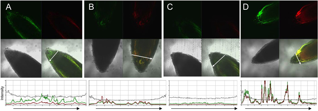
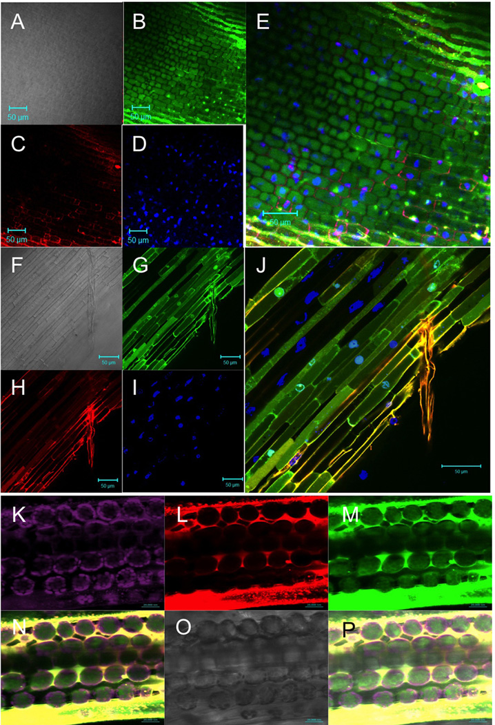
A wide diversity of pathogens and mutualists of plant and animal hosts, including oomycetes and fungi, produce effector proteins that enter the cytoplasm of host cells. A major question has been whether or not entry by these effectors can occur independently of the microbe or requires machinery provided by the microbe. Numerous publications have documented that oomycete RxLR effectors and fungal RxLR-like effectors can enter plant and animal cells independent of the microbe. A recent reexamination of whether the RxLR domain of oomycete RxLR effectors is sufficient for microbe-independent entry into host cells concluded that the RxLR domains of Phytophthora infestans Avr3a and of P. sojae Avr1b alone are NOT sufficient to enable microbe-independent entry of proteins into host and nonhost plant and animal cells. Here, we present new, more detailed data that unambiguously demonstrate that the RxLR domain of Avr1b does show efficient and specific entry into soybean root cells and also into wheat leaf cells, at levels well above background nonspecific entry. We also summarize host cell entry experiments with a wide diversity of oomycete and fungal effectors with RxLR or RxLR-like motifs that have been independently carried out by the seven different labs that coauthored this letter. Finally we discuss possible technical reasons why specific cell entry may have been not detected by Wawra et al. (2013).


Figures

Fig. 1
Fig. 1
Specific microbe-independent entry of Avr1bNt-GFP fusions into soybean root cells. A, Avr1b N-terminus (residues 1–50 of the mature protein) fused to GFP mixed with mCherry protein. B, Avr1b N-terminus (residues 1–50) with RSLR->AAAA and RFLR->AAAA mutations, fused to GFP mixed with mCherry protein. C and D, GFP protein (no fusions) mixed with mCherry protein. Purified fusion proteins (0.5 mg/ml each) were incubated with soybean (Williams) root tips, for 12 h in the dark at 23°C in phosphate-buffered saline (10 mM Na phosphate, 138 mM NaCl, 2.7 mM KCl, adjusted to pH 6.8 with HCl; Sigma P5368), then washed four times for 1 h each in PBS pH 6.8 at 23°C. Proteins all carried C-terminal his6 tags and were purified as described (Kale et al., 2010). The roots were imaged with a Zeiss LSM 510 confocal microscope, with an excitation wavelength of 488 nm and emission window of 505–530 nm for GFP or an excitation wavelength of 543 nm and emission window of >560 nm for mCherry. Master gain was set to 500 for A-C and to 650 for D. In each figure section, the upper left panel shows the GFP image, the upper right the mCherry image, the middle left, the light image, the middle right the overlay of GFP, mCherry and light images, AND the bottom panel shows an intensity scan of the GFP (green), mCherry (red) and light (gray) channels along the transect shown by the white arrow in the overlay image. The intensity scale for the scans are all on the same scale. The experiments shown in this figure were conducted at Oregon State University.
Fig. 2
Fig. 2
Specific microbe-independent entry of Avr1bNt proteins into soybean root cells and wheat leaf cells. A to E, Soybean root entry by Avr1b N-terminus (residues 1–50) labeled by Dylight488, and counter stained with propidium iodide and 4',6-diamidino-2-phenylindole (DAPI). F to J, Soybean root entry by Avr1b N-terminus (residues 1–50) labeled by Dylight488, mixed with Avr1b N-terminus with RFLR->qFLR mutation labeled with DyLight550, and counter stained with DAPI. A,F, light image; B,G, Dylight488 image; C, propidium image; H, Dylight550 image; D and I, DAPI image; E and J, overlay of the three fluorescent images. In A to J, purified fusion proteins (0.4 mg/ml each) were incubated with soybean (Williams) root tips, for 2 hours at 25°C in PBS buffer adjusted to pH 7.2, then washed for 15 min in formalin (PBS + 10% formaldehyde) containing 0.2 µg/ml propidium iodide (A to E only) and 0.4 µg/ml DAPI. K to P, Wheat leaf cell entry by Avr1b N-terminus (residues 1–50) labeled by Dylight488 mixed with Avr1b N-terminus (residues 1–50) with RFLR->qFLR mutation labeled by Dylight550. Purified fusion proteins (0.4 mg/ml each) in PBS buffer adjusted to pH 7.2 were infiltrated with a blunt syringe into ~9 day old wheat seedling leaves. Leaves were imaged after 6 h without washing. K, chloroplast fluorescence (excitation 488 nm; emission meta filter 675–715 nm); L, Dylight550 image (excitation 543 nm; emission window 585–615 nm; gain 600 to 654; digital offset −0.1 to 0); M, Dylight488 image (excitation 488 nm; emission window 505–530 nm; gain 580 to 610; digital offset −0.1 to 0); N, overlay of K to M; O, light image; overlay of N and O. Labeling of proteins with Dylight dyes was as described (Sun et al., 2013). The experiments shown in this figure were conducted at Virginia Tech using a Zeiss LSM 510 Meta confocal microscope.

References

    1. Anderson RG, Casady MS, Fee RA, Vaughan MM, Deb D, Fedkenheuer K, Huffaker A, Schmelz EA, Tyler BM, McDowell JM. Homologous RXLR effectors from Hyaloperonospora arabidopsidis and Phytophthora sojae suppress immunity in distantly related plants. Plant J. 2012;72:882–893. - PubMed
    1. Boothroyd JC, Dubremetz JF. Kiss and spit: the dual roles of Toxoplasma rhoptries. Nat Rev Microbiol. 2008;6:79–88. - PubMed
    1. Chang M, Chou JC, Lee HJ. Cellular internalization of fluorescent proteins via arginine-rich intracellular delivery peptide in plant cells. Plant & cell physiology. 2005;46:482–488. - PubMed
    1. Dou D, Kale SD, Wang X, Jiang RHY, Bruce NA, Arredondo FD, Zhang X, Tyler BM. RXLR-mediated entry of Phytophthora sojae effector Avr1b into soybean cells does not require pathogen-encoded machinery. Plant Cell. 2008;20:1930–1947. - PMC - PubMed
    1. Ellis J, Catanzariti AM, Dodds P. The problem of how fungal and oomycete avirulence proteins enter plant cells. Trends Plant Sci. 2006;11:61–63. - PubMed

Publication types

MeSH terms

LinkOut - more resources