. 2013 May;37(5):685-98.
doi: 10.1097/PAS.0b013e31827f9c5e.

Epithelioid GBMs show a high percentage of BRAF V600E mutation

Affiliations

Epithelioid GBMs show a high percentage of BRAF V600E mutation

Bette Kay Kleinschmidt-DeMasters et al. Am J Surg Pathol. 2013 May.

Abstract

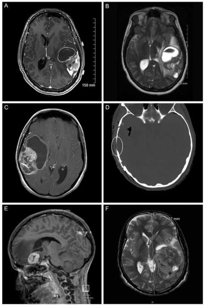
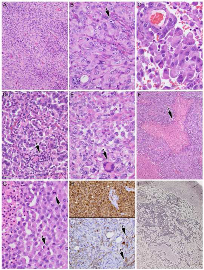
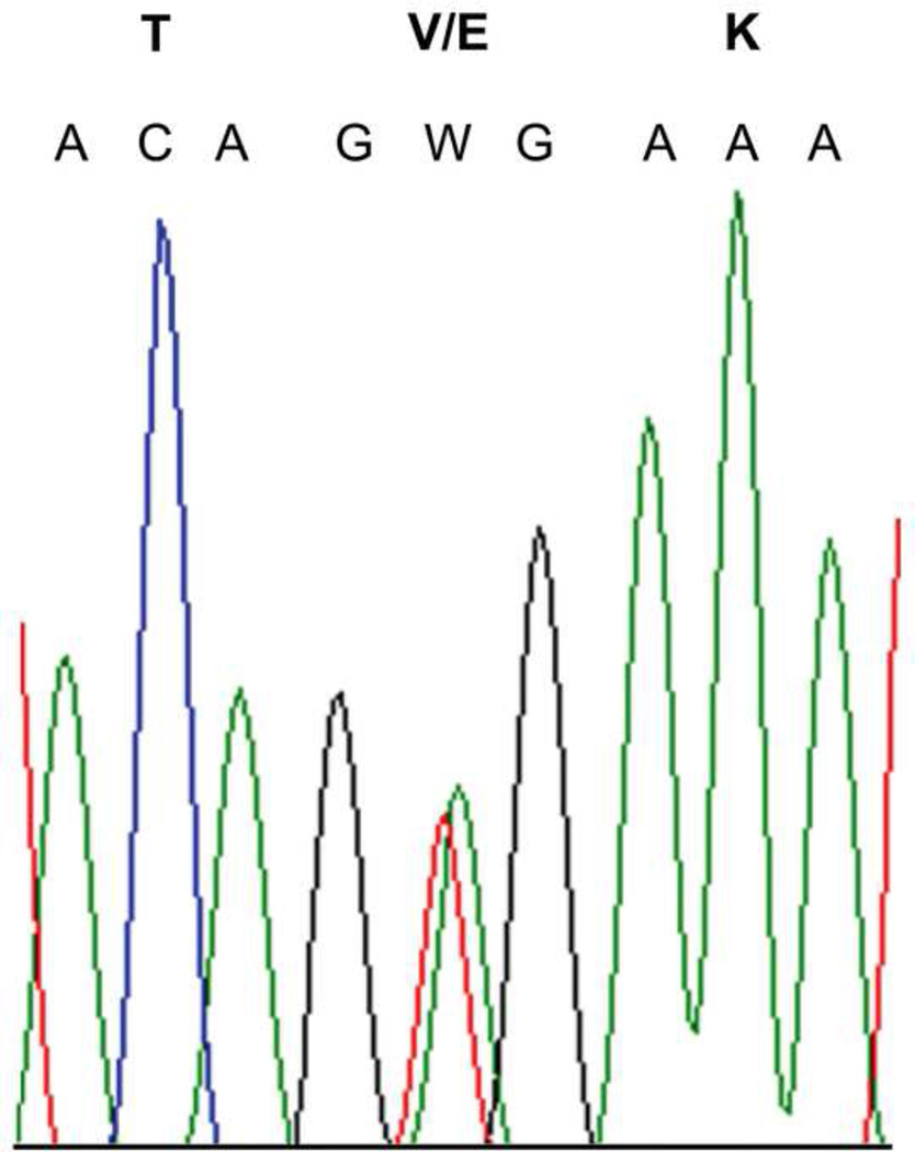
BRAF V600E mutation has been identified in up to 2/3 of pleomorphic xanthoastrocytomas (PXAs), World Health Organization grade II, as well as in varying percentages of PXAs with anaplastic features (PXA-A), gangliogliomas, extracerebellar pilocytic astrocytomas, and, rarely, giant cell glioblastoma multiforme (GC-GBMs). GC-GBMs and epithelioid GBMs (E-GBMs) can be histologically challenging to distinguish from PXA-A. We undertook this study specifically to address whether these 2 tumor types also showed the mutation. We tested our originally reported cohort of 8 E-GBMs and 2 rhabdoid GBMs (R-GBM) as well as 5 new E-GBMs (1 pediatric, 4 adult) and 9 GC-GBMs (2 pediatric, 7 adult) (n=24) for BRAF V600E mutational status. Twenty-one of 24 had sufficient material for IDH-1 immunostaining, which is usually absent in PXAs, PXA-As, and primary GBMs but present in secondary GBMs. Patients ranged in age from 4 to 67 years. BRAF V600E mutation was identified in 7/13 of E-GBMs, including 3 of our original cases; patients with mutation were aged 10 to 50 years. None of the 9 GC-GBMs or 2 R-GBMs manifested this mutation, including pediatric patients. The sole secondary E-GBM was the single case manifesting positive IDH-1 immunoreactivity. A high percentage of E-GBMs manifest BRAF V600E mutation, paralleling PXAs. All R-GBMs and GC-GBMs were negative, although larger multi-institutional cohorts will have to be tested to extend this result. BRAF V600E mutational analyses should be performed on E-GBMs, particularly in all pediatric and young-aged adults, given the potential for BRAF inhibitor therapy in this subset of GBM patients.


Conflict of interest statement

DISCLOSURE/CONFLICT OF INTEREST

No conflicts of interest to report.

Figures

Figure 1
Figure 1
A. Magnetic resonance imaging (MRI) scan, axial, T1-weighted with gadolinium enhancement shows an E-GBM with complex cystic and solid enhancing areas; none of the 5 new E-GBMs cases in this study showed a simple mural nodule-cyst configuration. Case 14 illustrated. B. MRI, axial, T2-weighted scan, better highlights the bright signal in the cystic portions of this same E-GBM, as well as the surrounding edema and midline generated by this high grade tumor. Case 14 illustrated. C. MRI scan, axial, T1-weighted with gadolinium, from another E-GBM shows an even larger cystic component in this example. Case 12 illustrated. D. Computerized tomographic scan, axial, shows the nearby bony thinning (encircled) in this same patient, suggesting a more longstanding tumor had been present. Despite this radiographic feature, low grade tumor areas were not identified in the resection specimen. Case 12 illustrated. E. MRI scan, sagittal, T1-weighted with gadolinium, shows a small cystic component and more solid, relatively well-demarcated enhancing component in this pediatric patient with E-GBM; note the significant surrounding edema (dark intensity signal). Case 13 illustrated. F. MRI scan, axial, T2-weighted, highlights the massive size of some of these E-GBMs, as measured on the scan, as well as the extent of midline shift that can be produced. Case 11 illustrated.
Figure 2
Figure 2
A. E-GBMs were characterized by relatively monotonous sheets of small- to moderate-sized epithelioid cells with eosinophilic cytoplasm with rounded cell borders and a paucity of stellate cytoplasmic processes. Case 14 illustrated, hematoxylin and eosin (H&E), 200X. B. At higher power magnification, prominent nucleoli (arrowhead) and abundant eosinophilic cytoplasm could be seen, mimicking a rhabdoid phenotype. Case 14 illustrated, H&E, 400X. C. Discohesive tumor cells could lead to areas almost identical to metastatic carcinoma or melanoma. Case 12 illustrated, H&E, 600X. D. Variable numbers of accompanying non-neoplastic lymphocytes (arrow) add to the diagnostic overlap, at least focally, with metastatic carcinoma or melanoma. Case 15 illustrated, H&E, 400X. E. E-GBMs in most cases showed almost complete cellular monotony (cases 11, 12, 15), although slightly more variation in cellular size and scattered larger, sometimes multinucleated (arrowhead) cells could be identified. Case 13 illustrated, H&E, 400X. F. Necrosis (arrow) was identified in 4 of 5 new E-GBMs. Case 11 illustrated, H&E 100X. G. Multiple mitotic figures (arrows) were found in all examples; note necrosis at higher magnification from this same patient at left. Case 11 illustrated, H&E 600X. H. Strong diffuse S100 immunoreactivity (top) with more variable and patchy GFAP immunostaining (bottom) is characteristic of E-GBMs. Note spindled morphology of some cells on GFAP (arrows). Case 14 illustrated, both 200X, immunohistochemistry with light hematoxylin counterstain for S100 protein and glial fibrillary acidic protein. I. Foci with increased reticulin fiber deposition were often found in E-GBMs. Case 15 illustrated, Gomori’s reticulum stain, 40X.
Figure 3
Figure 3
Electropherogram demonstrating a representative example of c.1799T>A (p.V600E) mutation identified in 4 of 4 new E-GBM cases, with nucleotide and amino acid designations indicated. Case 11 illustrated.

References

    1. Dougherty MJ, Santi M, Brose MS, et al. Activating mutations in BRAF characterize a spectrum of pediatric low-grade gliomas. Neuro Oncol. 2010;12:621–630. - PMC - PubMed
    1. Dias-Santagata D, Lam Q, Vernovsky K, et al. BRAF V600E mutations are common in pleomorphic xanthoastrocytoma: diagnostic and therapeutic implications. PLoS One. 2011;6:e17948. - PMC - PubMed
    1. Schindler G, Capper D, Meyer J, et al. Analysis of BRAF V600E mutation in 1,320 nervous system tumors reveals high mutation frequencies in pleomorphic xanthoastrocytoma, ganglioglioma and extra-cerebellar pilocytic astrocytoma. Acta Neuropathol. 2011;121:397–405. - PubMed
    1. Balss J, Meyer J, Mueller W, et al. Analysis of the IDH1 codon 132 mutation in brain tumors. Acta Neuropathol. 2008;116:597–602. - PubMed
    1. Ichimura k, Pearson DM, Kocialkowski S, et al. IDH1 mutations are present in the majority of common adult gliomas but rare in primary glioblastomas. Neuro Oncol. 2009;11(4):341–347. - PMC - PubMed

MeSH terms

Substances