. 2013 Jun;87(12):6644-59.
doi: 10.1128/JVI.03311-12. Epub 2013 Apr 3.

Vesicular stomatitis virus variants selectively infect and kill human melanomas but not normal melanocytes

Affiliations

Vesicular stomatitis virus variants selectively infect and kill human melanomas but not normal melanocytes

Guido Wollmann et al. J Virol. 2013 Jun.

Abstract

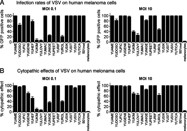
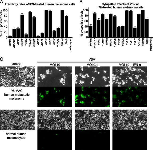
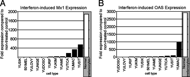
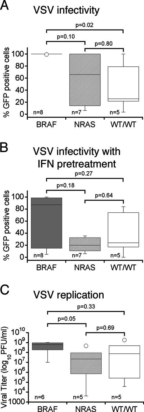
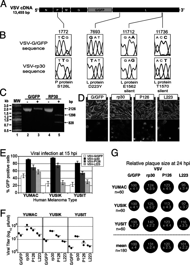
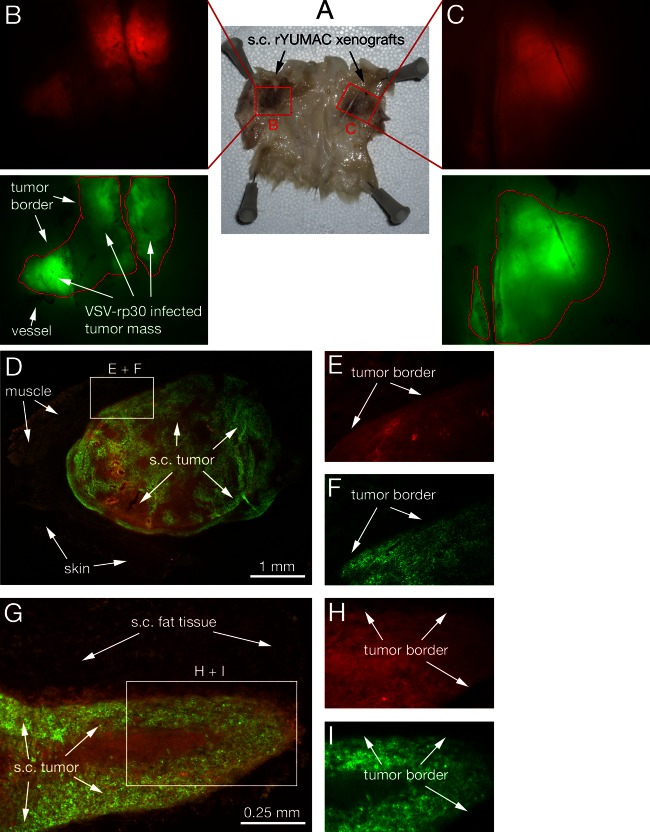
Metastatic malignant melanoma remains one of the most therapeutically challenging forms of cancer. Here we test replication-competent vesicular stomatitis viruses (VSV) on 19 primary human melanoma samples and compare these infections with those of normal human melanocyte control cells. Even at a low viral concentration, we found a strong susceptibility to viral oncolysis in over 70% of melanomas. In contrast, melanocytes displayed strong resistance to virus infection and showed complete protection by interferon. Several recombinant VSVs were compared, and all infected and killed most melanomas with differences in the time course with increasing rates of melanoma infection, as follows: VSV-CT9-M51 < VSV-M51 < VSV-G/GFP < VSV-rp30. VSV-rp30 sequencing revealed 2 nonsynonymous mutations at codon positions P126 and L223, both of which appear to be required for the enhanced phenotype. VSV-rp30 showed effective targeting and infection of multiple subcutaneous and intracranial melanoma xenografts in SCID mice after tail vein virus application. Sequence analysis of mutations in the melanomas used revealed that BRAF but not NRAS gene mutation status was predictive for enhanced susceptibility to infection. In mouse melanoma models with specific induced gene mutations including mutations of the Braf, Pten, and Cdkn2a genes, viral infection correlated with the extent of malignant transformation. Similar to human melanocytes, mouse melanocytes resisted VSV-rp30 infection. This study confirms the general susceptibility of the majority of human melanoma types for VSV-mediated oncolysis.


Figures

Fig 1
Fig 1
VSV-rp30 infection of 17 short-term human melanoma cultures. Cultures of human melanoma from primary and metastatic sites were infected with VSV-rp30 at a low (0.1) or high (10) MOI with VSV-rp30 and analyzed 36 h later for viral infection (A) displayed as percentage of GFP-expressing cells and for rates of cytopathic effects (B). Viral infection was correlated with cytopathic effects.
Fig 2
Fig 2
Oncolytic action of VSV on human melanoma. (A) Representative collection of 5 human melanoma cultures infected with VSV-rp30 at an MOI of 0.1 for 36 h. EthD-1 (red) labels the nuclei of dead or dying cells. Fluorescence imaging reveals a strong correlation between the number of infected cells (green) and dead cells (red) and the appearance of cytopathic effects (phase-contrast images). (B) Bar graph showing quantification of cytotoxicity of 7 human melanoma cultures infected with VSV-rp30 at an MOI of 0.1 using EthD-1 cell death staining.
Fig 3
Fig 3
IFN pretreatment retards VSV-rp30 infection and cytolytic rates on some human melanomas. (A) The presence of IFN reduced infection in about one half of melanoma samples tested at 36 hpi. (B) The other half of the melanoma samples displayed cytopathic effects after VSV-rp30 infection even in the presence of IFN. (C) Panel of representative images of YUMAC melanoma cells and normal human melanocytes (control cells) taken 24 h after VSV-rp30 inoculation at high (10) or low (0.1) MOI and in the presence of IFN-α. Phase-contrast images show normal cell morphology or cytopathic effects. GFP expression indicates viral infection.
Fig 4
Fig 4
Reduced induction of interferon-stimulated genes in melanoma compared to normal melanocytes. Quantitative RT-PCR was applied for gene expression studies on control and IFN-αA/D-treated cultures of 11 human melanoma samples and normal human melanocytes. Two specific interferon-stimulated genes (ISGs), Mx1 (A) and OAS (B), were analyzed. β-Actin was used as a reference for each cell type to normalize expression levels. Data reflect the fold induction relative to nontreated control cultures. Normal melanocytes showed induction levels of several thousandfold for both ISGs. In contrast, melanoma tumor cells showed significantly reduced induction of ISGs.
Fig 5
Fig 5
Replication and oncolytic effects of different VSVs in melanoma cells. Viral replication was quantified at 7 (A) and 24 (B) hours following virus inoculation at an MOI of 0.1. For most melanoma cells, infection of VSV-rp30 (black diamonds) resulted in higher titers than VSV-G/GFP (white diamonds), with a particularly significant difference at the early time point. Viral replication in normal melanocytes yielded significantly smaller amounts of VSV progeny. (C) Comparison of low MOI (0.1) infection of different VSV (VSV-G/GFP, VSV-rp30, VSV-M51, and VSV-CT9-M51) on 5 different human melanoma cultures at 24 hpi. VSVrp30 showed the strongest cytopathic effect on melanoma cells. Displayed is the percentage of cytopathic effects compared to noninfected controls. This was based on analysis of duplicate cultures and 10 microscopic fields per cell type and condition. Horizontal red bars indicate the mean values of 5 melanoma cultures combined.
Fig 6
Fig 6
Relationship between BRAF and NRAS mutation status and VSV susceptibility. Box plots of human melanoma cells are grouped according to their melanoma-associated BRAF (dark gray panels) and NRAS (light gray panels) mutation status. WT/WT (white panels) indicates wild-type gene status for both BRAF and NRAS. Parameters assessed for correlation were (i) infectivity at low MOI (0.1) (A), (ii) infectivity after IFN pretreatment (B), and (iii) viral replication (C). All melanoma cells with BRAF mutation show a strong susceptibility for VSV infection, whereas NRAS-mutated cells and wild-type cells vary in their susceptibility. No association was found for RAS mutation and VSV susceptibility. Horizontal bars within a box indicate group means; statistically significant difference is defined at P values of <0.05 using the Mann-Whitney test.
Fig 7
Fig 7
VSV-rp30 sequencing and mutation identification. Sequencing of the entire genome for both VSV-G/GFP and VSV-rp30 was performed using genomic-length cDNAs. (A) Diagram illustrating the parent VSV-G/GFP genome and the positions of the four mutations (C1772T, G7693T, A11712G, and C11736T) identified in the glioblastoma cell-adapted variant VSV-rp30. (B) Sections of the sequencing chromatographs showing both the parent VSV-G/GFP (above) and the mutated VSV-rp30 codons (below). Altered nucleotides are shown in bold type. The affected protein and corresponding amino acid positions are shown below each pair of chromatographs. Nucleotide numbering is based on the VSV-G/GFP sequence (GenBank accession number FJ478454). (C) BsrDI restriction digestion of a 2.1-kb PCR product encompassing the C1772T mutation site identified in the P gene of VSV-rp30. VSV-rp30 PCR product incubated in the presence (+) of BsrDI resulting in the complete digestion of the product into the predicted 1.3-kb and 0.8-kb fragments (lane 5), indicating that all of the VSV-rp30 product contained the C1772T mutation recognized by BsrDI. (D) Representative panel of micrographs showing phase-contrast and corresponding GFP fluorescence of YUSIK melanoma tissue infected with VSV-G/GFP, VSV-rp30, VSV-P126, and VSV-L223 at an MOI of 0.1 at 15 hpi. (E) Graph depicting infection rates using GFP expression analysis for each VSV variant on three different human melanomas (mean ± SEM; n = 10 microscopic fields per condition). (F) Comparison of replication of VSV variants on 3 different human melanoma samples. Horizontal lines show means of duplicate titer determinations. (G) Comparison of plaque sizes. Black circles indicate mean (± SEM) relative plaque sizes compared to control VSV-G/GFP plaque diameters (n = 60 per condition). The “mean” row shows the combined mean relative plaque sizes from all three melanoma cells tested.
Fig 8
Fig 8
VSV-rp30 infection and cytopathic rates on mouse melanoma with defined mutation pattern. (A) Oncogenically transformed mouse melanocytes expressing combinations of Braf, Pten, Cdkn2a, or beta catenin and normal mouse melanocytes were infected by VSV-rp30 at an MOI of 0.1. Infection rates and cytopathic effect rates were quantified at 24 h postinoculation. Cells with more mutations showed a trend for greater susceptibility to VSV-rp30 infection than cells with fewer mutations, with normal mouse melanocytes showing very limited signs of infection. Gray shading in boxes indicates cells with three mutations. (B) Representative micrographs showing phase-contrast and GFP fluorescence images of infected cultures from cell type 2901, with three mutations, compared to cell type 1111, with two mutations, and normal mouse melanocytes.
Fig 9
Fig 9
Intravenous VSV-rp30 targets multiple subcutaneous melanoma xenografts. SCID mice bearing multiple subcutaneous rYUMAC melanoma xenografts were given a single tail vein injection of VSV-rp30 (1 × 10⁸ PFU/100 μl). (A) Skin of the back and flank section with bilateral xenografts, with the subcutaneous (s.c.) side facing up. Human YUMAC melanoma cells modified to express red fluorescent protein (RFP) allow tracing of tumor masses surrounded by normal tissue, as illustrated by stereomicroscope images (B and C). Green filter images confirm VSV-rp30 infection restricted to red tumor masses at 5 dpi. Microsections of individual tumors reveal details of close adherence of viral intratumoral spread to the tumor border, as seen in green-red merged low-magnification images (D and G) and high-magnification details (E, F, H, I).
Fig 10
Fig 10
Targeting of intracranial human melanoma xenografts by intravenous VSV-rp30. (A to E) Melanin pigment-filled human melanoma YUSIK cells were injected stereotactically in the striatum of SCID mice. Border of tumor mass (A) and isolated cell clusters surrounded by brain parenchyma (C) were easily traceable in bright-field observation of brain microsections. A single injection of 108 PFU VSV-rp30 led to complete infection of large tumor masses (B) as well as isolated tumor cells (D) within 2 days, with limited infection of surrounding brain tissue; the cellularity of normal brain tissue is indicated by DAPI nuclear stain (E). Alternatively, the amelanotic human melanoma YUMAC tissue was stably transfected to express red fluorescent protein for easy tracing of tumor cells injected into the brain of SCID mice (F to J). Short-term brain melanoma xenografts (7 days) were established through stereotactic injection into the striatum of SCID mice (F, H). Tail vein injection of VSV-rp30 led to complete infection of the red tumor cells within 2 days (G, H). DAPI stain illustrating the cellularity of the brain parenchyma. Red YUMAC tumor-bearing mice not receiving VSV-rp30 via tail vein were monitored for an additional 3 weeks, and brains were harvested and analyzed for progressed tumor growth (K). Detail sections (L) display high cellularity via DAPI stain (M) and negative signal for GFP fluorescence in the absence of virus (N).

Similar articles

Cited by

References

    1. Mocellin S, Pasquali S, Rossi CR, Nitti D. 2010. Interferon alpha adjuvant therapy in patients with high-risk melanoma: a systematic review and meta-analysis. J. Natl. Cancer Inst. 102:493–501 - PubMed
    1. Lee B, Mukhi N, Liu D. 2012. Current management and novel agents for malignant melanoma. J. Hematol. Oncol. 5:3 doi:10.1186/1756-8722-5-3 - DOI - PMC - PubMed
    1. Carlino MS, Fogarty GB, Long GV. 2012. Treatment of melanoma brain metastases: a new paradigm. Cancer J. 18:208–212 - PubMed
    1. Sloan AE, Nock CJ, Einstein DB. 2009. Diagnosis and treatment of melanoma brain metastasis: a literature review. Cancer Control 16:248–255 - PubMed
    1. Trinh VA, Hwu WJ. 2012. Ipilimumab in the treatment of melanoma. Expert Opin. Biol. Ther. 12:773–782 - PubMed

Publication types

Substances