. 2013 Jun;87(12):6604-14.
doi: 10.1128/JVI.00009-13. Epub 2013 Apr 3.

Tropism of and innate immune responses to the novel human betacoronavirus lineage C virus in human ex vivo respiratory organ cultures

Affiliations

Tropism of and innate immune responses to the novel human betacoronavirus lineage C virus in human ex vivo respiratory organ cultures

Renee W Y Chan et al. J Virol. 2013 Jun.

Erratum in

Abstract

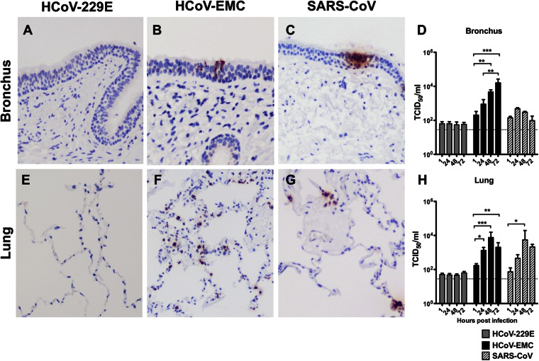
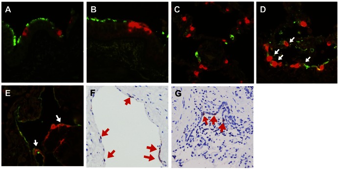
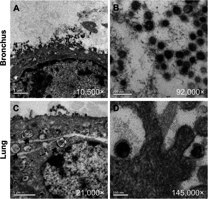
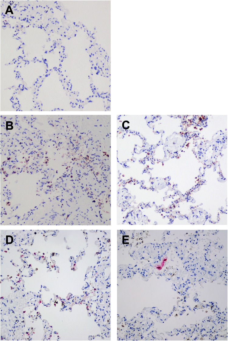
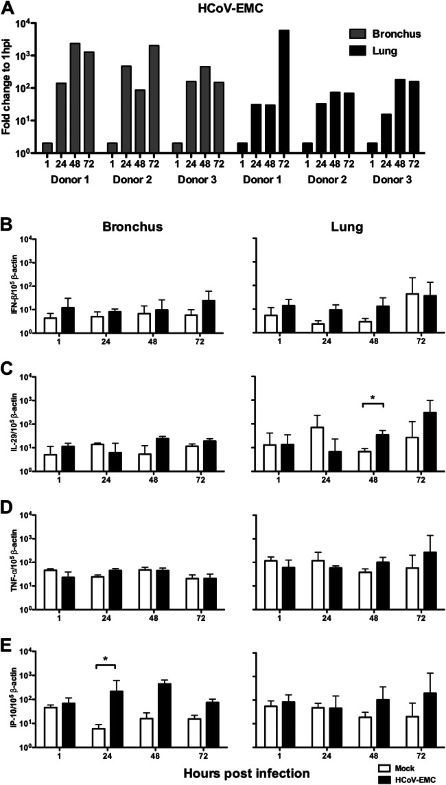
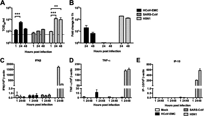
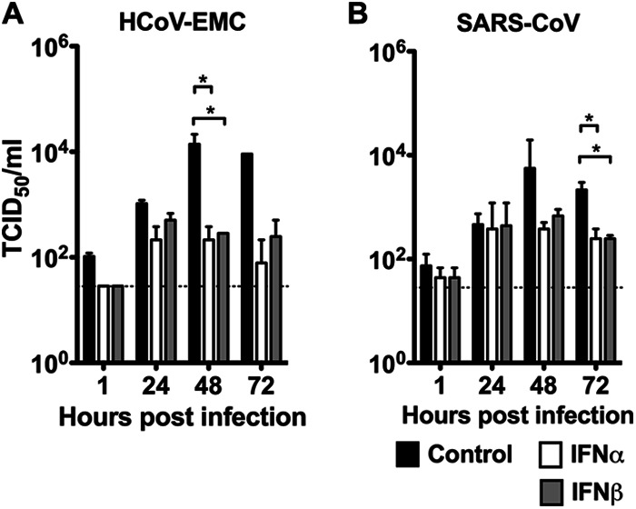
Since April 2012, there have been 17 laboratory-confirmed human cases of respiratory disease associated with newly recognized human betacoronavirus lineage C virus EMC (HCoV-EMC), and 7 of them were fatal. The transmissibility and pathogenesis of HCoV-EMC remain poorly understood, and elucidating its cellular tropism in human respiratory tissues will provide mechanistic insights into the key cellular targets for virus propagation and spread. We utilized ex vivo cultures of human bronchial and lung tissue specimens to investigate the tissue tropism and virus replication kinetics following experimental infection with HCoV-EMC compared with those following infection with human coronavirus 229E (HCoV-229E) and severe acute respiratory syndrome coronavirus (SARS-CoV). The innate immune responses elicited by HCoV-EMC were also investigated. HCoV-EMC productively replicated in human bronchial and lung ex vivo organ cultures. While SARS-CoV productively replicated in lung tissue, replication in human bronchial tissue was limited. Immunohistochemistry revealed that HCoV-EMC infected nonciliated bronchial epithelium, bronchiolar epithelial cells, alveolar epithelial cells, and endothelial cells. Transmission electron microscopy showed virions within the cytoplasm of bronchial epithelial cells and budding virions from alveolar epithelial cells (type II). In contrast, there was minimal HCoV-229E infection in these tissues. HCoV-EMC failed to elicit strong type I or III interferon (IFN) or proinflammatory innate immune responses in ex vivo respiratory tissue cultures. Treatment of human lung tissue ex vivo organ cultures with type I IFNs (alpha and beta IFNs) at 1 h postinfection reduced the replication of HCoV-EMC, suggesting a potential therapeutic use of IFNs for treatment of human infection.


Figures

Fig 1
Fig 1
Tissue tropism of HCoV-EMC in human bronchus and lung. Bronchial (A to C) and lung (E to G) tissues were infected with HCoV-229E, HCoV-EMC, and SARS-CoV. At 24 hpi, the tissues were fixed and tissue sections were stained for human coronavirus N protein (reddish brown), as described in Materials and Methods. Viral replication kinetics in ex vivo cultures of bronchial (D) and lung (H) biopsy tissue specimens infected with 106 TCID50s/ml of coronaviruses were determined by virus titration at 37°C. The bar charts show the mean and the standard error of the mean of the virus titer pooled from at least three independent experiments. Asterisks indicate a statistically significant increase in viral yield compared to that at 1 hpi: *, P < 0.05; **, P < 0.005; ***, P < 0.0005.
Fig 2
Fig 2
Cellular localization of HCoV-EMC in lung tissue. HCoV-EMC was stained with Vector Red (red) and cell markers conjugated with FITC (green) for detection of β-tubulin (ciliated cell marker) (A) and MUC5AC (goblet cell marker) (B) in bronchial tissue and CD68 (macrophage marker) (C), AE1/AE3 (epithelial cell marker) (D), and podoplanin (type I pneumocyte marker) (E) in lung tissue at 24 hpi. White arrows in panels D and E denote cells with costaining. (F) Cellular tropism of HCoV-EMC in lung tissue. Human coronavirus N protein (stained in reddish brown and marked with red arrows) was identified in endothelial cells at 24 hpi. (G) Bronchiolar epithelial cells at 48 hpi.
Fig 3
Fig 3
Transmission electron microscopy locating the budding site of HCoV-EMC in the human bronchus and lung. Budding of virions from bronchial epithelial cells (A, B) and alveolar epithelial cells (C, D) at 48 hpi. Black arrows, type II alveolar epithelial cells with lamellar bodies.
Fig 4
Fig 4
Apoptotic cells identified in human lung tissue ex vivo culture upon HCoV-EMC and SARS-CoV infection. (A to C) Ex vivo culture of lung tissue mock infected (A) or infected with HCoV-EMC (B) or SARS-CoV (C) at 48 hpi. The reddish brown stain identifies the presence of cleaved caspase 3. (D and E) Costaining of HCoV-EMC (D) and SARS-CoV (E) antigen (pink stain) with cleaved caspase 3 (reddish brown stain).
Fig 5
Fig 5
Human HCoV-EMC gene expression and the major cytokine and chemokine expression in bronchial and lung tissues in response to infection. (A) Expression of HCoV-EMC genes expressed as the fold change at 24, 48, and 72 hpi compared with that at 1 hpi. Gray bars, data from ex vivo bronchial tissue from three donors; black bars, data from ex vivo lung tissue from three donors. (B to E) IFN-β (B), IL-29 (C), TNF-α (D), and IP-10 (E) expression from mock-infected and infected ex vivo cultures at 1, 24, 48, and 72 hpi. The graph shows the mean and standard error of mean numbers of copies of mRNA per 105 β-actin copies from three representative experiments. *, P < 0.05.
Fig 6
Fig 6
Viral replication and cytokine and chemokine expression of HCoV-EMC in A549 cells. (A) A549 cells infected with HCoV-EMC, SARS-CoV, and influenza H5N1 virus (MOI, 1). Viral replication was determined by TCID50 assay. The bar chart shows the mean and the standard error of mean titer of virus pooled from three independent experiments. Asterisks indicate a statistically significant increase in viral yield compared to that at 1 hpi: **, P < 0.05; ***, P < 0.0005. (B) Expression of HCoV-EMC, SARS-CoV, and influenza H5N1 virus genes expressed as the fold change compared with that at 1 hpi. (C to E) IFN-β (C), TNF-α (D), and IP-10 (E) gene expression from mock-infected and HCoV-EMC-, SARS-CoV-, and influenza H5N1 virus-infected A549 cells at 1, 24, and 48 hpi. The graph shows the mean and standard error of the mean numbers of copies of mRNA per 105 β-actin copies from three representative experiments.
Fig 7
Fig 7
Interferon treatments suppressed HCoV-EMC and SARS CoV replication in human lung ex vivo culture. The human lung ex vivo cultures were infected with 106 TCID50s/ml of HCoV-EMC or SARS-CoV for 1 h at 37°C. A control culture consisting of F12K medium plus 1% PS (black bars) or culture medium with IFN-α (white bars) or IFN-β (gray bars) at a concentration of 1,000 U/ml in F12K medium plus 1% PS was used to replenish the medium at 1 hpi. Supernatants were collected from infected cultures at 1, 24, 48, and 72 hpi and titrated in Vero cells. Replication kinetics of HCoV-EMC (A) and SARS-CoV (B) were plotted to show the inhibitory effect of IFNs on virus replication posttreatment. *, a statistically significant difference (P < 0.05) at the same time point between control and IFN treatments.

References

    1. Cheung CY, Poon LL, Ng IH, Luk W, Sia SF, Wu MH, Chan KH, Yuen KY, Gordon S, Guan Y, Peiris JS. 2005. Cytokine responses in severe acute respiratory syndrome coronavirus-infected macrophages in vitro: possible relevance to pathogenesis. J. Virol. 79:7819–7826 - PMC - PubMed
    1. Law HK, Cheung CY, Ng HY, Sia SF, Chan YO, Luk W, Nicholls JM, Peiris JS, Lau YL. 2005. Chemokine up-regulation in SARS-coronavirus-infected, monocyte-derived human dendritic cells. Blood 106:2366–2374 - PMC - PubMed
    1. de Lang A, Baas T, Smits SL, Katze MG, Osterhaus AD, Haagmans BL. 2009. Unraveling the complexities of the interferon response during SARS-CoV infection. Future Virol. 4:71–78 - PMC - PubMed
    1. Totura AL, Baric RS. 2012. SARS coronavirus pathogenesis: host innate immune responses and viral antagonism of interferon. Curr. Opin. Virol. 2:264–275 - PMC - PubMed
    1. Sims AC, Baric RS, Yount B, Burkett SE, Collins PL, Pickles RJ. 2005. Severe acute respiratory syndrome coronavirus infection of human ciliated airway epithelia: role of ciliated cells in viral spread in the conducting airways of the lungs. J. Virol. 79:15511–15524 - PMC - PubMed

Publication types

MeSH terms