. 2013 Jun;24(11):1801-11, S1-4.
doi: 10.1091/mbc.E12-11-0806. Epub 2013 Apr 3.

Sumoylation of AMPKβ2 subunit enhances AMP-activated protein kinase activity

Affiliations

Sumoylation of AMPKβ2 subunit enhances AMP-activated protein kinase activity

Teresa Rubio et al. Mol Biol Cell. 2013 Jun.

Abstract

AMP-activated protein kinase (AMPK) is a sensor of cellular energy status. It is a heterotrimer composed of a catalytic α and two regulatory subunits (β and γ). AMPK activity is regulated allosterically by AMP and by the phosphorylation of residue Thr-172 within the catalytic domain of the AMPKα subunit by upstream kinases. We present evidence that the AMPKβ2 subunit may be posttranslationally modified by sumoylation. This process is carried out by the E3-small ubiquitin-like modifier (SUMO) ligase protein inhibitor of activated STAT PIASy, which modifies the AMPKβ2 subunit by the attachment of SUMO2 but not SUMO1 moieties. Of interest, AMPKβ1 is not a substrate for this modification. We also demonstrate that sumoylation of AMPKβ2 enhances the activity of the trimeric α2β2γ1 AMPK complex. In addition, our results indicate that sumoylation is antagonist and competes with the ubiquitination of the AMPKβ2 subunit. This adds a new layer of complexity to the regulation of the activity of the AMPK complex, since conditions that promote ubiquitination result in inactivation, whereas those that promote sumoylation result in the activation of the AMPK complex.


Figures
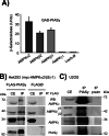
FIGURE 1:
FIGURE 1:
AMPK interacts physically with PIASy. (A) Yeast two-hybrid analysis. Yeast CTY10.5d cells transformed with plasmid pBTM116 (empty, ∅), pBTM-AMPKα2, pBTM-AMPKβ2, or pBTM-AMPKγ1 were cotransformed with plasmid pACT2-PIASy. Protein interaction was estimated by using the yeast two-hybrid system by measuring the β-galactosidase activity. Values correspond to means from four to six different transformants (bars, ±SD). (B) The three subunits of the AMPK complex coimmunoprecipitate with PIASy. Protein extracts (1.2 mg) were prepared from HEK293 cells cotransfected with plasmids pCMV-myc-AMPKα2/β2/γ1 and pFLAG-PIASy or pFLAG (empty, ∅). One microliter of anti-FLAG was used to immunoprecipitate the extracts (IP). Pelleted proteins and proteins in the input crude extracts (CE; 40 μg) were analyzed by SDS–PAGE and immunodetected (WB) with anti-AMPKα, anti-AMPKβ, anti-AMPKγ, and anti-PIASy antibodies. Molecular-size markers are indicated. (C) Human U2OS cells were treated with 6 μM MG132 for 8 h to prevent the degradation of endogenous PIASy. Then protein extracts (1.2 mg) were immunoprecipitated with 1 μl of anti-PIASy (IP PIASy) or preimmune serum (IP preim). Pelleted proteins and proteins in the input crude extracts (CE; 40 μg) were analyzed by SDS–PAGE and immunodetected as in B.
FIGURE 2:
FIGURE 2:
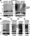
PIASy promotes the sumoylation of AMPKβ2 subunit. (A) PIASy promotes the SUMO2-dependent sumoylation of AMPKβ2 but does not affect AMPKα2. HEK293 cells were cotransfected with plasmids pCMV-6xHis-SUMO1 or pCMV-6xHis-SUMO2, pCMV-myc-AMPKα2/β2/γ1, and pFLAG-PIASy or pFLAG (empty). Cell extracts were obtained and sumoylated proteins purified by metal-affinity chromatography as described in Materials and Methods. Clarified extract (CE; 40 μg) and the material bound to the metal-affinity chromatography column (Bound) was analyzed by SDS–PAGE and Western blotting using anti-AMPKα (left) and anti-AMPKβ (right) antibodies. (B) PIASy does not promote the sumoylation of the AMPKβ1 subunit. HEK293 cells were transfected with plasmids pCMV-6xHis-SUMO1 or pCMV-6xHis-SUMO2, pCMV-myc-AMPKα2/γ1, pCMV-myc-AMPKβ1 instead of AMPKβ2, and pFLAG-PIASy or pFLAG (empty). Cell extracts were analyzed as described using anti-AMPKβ antibody. (C) Sumoylation of AMPKβ2 is dependent on PIASy activity. HEK293 cells were cotransfected with plasmids pCMV-6xHis-SUMO2, pCMV-myc-AMPKα2/β2/γ1, and pFLAG-PIASy or pFLAG-PIASy-CI (expressing a form of PIASy with reduced catalytic activity) or pFLAG (empty). Cell extracts were analyzed as described using anti-AMPKβ antibody. (D) Human U2OS cells were transfected with plasmids pCMV-6xHis-SUMO2 and pFLAG-PIASy or pFLAG (empty) and treated with 6 μM MG132 for 8 h. Cell extracts were analyzed as described using anti-AMPKβ antibody. (E) Sumoylation of free AMPKβ2 subunit is independent of PIASy expression. HEK293 cells were cotransfected with plasmids pCMV-6xHis-SUMO2, pCMV-myc-AMPKβ2, and pFLAG-PIASy or pFLAG (empty). Cell extracts were analyzed as described using anti-AMPKβ antibody. Molecular-size markers are indicated on the side; the presence of poly/multisumoylated proteins is indicated.
FIGURE 3:
FIGURE 3:
Mutation in consensus sumoylation sites of AMPKβ2 does not decrease its modification. HEK293 cells were cotransfected with plasmids pCMV-6xHis-SUMO2, pFLAG-PIASy or pFLAG (empty), pCMV-myc-AMPKα2/γ1 and pCMV-myc-AMPKβ2 or pCMV-myc-AMPKβ2 K71R, or pCMV-myc-AMPK K167R or the double mutant pCMV-myc-AMPKβ2 K71R, K167R. Cell extracts were analyzed as described in Figure 2 by using anti-AMPKβ antibody. Molecular-size markers are indicated on the left. Presence of poly/multisumoylated proteins is indicated.
FIGURE 4:
FIGURE 4:
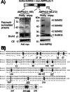
PIASy is able to promote the sumoylation of a C-terminal fragment of AMPKβ2. (A) HEK293 cells were cotransfected with pCMV-6xHis-SUMO2, pCMV-myc-AMPKα2/γ1, and pCMV-myc-AMPKβ2 (1–185), expressing the N-terminal domain, or pCMV-myc-AMPKβ2 (186–272), expressing the C-terminal domain, and pFLAG-PIASy or pFLAG (empty). Cell extracts were analyzed as described in Figure 2 by using anti-AMPKβ or anti-myc antibodies. The calculated size of SUMO-modified forms is indicated. Asterisk indicates possible degradation products. ASC, association with Snf1 complex domain; GBD, glycogen-binding domain. The presence of poly/multisumoylated proteins is indicated. (B) Alignment of human AMPKβ1 vs. AMPKβ2 protein sequences. The lysine residues in AMPKβ2 are indicated in a black box. Lysine residues mutated to arginine analyzed in this work are underlined.
FIGURE 5:
FIGURE 5:
PIASy-dependent sumoylation of AMPKβ2 K203R and K262R mutants. HEK293 cells were transfected with plasmids pCMV-6xHis-SUMO2, pFLAG-PIASy (+) or pFLAG (–), pCMV-myc-AMPKα2/γ1, and pCMV-myc-AMPKβ2 or pCMV-myc-AMPKβ2 K203R or pCMV-myc-AMPKβ2 K262R mutants. Cell extracts were analyzed as described in Figure 2 by using anti-AMPKβ antibodies. The presence of poly/multisumoylated proteins is indicated.
FIGURE 6:
FIGURE 6:
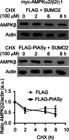
Degradation rates of AMPKβ2 in the presence of PIASy and SUMO2. HEK293 cells were cotransfected with plasmids pCMV-myc-AMPKα2/β2/γ1, pCMV-6xHis-SUMO2, and pFLAG-PIASy or pFLAG (empty). Twenty-four hours after transfection, cycloheximide (CHX; 100 μM) was added to the cultures, and cell extracts were analyzed by Western blotting at the indicated times, using anti-AMPKβ (top) and anti-actin (bottom) antibodies. The relative intensity of the AMPKβ2 bands with respect to the corresponding actin band is plotted as a function of time. Diagram shows means of three independent experiments; bars, SD.
FIGURE 7:
FIGURE 7:
PIASy-dependent sumoylation of AMPKβ2 promotes its aggregation. (A) HEK293 cells were cotransfected with plasmids expressing the AMPK subunits (pCMV-myc-AMPKα2/β2/γ1), pCMV-6xHis-SUMO2, and pFLAG-PIASy or pFLAG empty vector. The subcellular localization of AMPKβ2 subunit was carried out as described in Materials and Methods by using anti-AMPKβ as a primary and anti-rabbit Alexa Fluor 488 as a secondary antibody. The same samples were also immunodetected with anti-FLAG as a primary and anti-mouse Texas red as a secondary antibody to determine the localization of PIASy. All the samples were treated with Topro3 to stain the nucleus, and the three images were subjected to a merge analysis. (B) Similar samples were used to immunodetect AMPKβ2 subunit using anti-myc as a primary and anti-mouse Texas red as a secondary antibody and also to immunodetect SUMO-modified proteins using anti-SUMO2 as a primary and Alexa Fluor 488 as a secondary antibody. (C) Quantification of cells expressing AMPKβ2 and showing a granular pattern of intracellular inclusions. One hundred cells expressing AMPKβ2 from each of the indicated conditions were used to estimate the proportion of cells with intracellular inclusions. Values are mean ± SD; statistical significance was considered at *p < 0.05, **p < 0.01, and ***p < 0.001.
FIGURE 8:
FIGURE 8:
PIASy-dependent sumoylation of AMPKβ2 enhances the activity of the AMPK complex. (A) HEK293 cells were cotransfected with pCMV-myc-AMPKα2/γ1 and pCMV-myc-AMPKβ2 or pCMV-myc-AMPKβ2 K262R and pCMV-6xHis-SUMO2 and pFLAG-PIASy or pFLAG (empty). Twenty-four hours after transfection, cells were treated with 5 mM phenformin for different times. Cells extracts were analyzed by Western blotting using anti–pThr-172AMPKα, anti-AMPKα, anti–pSer-79ACC, and anti-pACC antibodies. The relative intensity of the phosphorylated bands with respect to the amount of the corresponding total protein was plotted as a function of time (time 0 and after 60 min of the treatment). Values are means from three independent assays (bars, ±SD). Statistical significance was considered at *p < 0.05. (B) Cells expressing a combination of plasmids pCMV-HA-AMPKα2, pCMV-myc-AMPKβ2 (WT or K262R), and pCMV-HA-AMPKγ1 were cotransfected or not with plasmids pFLAG-PIASy and pCDNA3–6His-SUMO2. Cell extracts were immunoprecipitated using anti-myc antibody to pull-down the AMPK complex and the in vitro kinase activity measured in the immunoprecipitates as indicated in Materials and Methods. Values indicate the 32P incorporation into GST-ACC relative to the levels of catalytic AMPKα2 in the immunoprecipitates. Statistical significance was considered at *p < 0.05.
FIGURE 9:
FIGURE 9:
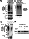
Sumoylation and ubiquitination of AMPKβ2 are antagonistic processes. (A) The coexpression of HA-ubiquitin decreases the levels of PIASy-dependent sumoylated AMPKβ2 protein. HEK293 cells were cotransfected with plasmids pCMV-6xHis-SUMO2, pCMV-myc-AMPKα2/β2/γ1, pFLAG-PIASy, and pCMV-HA-ubiquitin or pCMV-HA (empty) vector. Cell extracts were analyzed as described in Figure 2 by using anti-AMPKβ antibody. (B) The coexpression of HA-SUMO2 decreases the levels of ubiquitinated AMPKβ2 protein. HEK293 cells were cotransfected with plasmids pCMV-6xHis-ubiquitin and pCMV-myc-AMPKα2/β2/γ1, in the presence or absence of pFLAG-PIASy and pCMV-HA-SUMO2. Twenty-four hours after transfection, cells were treated with 6 μM MG132 during 8 h. Then cell extracts were analyzed as described in Figure 2 by using anti-AMPKβ antibodies. (C) Ubiquitination of AMPKβ2 K262R is less efficient than for wild-type protein. HEK293 cells were cotransfected with plasmids pCMV-6xHis-ubiquitin, pCMV-myc-AMPKα2/γ1, and pCMV-myc-AMPKβ2 or pCMV-myc-AMPKβ2K262R mutant. Cells were treated with 6 μM MG132 during 8 h, and cells extracts were analyzed as described in Figure 2 by using anti-AMPKβ antibody. (D) CIDEA-dependent degradation of AMPKβ2 is prevented to some degree in AMPKβ2 K262R mutant. HEK293 cells were cotransfected with pCMV-myc-AMPKα2/γ1 and pCMV-myc-AMPKβ2 or pCMV-myc-AMPKβ2 K262R mutant and FLAG-CIDEA or pFLAG empty vector. Twenty-four hours after transfection, cell extracts (40 μg) were analyzed by SDS–PAGE and immunodetected with anti-AMPKβ and anti-tubulin antibodies.

Similar articles

Cited by

References

    1. Al-Hakim AK, Zagorska A, Chapman L, Deak M, Peggie M, Alessi DR. Control of AMPK-related kinases by USP9X and atypical Lys(29)/Lys(33)-linked polyubiquitin chains. Biochem J. 2008;411:249–260. - PubMed
    1. Anderson DD, Eom JY, Stover PJ. Competition between sumoylation and ubiquitination of serine hydroxymethyltransferase 1 determines its nuclear localization and its accumulation in the nucleus. J Biol Chem. 2012;287:4790–4799. - PMC - PubMed
    1. Bateman A. The structure of a domain common to archaebacteria and the homocystinuria disease protein. Trends Biochem Sci. 1997;22:12–13. - PubMed
    1. Bergink S, Jentsch S. Principles of ubiquitin and SUMO modifications in DNA repair. Nature. 2009;458:461–467. - PubMed
    1. Carling D, Thornton C, Woods A, Sanders MJ. AMP-activated protein kinase: new regulation, new roles. Biochem J. 2012;445:11–27. - PubMed

Publication types

MeSH terms

Substances