

The UPR in atherosclerosis
- PMID: 23553213
- PMCID: PMC3967405
- DOI: 10.1007/s00281-013-0372-x
The UPR in atherosclerosis
Abstract
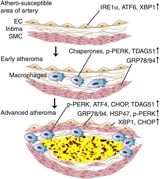
Multiple systemic factors and local stressors in the arterial wall can disturb the functions of endoplasmic reticulum (ER), causing ER stress in endothelial cells (ECs), smooth muscle cells (SMCs), and macrophages during the initiation and progression of atherosclerosis. As a protective response to restore ER homeostasis, the unfolded protein response (UPR) is initiated by three major ER sensors: protein kinase RNA-like ER kinase (PERK), inositol-requiring protein 1α (IRE1α), and activating transcription factor 6 (ATF6). The activation of the various UPR signaling pathways displays a temporal pattern of activation at different stages of the disease. The ATF6 and IRE1α pathways that promote the expression of protein chaperones in ER are activated in ECs in athero-susceptible regions of pre-lesional arteries and before the appearance of foam cells. The PERK pathway that reduces ER protein client load by blocking protein translation is activated in SMCs and macrophages in early lesions. The activation of these UPR signaling pathways aims to cope with the ER stress and plays a pro-survival role in the early stage of atherosclerosis. However, with the progression of atherosclerosis, the extended duration and increased intensity of ER stress in lesions lead to prolonged and enhanced UPR signaling. Under this circumstance, the PERK pathway induces expression of death effectors, and possibly IRE1α activates apoptosis signaling pathways, leading to apoptosis of macrophages and SMCs in advanced lesions. Importantly, UPR-mediated cell death is associated with plaque instability and the clinical progression of atherosclerosis. Moreover, UPR signaling is linked to inflammation and possibly to macrophage differentiation in lesions. Therapeutic approaches targeting the UPR may have promise in the prevention and/or regression of atherosclerosis. However, more progress is needed to fully understand all of the roles of the UPR in atherosclerosis and to harness this information for therapeutic advances.
Figures
Similar articles
-
Cholesterol-Induced Phenotypic Modulation of Smooth Muscle Cells to Macrophage/Fibroblast-like Cells Is Driven by an Unfolded Protein Response.Arterioscler Thromb Vasc Biol. 2021 Jan;41(1):302-316. doi: 10.1161/ATVBAHA.120.315164. Epub 2020 Oct 8. Arterioscler Thromb Vasc Biol. 2021. PMID: 33028096 Free PMC article.
-
Sustained IRE1 and ATF6 signaling is important for survival of melanoma cells undergoing ER stress.Cell Signal. 2014 Feb;26(2):287-94. doi: 10.1016/j.cellsig.2013.11.008. Epub 2013 Nov 12. Cell Signal. 2014. PMID: 24240056
-
A review on endoplasmic reticulum-dependent anti-breast cancer activity of herbal drugs: possible challenges and opportunities.J Drug Target. 2025 Feb;33(2):206-231. doi: 10.1080/1061186X.2024.2417189. Epub 2024 Oct 24. J Drug Target. 2025. PMID: 39404107 Review.
-
Mechanism of the induction of endoplasmic reticulum stress by the anti-cancer agent, di-2-pyridylketone 4,4-dimethyl-3-thiosemicarbazone (Dp44mT): Activation of PERK/eIF2α, IRE1α, ATF6 and calmodulin kinase.Biochem Pharmacol. 2016 Jun 1;109:27-47. doi: 10.1016/j.bcp.2016.04.001. Epub 2016 Apr 6. Biochem Pharmacol. 2016. PMID: 27059255
-
Unfolded protein response during cardiovascular disorders: a tilt towards pro-survival and cellular homeostasis.Mol Cell Biochem. 2021 Nov;476(11):4061-4080. doi: 10.1007/s11010-021-04223-0. Epub 2021 Jul 14. Mol Cell Biochem. 2021. PMID: 34259975 Review.
Cited by
-
AIP1-mediated stress signaling in atherosclerosis and arteriosclerosis.Curr Atheroscler Rep. 2015 May;17(5):503. doi: 10.1007/s11883-015-0503-z. Curr Atheroscler Rep. 2015. PMID: 25732743 Free PMC article. Review.
-
Pathological Crosstalk Between Oxidized LDL and ER Stress in Human Diseases: A Comprehensive Review.Front Cell Dev Biol. 2021 May 26;9:674103. doi: 10.3389/fcell.2021.674103. eCollection 2021. Front Cell Dev Biol. 2021. PMID: 34124059 Free PMC article. Review.
-
Genome-wide census of ATF4 binding sites and functional profiling of trait-associated genetic variants overlapping ATF4 binding motifs.PLoS Genet. 2023 Oct 31;19(10):e1011014. doi: 10.1371/journal.pgen.1011014. eCollection 2023 Oct. PLoS Genet. 2023. PMID: 37906604 Free PMC article.
-
Ab initio protein structure prediction: the necessary presence of external force field as it is delivered by Hsp40 chaperone.BMC Bioinformatics. 2023 Nov 7;24(1):418. doi: 10.1186/s12859-023-05545-0. BMC Bioinformatics. 2023. PMID: 37932669 Free PMC article.
-
How Similar Are Proteins and Origami?Biomolecules. 2022 Apr 21;12(5):622. doi: 10.3390/biom12050622. Biomolecules. 2022. PMID: 35625549 Free PMC article.
References
-
- Murray CJ, Lopez AD. Global mortality, disability, and the contribution of risk factors: Global Burden of Disease Study. Lancet. 1997;349(9063):1436–1442. - PubMed
-
- Rader DJ, Daugherty A. Translating molecular discoveries into new therapies for atherosclerosis. Nature. 2008;451(7181):904–913. - PubMed
-
- Minamino T, Komuro I, Kitakaze M. Endoplasmic reticulum stress as a therapeutic target in cardiovascular disease. Circ Res. 2010;107(9):1071–1082. - PubMed
-
- Ron D, Walter P. Signal integration in the endoplasmic reticulum unfolded protein response. Nat Rev Mol Cell Biol. 2007;8(7):519–529. - PubMed
Publication types
MeSH terms
Grants and funding
LinkOut - more resources
Full Text Sources
Other Literature Sources
Medical
Research Materials
