. 2014 May;219(3):983-94.
doi: 10.1007/s00429-013-0548-0. Epub 2013 Apr 4.

Changes in cerebral morphometry and amplitude of low-frequency fluctuations of BOLD signals during healthy aging: correlation with inhibitory control

Affiliations

Changes in cerebral morphometry and amplitude of low-frequency fluctuations of BOLD signals during healthy aging: correlation with inhibitory control

Sien Hu et al. Brain Struct Funct. 2014 May.

Abstract

Aging is known to be associated with changes in cerebral morphometry and in regional activations during resting or cognitive challenges. Here, we investigated the effects of age on cerebral gray matter (GM) volumes and fractional amplitude of low-frequency fluctuation (fALFF) of blood oxygenation level-dependent signals in 111 healthy adults, 18-72 years of age. GM volumes were computed using voxel-based morphometry as implemented in Statistical Parametric Mapping, and fALFF maps were computed for task-residuals as described in Zhang and Li (Neuroimage 49:1911-1918, 2010) for individual participants. Across participants, a simple regression against age was performed for GM volumes and fALFF, respectively, with quantity of recent alcohol use as a covariate. At cluster level p < 0.05, corrected for family-wise error of multiple comparisons, GM volumes declined with age in prefrontal/frontal regions, bilateral insula, and left inferior parietal lobule (IPL), suggesting structural vulnerability of these areas to aging. FALFF was negatively correlated with age in the supplementary motor area (SMA), pre-SMA, anterior cingulate cortex, bilateral dorsal lateral prefrontal cortex (DLPFC), right IPL, and posterior cingulate cortex, indicating that spontaneous neural activities in these areas during cognitive performance decrease with age. Notably, these age-related changes overlapped in the prefrontal/frontal regions including the pre-SMA, SMA, and DLPFC. Furthermore, GM volumes and fALFF of the pre-SMA/SMA were negatively correlated with the stop signal reaction time, in accord with our earlier work. Together, these results describe anatomical and functional changes in prefrontal/frontal regions and how these changes are associated with declining inhibitory control during aging.


Figures

Figure 1
Figure 1
Decrement of gray matter (GM) volume with aging, with the quantity of recent drinking accounted for in a linear regression. Areas include bilateral insula, putamen, pre-supplementary motor area (SMA) / SMA, mid cingulate cortex, right middle frontal gyrus, left precentral gyrus, and left inferior parietal lobule. All clusters survived at p<.05 corrected for family-wise error (FWE) of multiple comparisons. Color bar represents voxel T value.
Figure 2
Figure 2
Brain regions with age-related decrease in the fractional amplitude of low-frequency fluctuations (fALFF), controlling for the quantity of recent drinking. Areas include the bilateral inferior and middle temporal gyri, pre-supplementary motor area extending to anterior cingulate cortex and dorsal lateral prefrontal cortex, post cingulate cortex, left middle frontal gyrus, right inferior parietal lobule, left postcentral gyrus, and bilateral cuneus/precuneus. All clusters survived at p<.05 corrected for family-wise error (FWE) of multiple comparisons. Color bar represents voxel T value.
Figure 3
Figure 3
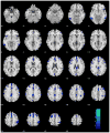
Areas overlapped in the regression of age with GM volumes and with fALFF, including the anterior pre-supplementary motor area (SMA) and posterior pre-SMA extending to SMA, bilateral dorsal lateral prefrontal cortices, bilateral middle frontal gyri, and mid cingulate cortex. All clusters survived at p<.05 corrected for family-wise error (FWE) of multiple comparisons. The inset shows the two clusters (blue), anterior pre-SMA and posterior pre-SMA/SMA, in a sagittal section at x=2.
Figure 4
Figure 4
Negative correlation of SSRT with (a) the GM volume of anterior pre-supplementary motor area (SMA), and (b) the fALFF of the combination of two clusters in the pre-SMA/SMA. Each data point represents one subject.

References

    1. Ashburner J, Friston KJ. Nonlinear spatial normalization using basis functions. Hum Brain Mapp. 1999;7:254–266. - PMC - PubMed
    1. Ashburner J, Friston KJ. Voxel-based morphometry - The methods. Neuroimage. 2000;11:805–821. - PubMed
    1. Bednarski SR, Erdman E, Luo X, Zhang S, Hu S, Li CSR. Neural Processes of an Indirect Analog of Risk Taking in Young Nondependent Adult Alcohol Drinkers-An fMRI Study of the Stop Signal Task. Alcohol Clin Exp Res. 2012;36:768–779. - PMC - PubMed
    1. Bergfield KL, Hanson KD, Chen KW, Teipel SJ, Hampel H, Rapoport SI, Moeller JR, Alexander GE. Age-related networks of regional covariance in MRI gray matter: Reproducible multivariate patterns in healthy aging. Neuroimage. 2010;49:1750–1759. - PMC - PubMed
    1. Birn RM, Diamond JB, Smith MA, Bandettini PA. Separating respiratory-variation-related neuronal-activity-related fluctuations in fluctuations from fMRI. Neuroimage. 2006;31:1536–1548. - PubMed

Publication types

LinkOut - more resources