Review
. 2013 Jun;33(6):547-54.
doi: 10.1002/pd.4097. Epub 2013 Apr 1.

Noninvasive fetal genome sequencing: a primer

Affiliations
Review

Noninvasive fetal genome sequencing: a primer

Matthew W Snyder et al. Prenat Diagn. 2013 Jun.

Abstract

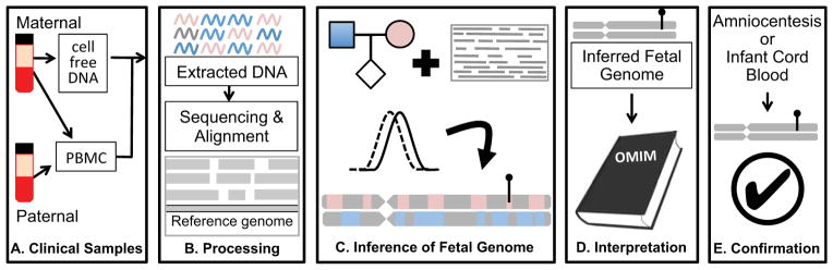
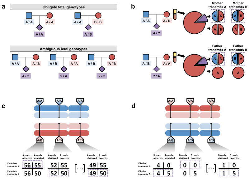
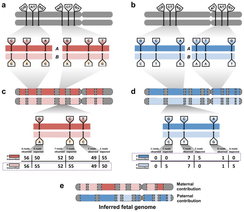
We recently demonstrated whole genome sequencing of a human fetus using only parental DNA samples and plasma from the pregnant mother. This proof-of-concept study demonstrated how samples obtained noninvasively in the first or second trimester can be analyzed to yield a highly accurate and substantially complete genetic profile of the fetus, including both inherited and de novo variation. Here, we revisit our original study from a clinical standpoint, provide an overview of the scientific approach, and describe opportunities and challenges along the path toward clinical adoption of noninvasive fetal whole genome sequencing.


Conflict of interest statement

Conflict of interest disclosures

J.S. is a member of the scientific advisory board or serves as a consultant for Ariosa Diagnostics, Stratos Genomics, Good Start Genetics, and Adaptive Biotechnologies. A provisional patent application has been deposited for aspects of these methods (M.W.S., J.O.K., and J.S.; “Non-invasive whole genome sequencing of a human fetus”; 61/651,356)

Figures

Figure 1
Figure 1
Overview of noninvasive fetal whole genome sequencing. (a). Sample collection. Parental blood samples are collected in the first or second trimester. After centrifugation, parental DNA is extracted from peripheral blood mononuclear cells (PBMC) or buffy coat, while cfDNA is isolated from the maternal plasma. (b). Sample processing. Extracted DNA is amplified for library preparation and sequenced to high depth. Reads are aligned to a reference genome to identify variant alleles carried by one or both parents. (c). Inference of fetal genome. A statistical model combines known parental genotypes and alleles observed in cfDNA reads to predict fetal inheritance. High-impact mutations, whether inherited or de novo, are identified (lollypop). (d). Interpretation. Identified variants are compared to catalogs of known disease-associated mutations. (e). Confirmation. A subset of clinically actionable predicted mutations is confirmed with conventional procedures such as amniocentesis. Accuracy of genome inference can be assessed post-hoc with DNA extracted from cord blood after delivery.
Figure 2
Figure 2
Inference of the fetal genome on a site-by-site basis. (a). Observed parental genotypes at a given site constrain the possible fetal genotypes. At the vast majority of sites, both parents are homozygotes and the fetal genotype is unambiguous. (b). Expected cfDNA makeup in the maternal plasma at maternal (top) and paternal (bottom) heterozygous sites. At these sites, either allele could be transmitted, yielding different expected allelic fractions. (c). Schematic of inference of fetal inheritance at maternal heterozygous sites. Numbers shown assume a constant sequencing depth of 100X at each site and do not represent real data. After sequencing the cfDNA, observed allele counts are compared to expected allele counts at each site, and in each case, the more likely scenario is chosen (purple boxes). (d). Schematic of inference of fetal inheritance at paternal heterozygous sites. Numbers are presented as in (c). At each site, presence of the B allele is unexpected in cases where the father transmits the “A” allele. At the rightmost site, the single observed “B” allele could be evidence of true “B” transmission or an error introduced during the sequencing process.
Figure 3
Figure 3
Inference of the fetal genome from haplotype blocks. (a). Phasing of maternal heterozygous sites into haplotype blocks (red bars). Haplotype blocks contain dozens or hundreds of such sites and cover over 300 kilobases on average. A single chromosome may have over 100 haplotype blocks; contiguity between blocks is not defined. Approximately 90% of all heterozygous sites are incorporated into haplotype blocks. Sites shown do not represent real data. (b). Phasing of paternal heterozygous sites into haplotype blocks (blue bars). Paternal and maternal blocks may overlap but are independently defined. (c). Schematic of inference of fetal inheritance of maternal haplotype blocks. Numbers shown assume a constant sequencing depth of 100X at each. After sequencing the cfDNA, evidence of deviations from expected allele counts is aggregated over each site in haplotype blocks “A” and “B”, and the more likely block is predicted. Block-level predictions in turn determine predictions at each contained site. The site in the center of the block would be incorrectly predicted if sites were considered independently; its inclusion in a haplotype block mitigates sampling noise and corrects the prediction. (d). Schematic of inference of fetal inheritance of paternal haplotype blocks. Numbers are presented as in (c). The observed “G” allele at the rightmost site, likely to cause an incorrect prediction if sites were considered independently, is now correctly identified as an error introduced during the sequencing process rather than evidence of transmission of the “G” allele. (e). The inferred fetal genome is a composite of the parental haplotype blocks.

Similar articles

Cited by

References

    1. Merkatz IR, Nitowsky HM, Macri JN, Johnson WE. An association between low maternal serum alpha-fetoprotein and fetal chromosomal abnormalities. Am J Obstet Gynecol. 1984;148:886–894. - PubMed
    1. ACOG Committee on Practice Bulletins ACOG Practice Bulletin No. 77: screening for fetal chromosomal abnormalities. Obstet Gynecol. 2007;109:217–227. - PubMed
    1. Lapaire O, Holzgreve W, Oosterwijk JC, Brinkhaus R, Bianchi DW. Georg Schmorl on Trophoblasts in the Maternal Circulation. Placenta. 2007;28:1–5. - PubMed
    1. Schröder J, De la Chapelle A. Fetal lymphocytes in the maternal blood. Blood. 1972;39:153–162. - PubMed
    1. Herzenberg LA, Bianchi DW, Schröder J, Cann HM, Iverson GM. Fetal cells in the blood of pregnant women: detection and enrichment by fluorescence-activated cell sorting. Proc Natl Acad Sci USA. 1979;76:1453–1455. - PMC - PubMed

Publication types