. 2013 Jul;61(7):1029-40.
doi: 10.1002/glia.22491. Epub 2013 Apr 2.

Nerve injury induces glial cell line-derived neurotrophic factor (GDNF) expression in Schwann cells through purinergic signaling and the PKC-PKD pathway

Affiliations

Nerve injury induces glial cell line-derived neurotrophic factor (GDNF) expression in Schwann cells through purinergic signaling and the PKC-PKD pathway

Pin Xu et al. Glia. 2013 Jul.

Abstract

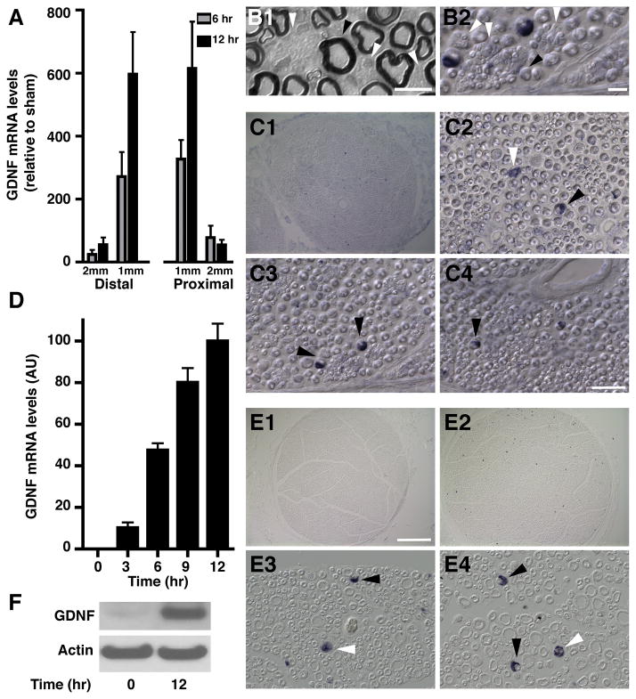
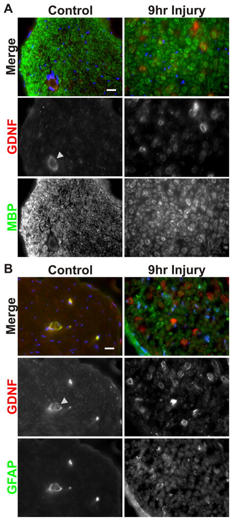
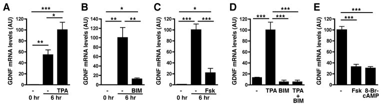
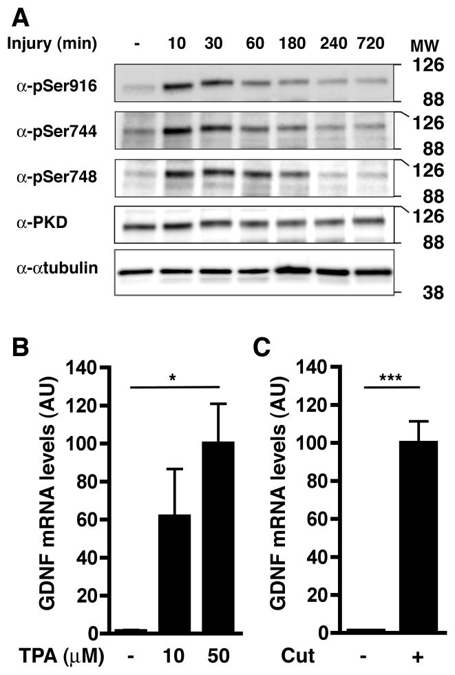
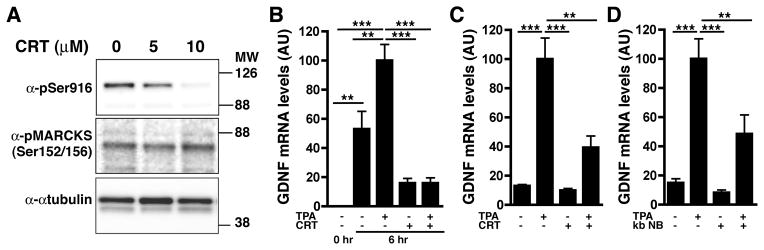
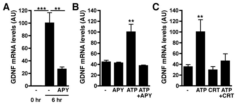
Upon peripheral nerve injury, specific molecular events, including increases in the expression of selected neurotrophic factors, are initiated to prepare the tissue for regeneration. However, the mechanisms underlying these events and the nature of the cells involved are poorly understood. We used the injury-induced upregulation of glial cell-derived neurotrophic factor (GDNF) expression as a tool to gain insights into these processes. We found that both myelinating and nonmyelinating Schwann cells are responsible for the dramatic increase in GDNF expression after injury. We also demonstrate that the GDNF upregulation is mediated by a signaling cascade involving activation of Schwann cell purinergic receptors, followed by protein kinase C signaling which activates protein kinase D (PKD), which leads to increased GDNF transcription. Given the potent effects of GDNF on survival and repair of injured peripheral neurons, we propose that targeting these pathways may yield therapeutic tools to treat peripheral nerve injury and neuropathies.


Figures

Fig. 1
Fig. 1. Nerve injury induces GDNF mRNA in Schwann cells
A) GDNF mRNA levels in adjacent 1 mm segments both proximal and distal to the injury site in sciatic nerve 6 and 12 hours post-axotomy were measured using real time q-RT-PCR. Values are expressed relative to non-axotomized sham using 18S rRNA as normalizer. B) Images of sections of plastic embedded (B1) and frozen (B2) sciatic nerves illustrating the distinct morphology of myelinating (black arrowheads) and non-myelinating (white arrowheads) Schwann cells. The scale bars represent 20 μm for B1 and 150μm for B2. C) In situ hybridization on frozen sections of adult rat sciatic nerve immediately adjacent to the site of injury, 12 hours post axotomy, show GDNF mRNA is restricted to Schwann cells. B1 shows a low magnification image, B2–4 show representative fields at high magnification to illustrate the morphology of GDNF-expressing cells. Black arrowheads in B2–4 indicate myelinating Schwann cells, white arrowheads indicate non-myelinating Schwann cells. The scale bar represents 200 μm for C1 and 50 μm for C2–4. D) GDNF mRNA levels measured in ex vivo nerve segments at 0, 3, 6, 9, and 12 hours post-explant. Values were normalized to the maximum expression at 12 hours. ANOVA showed that most time points are significantly different from the others with p < 0.001, except for 0 hr vs. 3 hr and 9 hr vs. 12 hr (p > 0.05). E) In situ hybridization for GDNF performed on sections prepared from paraffin embedded ex vivo sciatic nerve explants. D1: low magnification image of ex vivo nerve segment at time 0. D2: low magnification image of GDNF expression in an ex vivo nerve segment at 12 hours post-explant. D3–4: high magnification view of representative fields of ex vivo nerve sections 12 hours post-explant. Black arrowheads indicate myelinating Schwann cells, white arrowheads indicate non-myelinating Schwann cells. The scale bar represents 200 μm for D1–2 and 50 μm for D3–4. F) Western blot analysis of ex vivo nerve segments at 0 and 12 hours post-explant shows that the levels of GDNF protein are increased after explantation. Actin levels were measured to control for loading.
Fig. 2
Fig. 2. Nerve injury increases GDNF protein expression in myelinating Schwann cells
A) Immunofluorescence analysis shows that ex vivo nerve segments 9 hours post-explantation have higher GDNF protein levels associated with myelinating (MBP+) Schwann cells compared to freshly dissected nerve segments. The arrowhead shows blood vessels, which display non-specific staining (see also Panel B). Scale bar = 20μm. B) Immunofluorescence analysis shows that staining for GFAP, a marker for non-myelinating Schwann cells, is widely increased in ex vivo nerve segments at 9 hours post-explantation compared to freshly dissected nerve segments. As a result, changes in GDNF protein levels in non-myelinating Schwann cells could not be validated. Scale bar = 20μm.
Fig. 3
Fig. 3. PKC up-regulates and PKA downregulates GDNF expression in sciatic nerve segments and cultured Schwann cells
A) The PKC activator TPA enhances the upregulation of GDNF expression in ex vivo sciatic nerve segments after 6hr of culture. B) PKC inhibition by Bisindolylmaleimide I hydrochloride (BIM) blocks the injury-induced increase of GDNF expression in ex vivo nerve segments. C) Forskolin reduces the injury-induced increase of GDNF expression in ex vivo nerve segments. D) TPA treatment induces GDNF expression in Schwann cell cultures, an effect that is blocked by BIM. E) Forskolin and 8-Br-cAMP treatments reduce GDNF expression by Schwann cells.
Fig. 4
Fig. 4. PKC becomes activated in the injured nerve and PKC activation is sufficient to induce GDNF expression in intact peripheral nerve
A) Western blot depicting the rapid site-specific phosphorylation of protein kinase D, a PKC substrate, at Ser916, Ser744, and Ser748 in ex vivo nerve segments at the indicated times following culture. Levels of total PKD are unchanged by nerve injury. α-tubulin was used as loading control. B) Exposure of uninjured nerve to GelFoam® containing the PKC activator TPA for 6 hr induces GDNF expression. C) Exposure of sciatic nerve to GelFoam® soaked with vehicle (DMSO) does not interfere with GDNF induction in the 2 mm segment adjacent to the injury site.
Fig. 5
Fig. 5. PKD activation, downstream of PKC, is necessary for induction of GDNF expression by nerve injury
A) Inhibition of PKD using CRT0066101 blocks PKD catalytic activity as revealed by the inhibition of its autophosphorylation (pSer916) but has no impact on the phosphorylation of MARCKS, another target of PKC. α-tubulin was used as a control for loading. B) TPA treatment of ex vivo nerve segments increases GDNF expression to levels above those induced by injury alone. Both the injury and TPA-induced effects are blocked by CRT0066101. C) The TPA-induced increase in Schwann cell GDNF expression is significantly reduced by PKD blockade using CRT0066101 (CRT). D) Blocking PKD activity using the inhibitor kb NB 142-70 (kb NB) greatly diminishes the TPA-induced increase in GDNF expression in cultured Schwann cells.
Fig. 6
Fig. 6. Purinergic signaling induces GDNF expression in explants and Schwann cells through PKD activation
A) Induction of GDNF expression by injury in ex vivo nerve segments is reduced by the presence of apyrase (APY), an ectonucleotidase that degrades ATP and ADP. B) ATP stimulates GDNF expression by Schwann cells, an effect that is blocked by apyrase (APY). C) Inhibition of PKD by CRT0066101 (CRT) blocks the ATP-mediated increases in GDNF expression by Schwann cells.

Similar articles

Cited by

References

    1. Akkina SK, Patterson CL, Wright DE. GDNF rescues nonpeptidergic unmyelinated primary afferents in streptozotocin-treated diabetic mice. Exp Neurol. 2001;167(1):173–82. - PubMed
    1. Amadesi S, Grant AD, Cottrell GS, Vaksman N, Poole DP, Rozengurt E, Bunnett NW. Protein kinase D isoforms are expressed in rat and mouse primary sensory neurons and are activated by agonists of protease-activated receptor 2. J Comp Neurol. 2009;516(2):141–56. - PMC - PubMed
    1. Anitha M, Gondha C, Sutliff R, Parsadanian A, Mwangi S, Sitaraman SV, Srinivasan S. GDNF rescues hyperglycemia-induced diabetic enteric neuropathy through activation of the PI3K/Akt pathway. J Clin Invest. 2006;116(2):344–56. - PMC - PubMed
    1. Asaithambi A, Kanthasamy A, Saminathan H, Anantharam V, Kanthasamy AG. Protein kinase D1 (PKD1) activation mediates a compensatory protective response during early stages of oxidative stress-induced neuronal degeneration. Mol Neurodegener. 2011;6:43. - PMC - PubMed
    1. Bampton ET, Taylor JS. Effects of Schwann cell secreted factors on PC12 cell neuritogenesis and survival. J Neurobiol. 2005;63(1):29–48. - PubMed

Publication types

MeSH terms

LinkOut - more resources