. 2013 May 17;288(20):14463-14475.
doi: 10.1074/jbc.M112.437210. Epub 2013 Apr 3.

Metabolomic profiling reveals a role for caspase-2 in lipoapoptosis

Affiliations

Metabolomic profiling reveals a role for caspase-2 in lipoapoptosis

Erika Segear Johnson et al. J Biol Chem. .

Abstract

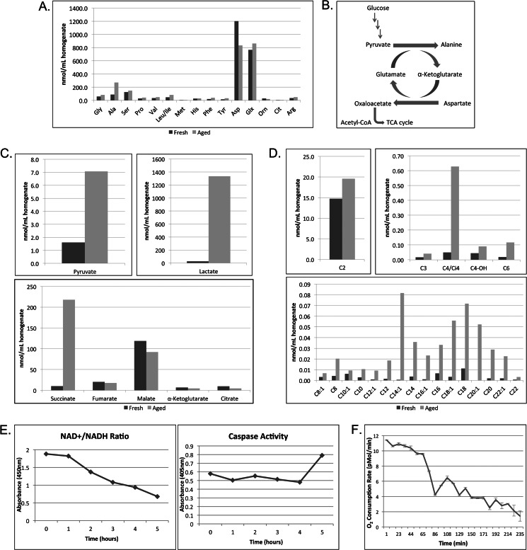
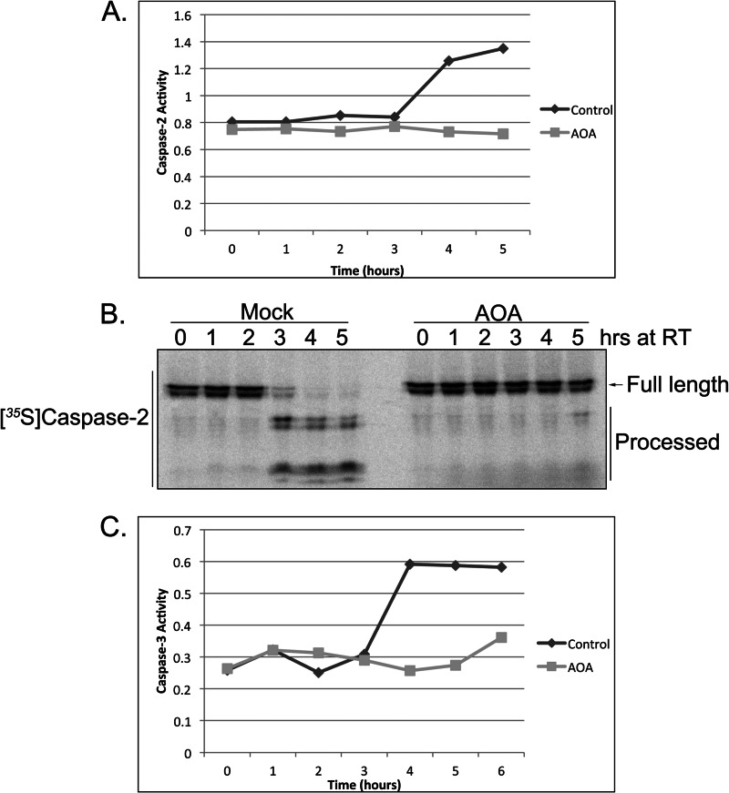
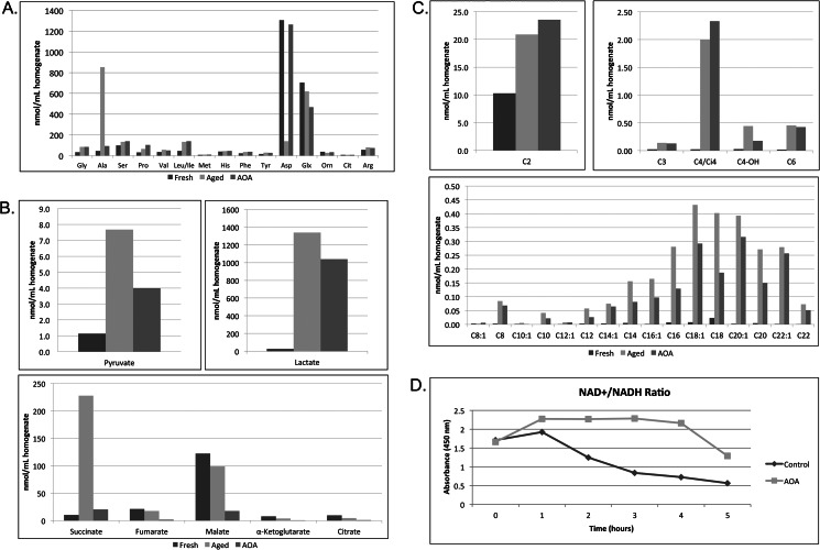
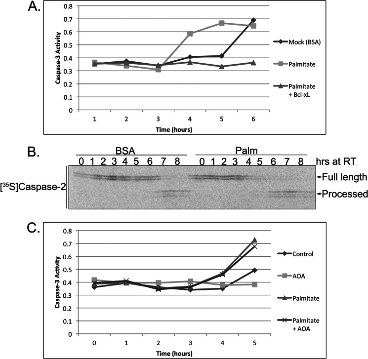
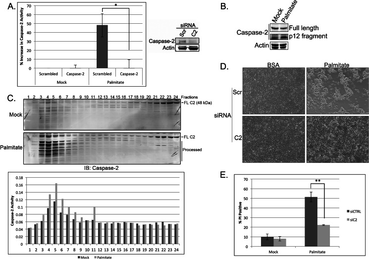
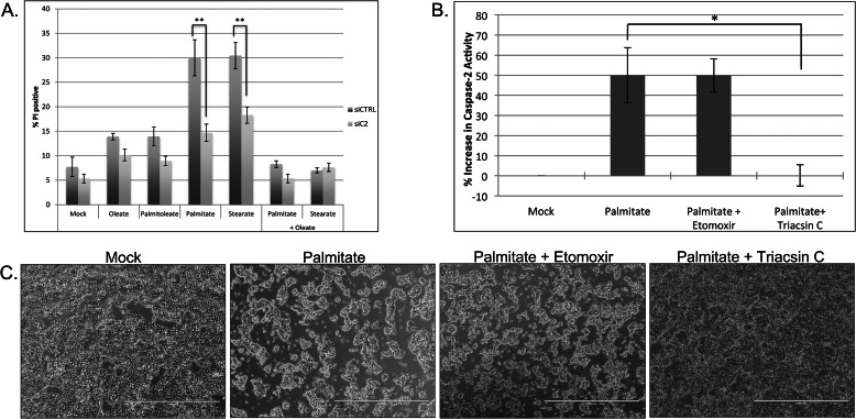
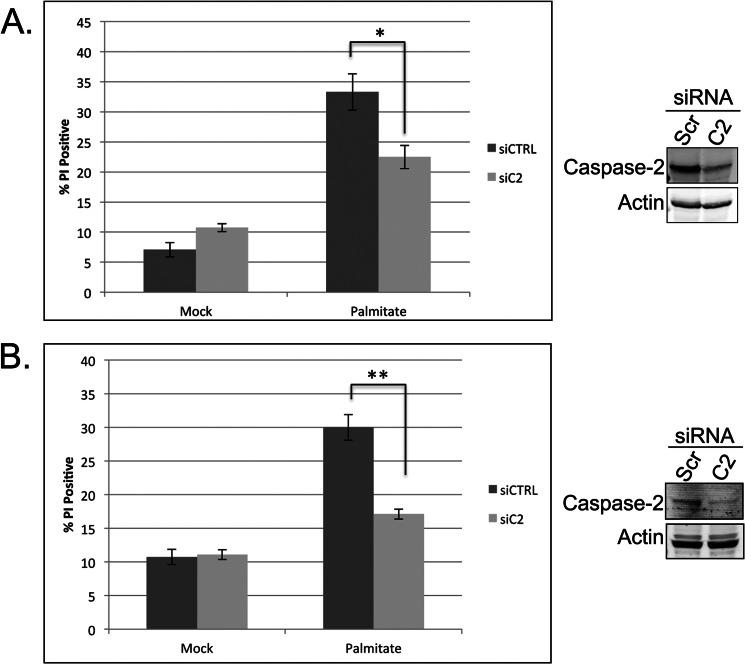
The accumulation of long-chain fatty acids (LCFAs) in non-adipose tissues results in lipid-induced cytotoxicity (or lipoapoptosis). Lipoapoptosis has been proposed to play an important role in the pathogenesis of several metabolic diseases, including non-alcoholic fatty liver disease, diabetes mellitus, and cardiovascular disease. In this report, we demonstrate a novel role for caspase-2 as an initiator of lipoapoptosis. Using a metabolomics approach, we discovered that the activation of caspase-2, the initiator of apoptosis in Xenopus egg extracts, is associated with an accumulation of LCFA metabolites. Metabolic treatments that blocked the buildup of LCFAs potently inhibited caspase-2 activation, whereas adding back an LCFA in this scenario restored caspase activation. Extending these findings to mammalian cells, we show that caspase-2 was engaged and activated in response to treatment with the saturated LCFA palmitate. Down-regulation of caspase-2 significantly impaired cell death induced by saturated LCFAs, suggesting that caspase-2 plays a pivotal role in lipid-induced cytotoxicity. Together, these findings reveal a previously unknown role for caspase-2 as an initiator caspase in lipoapoptosis and suggest that caspase-2 may be an attractive therapeutic target for inhibiting pathological lipid-induced apoptosis.

Keywords: Apoptosis; Caspase; Hepatocyte; Lipids; Metabolism.


Figures

FIGURE 1.
FIGURE 1.
Amino acid metabolism and the accumulation of succinate and long-chain fatty acid metabolites precede caspase activation in Xenopus egg extract. A, freshly prepared Xenopus egg extract (Fresh) or extract incubated at room temperature for 4 h (Aged) was analyzed for amino acid profile following protein precipitation. B, figure of carbon flux via transamination reactions in the egg extract. C, egg extracts treated as in A were analyzed for organic acids. D, egg extracts treated as in A were analyzed for acylcarnitines. E, samples of egg extract were collected at the indicated times and analyzed for NAD+/NADH ratio changes by spectrophotometric color change (kit supplied by BioVision). The same samples were used to measure caspase-3 activity using the caspase substrate Ac-DEVD-pNA. Substrate cleavage was measured spectrophotometrically at 405 nm. F, the O2 consumption rate of aging egg extracts was monitored for 4 h using the Seahorse Bioscience XF24 extracellular flux analyzer. The results represent the means ± S.D. (error bars) of three technical replicates. Orn, ornithine; Glx, glutamine/glutamic acid; Cit, citrulline.
FIGURE 2.
FIGURE 2.
Inhibition of amino acid transamination by AOA blocks caspase activation. A, egg extracts supplemented with AOA (10 mm) or buffer were analyzed for caspase-2 activity at the indicated time points using the BioVision caspase-2 colorimetric assay kit and measuring cleavage of the caspase-2 model substrate, VDVAD-pNA, spectrophotometrically. B, 35S-labeled caspase-2 was incubated in AOA- or mock-treated extracts, and samples were resolved by SDS-PAGE/PhosphorImager. C, egg extracts treated as in A were analyzed for caspase-3 activity at the indicated time points using the caspase substrate Ac-DEVD-pNA. RT, room temperature.
FIGURE 3.
FIGURE 3.
Treatment with AOA, which blocks caspase activation, prevents the underlying accumulation of long chain fatty acid metabolites. A, amino acid profiling was performed on egg extracts immediately following preparation (Fresh) or after room temperature incubation in the absence (Aged) or presence of 10 mm AOA (AOA). B, egg extracts treated as in A were analyzed for organic acids. C, egg extracts treated as in A were analyzed for acylcarnitines. D, samples of egg extract treated with and without 10 mm AOA were collected at the indicated times and analyzed for NAD+/NADH ratio changes by spectrophotometric color change (kit supplied by BioVision). Orn, ornithine; Glx, glutamine/glutamic acid; Cit, citrulline.
FIGURE 4.
FIGURE 4.
Palmitate accelerates caspase activation and overrides the protective effect of AOA. A, mock-treated (BSA) or palmitate-treated (BSA-conjugated palmitate; 4 mm) egg extracts were incubated in the presence or absence of Bcl-xL and analyzed for caspase-3 activity at the indicated time points. Caspase-3 activity was assessed using the caspase substrate Ac-DEVD-pNA, and cleavage was measured spectrophotometrically at 405 nm. B, 35S-labeled caspase-2 was incubated in mock- or palmitate (Palm)-treated extracts, and samples were resolved by SDS-PAGE/PhosphorImager. C, egg extracts were incubated with combinations of palmitate (4 mm) and AOA (10 mm), and caspase-3 activity was assessed using the caspase substrate Ac-DEVD-pNA. RT, room temperature.
FIGURE 5.
FIGURE 5.
Caspase-2 plays a central role in mediating lipoapoptosis in 293T cells. A, 293T cells were transfected with 50 nm scrambled (Scr) or caspase-2 (C2)-targeted siRNA. At 48 h post-transfection, cells were mock-treated with BSA or treated with 1 mm palmitate for 18 h, and caspase-2 activity was assessed using the BioVision caspase-2 colorimetric assay kit. Knockdown efficiency was determined by immunoblot. The results represent the means ± S.E. (error bars) for percent increase in activity compared with control siRNA-, mock-treated samples for three independent experiments. Statistical significance was determined using a two-tailed Student's t test. *, p < 0.05 versus control. B, 293T cells were treated with BSA or palmitate for 24 h and immunoblotted for caspase-2 and actin. The top caspase-2 panel is a short exposure (to show changes in full-length caspase-2 levels), and the bottom caspase-2 panel is a long exposure (to reveal the p12 fragment). C, 293T cells were treated with BSA or palmitate for 18 h before lysis by Dounce homogenization in hypotonic lysis buffer. Cell lysates were separated on a Superdex 200 column, and relevant fractions were immunoblotted (IB) for caspase-2. Full-length caspase-2 (FL C2) and processed caspase-2 are indicated. Fractions were also tested for caspase-2 activity by assessing cleavage of the caspase-2 model substrate, VDVAD-pNA, as part of the BioVision caspase-2 colorimetric assay kit. D, 293T cells were treated as in A, and at 24 h post-treatment, phase-contrast images were captured on an EVOS FL digital inverted microscope. E, 293T cells were treated as in A, and cell death was measured 24 h after palmitate treatment by propidium iodide staining and flow cytometric analysis. The results represent the means ± S.E. (error bars) for four independent experiments. Statistical significance was determined using a two-tailed Student's t test. **, p < 0.01 versus control. siCTRL, control siRNA; siC2, caspase-2 siRNA.
FIGURE 6.
FIGURE 6.
Caspase-2 mediates saturated fatty acid-induced lipoapoptosis via acyl-CoA cytotoxicity. A, 293T cells were transfected with 50 nm scrambled or caspase-2 (C2)-targeted siRNA. At 48 h post-transfection, cells were mock-treated with BSA or 1 mm fatty acids, and cell death was measured 24 h post-treatment by propidium iodide (PI) staining and flow cytometric analysis. The results represent the means ± S.E. (error bars) for four independent experiments. Statistical significance was determined using a two-tailed Student's t test. **, p < 0.01 versus control. B, 293T cells were mock-treated with BSA or treated with 1 mm palmitate with or without 200 μm etomoxir or 5 μm triacsin C. At 24 h post-treatment, phase-contrast images were captured on an EVOS FL digital inverted microscope. C, 293T cells were treated as in B. At 18 h post-treatment, caspase-2 activity was assessed using the BioVision caspase-2 colorimetric assay kit. The results represent the means ± S.E. (error bars) for percent increase in activity compared with mock-treated samples for four independent experiments. Statistical significance was determined using a two-tailed Student's t test. *, p < 0.05 versus control. siCTRL, control siRNA; siC2, caspase-2 siRNA. Scale bars represent 1000 μm.
FIGURE 7.
FIGURE 7.
Caspase-2 is required for LCFA-induced apoptosis in hepatocytes. A, HepG2 cells were transfected with 50 nm scrambled or caspase-2 (C2)-targeted siRNA. At 48 h post-transfection, cells were mock-treated with BSA or treated with 0.7 mm palmitate for 24 h, and cell death was measured by propidium iodide (PI) staining and flow cytometric analysis. Knockdown efficiency was determined by immunoblot. B, AML12 cells were treated with 50 nm scrambled (Scr) or mouse caspase-2-targeted SMARTpool siRNA. At 48 h post-transfection, cells were mock- or palmitate-treated and analyzed for cell death as in A. Knockdown efficiency was determined by immunoblot. The results represent the means ± S.E. (error bars) for three or more independent experiments. Statistical significance was determined using a two-tailed Student's t test. *, p < 0.05 versus control; **, p < 0.01 versus control. siCTRL, control siRNA; siC2, caspase-2 siRNA.

Similar articles

Cited by

References

    1. Frayn K. N. (2002) Adipose tissue as a buffer for daily lipid flux. Diabetologia 45, 1201–1210 - PubMed
    1. Lewis G. F., Carpentier A., Adeli K., Giacca A. (2002) Disordered fat storage and mobilization in the pathogenesis of insulin resistance and type 2 diabetes. Endocr. Rev. 23, 201–229 - PubMed
    1. Kusminski C. M., Shetty S., Orci L., Unger R. H., Scherer P. E. (2009) Diabetes and apoptosis: lipotoxicity. Apoptosis 14, 1484–1495 - PubMed
    1. Unger R. H., Clark G. O., Scherer P. E., Orci L. (2010) Lipid homeostasis, lipotoxicity and the metabolic syndrome. Biochim. Biophys. Acta 1801, 209–214 - PubMed
    1. Listenberger L. L., Schaffer J. E. (2002) Mechanisms of lipoapoptosis: implications for human heart disease. Trends Cardiovasc. Med. 12, 134–138 - PubMed

Publication types

LinkOut - more resources