Review
. 2013 Jun;22(6):693-724.
doi: 10.1002/pro.2261. Epub 2013 Apr 29.

A decade and a half of protein intrinsic disorder: biology still waits for physics

Affiliations
Review

A decade and a half of protein intrinsic disorder: biology still waits for physics

Vladimir N Uversky. Protein Sci. 2013 Jun.

Abstract

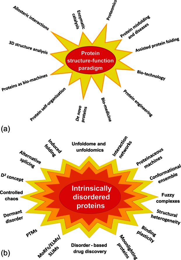
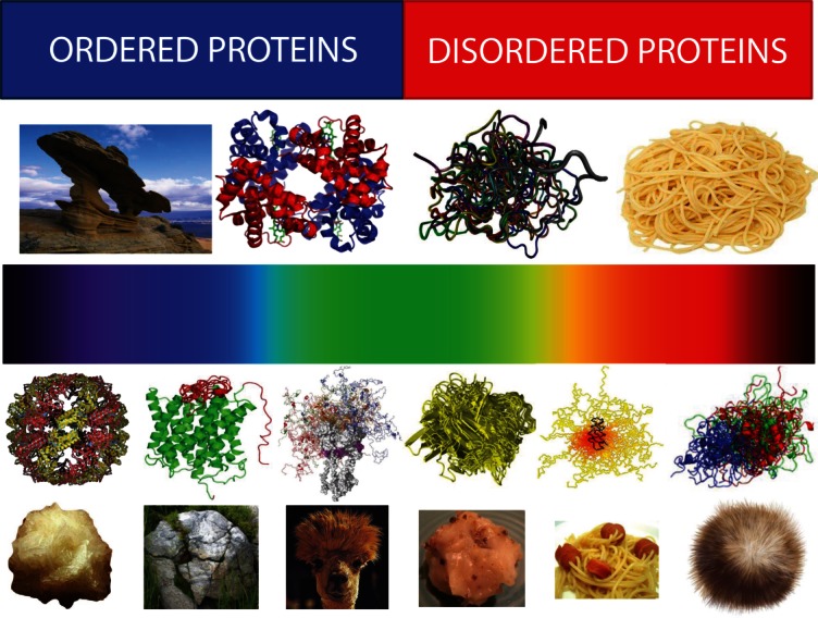
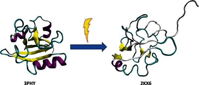
The abundant existence of proteins and regions that possess specific functions without being uniquely folded into unique 3D structures has become accepted by a significant number of protein scientists. Sequences of these intrinsically disordered proteins (IDPs) and IDP regions (IDPRs) are characterized by a number of specific features, such as low overall hydrophobicity and high net charge which makes these proteins predictable. IDPs/IDPRs possess large hydrodynamic volumes, low contents of ordered secondary structure, and are characterized by high structural heterogeneity. They are very flexible, but some may undergo disorder to order transitions in the presence of natural ligands. The degree of these structural rearrangements varies over a very wide range. IDPs/IDPRs are tightly controlled under the normal conditions and have numerous specific functions that complement functions of ordered proteins and domains. When lacking proper control, they have multiple roles in pathogenesis of various human diseases. Gaining structural and functional information about these proteins is a challenge, since they do not typically "freeze" while their "pictures are taken." However, despite or perhaps because of the experimental challenges, these fuzzy objects with fuzzy structures and fuzzy functions are among the most interesting targets for modern protein research. This review briefly summarizes some of the recent advances in this exciting field and considers some of the basic lessons learned from the analysis of physics, chemistry, and biology of IDPs.


Figures

Figure 1
Figure 1
A: Protein structure–function paradigm is the “Big Bang” created universe of the modern protein science. Some major directions based on the consideration of protein function as lock-and-key mechanism are shown. Modified from Ref.. B: Paradigm shift caused by the introduction of the protein intrinsic disorder concept opened a wide array of new directions in protein science. In essence, introduction of this concept can be considered as a scientific revolution that, according to Kuhn, “occurs when scientists encounter anomalies that cannot be explained by the universally accepted paradigm within which scientific progress has thereto been made” (http://en.wikipedia.org/wiki/Paradigm_shift).
Figure 2
Figure 2
Correlation between the intrinsic disorder content and proteome size for 3484 species from viruses, archaea, bacteria, and eukaryotes. Each symbol indicates a species. There are totally six groups of species: viruses expressing one polyprotein precursor (small red circles filled with blue), other viruses (small red circles), bacteria (small green circles), archaea (blue circles), unicellular eukaryotes (brown squares), and multicellular eukaryotes (pink triangles). Each viral polyprotein was analyzed as a single polypeptide chain, without parsing it into the individual proteins before predictions. The proteome size is the number of proteins in the proteome of that species and is shown in log base. The average fraction of disordered residues is calculated by averaging the fraction of disordered residues of each sequence over the all sequences of that species. Disorder prediction is evaluated by PONDR-VSL2B. Modified from Ref..
Figure 3
Figure 3
Structural heterogeneity of IDPs/IDPRs. Top half: Bi-colored view of functional proteins which are considered to be either ordered (folded, blue) or completely structure-less (disordered, red). Ordered proteins are taken as rigid rocks, whereas IDPs are considered as completely structure-less entities, kind of cooked noodles. Bottom half: A continuous emission spectrum representing the fact that functional proteins can extend from fully ordered to completely structure-less proteins, with everything in between. Intrinsic disorder can have multiple faces, can affect different levels of protein structural organization, and whole proteins, or various protein regions can be disordered to a different degree. Some illustrative examples includes ordered proteins that are completely devoid of disordered regions (rock-like type), ordered proteins with limited number of disordered regions (grass-on-the rock type), ordered proteins with significant amount of disordered regions (lhama/camel hair type), molten globule-like collapsed IDPs (greasy ball type), pre-molten globule-like extended IDPs (spaghetti-and-sausage type), and unstructured extended IDPs (hairball type).
Figure 4
Figure 4
Comparison of the ground state (left structure, PDB ID: 3PHY) and the transient light activated signaling state of the PYP (right structure, PDB ID: 2KX6). Ground state structure was determined by multidimensional NMR spectroscopy. This structure is in agreement with an earlier published 1.4 Å crystal structure, and modeled structure based on combined DEER, TR-SAXS/WAXS, and NMR data. It consists of an open, twisted, 6-stranded, antiparallel β-sheet, which is flanked by four α-helices on both sides.– On the contrary, the light-activated form is highly disordered. This structure satisfies DEER, SAXS/WAXS, and NMR data simultaneously.
Figure 5
Figure 5
Peculiarities of disorder evolution. A: Modern genetic code with information on the early and late codons (shown by light red and light blue colors, respectively) and disorder- and order-promoting residues (shown by red and blue colors, respectively). Codons with intermediate ages (i.e., those located between early and late codons) are shown by light pink color, whereas disorder-neutral residues are shown by pink color. B: Wavy pattern of the global disorder evolution. X-axis represents evolutionary time and Y-axis shows disorder content in proteins at given evolutionary time point. Here, primordial proteins are expected to be mostly disordered (left-hand side of the plot), proteins in LUA likely are mostly structured (center of the plot), whereas many protein in eukaryotes are either totally disordered or hybrids containing both ordered and disordered regions (right-hand side of the plot).

References

    1. Uversky VN. Natively unfolded proteins: a point where biology waits for physics. Protein Sci. 2002;11:739–756. - PMC - PubMed
    1. Fischer E. Einfluss der configuration auf die wirkung der enzyme. Ber Dt Chem Ges. 1894;27:2985–2993.
    1. Bernstein FC, Koetzle TF, Williams GJ, Meyer EF, Jr, Brice MD, Rodgers JR, Kennard O, Shimanouchi T, Tasumi M. The Protein Data Bank: a computer-based archival file for macromolecular structures. J Mol Biol. 1977;112:535–542. - PubMed
    1. Uversky VN, Dunker AK. Understanding protein non-folding. Biochim Biophys Acta. 2010;1804:1231–1264. - PMC - PubMed
    1. Kuhn TS. The structure of scientific revolutions. Chicago: University Of Chicago Press; 2012.