

Bisdemethoxycurcumin inhibits PDGF-induced vascular smooth muscle cell motility and proliferation
- PMID: 23554078
- PMCID: PMC3844678
- DOI: 10.1002/mnfr.201200852
Bisdemethoxycurcumin inhibits PDGF-induced vascular smooth muscle cell motility and proliferation
Abstract
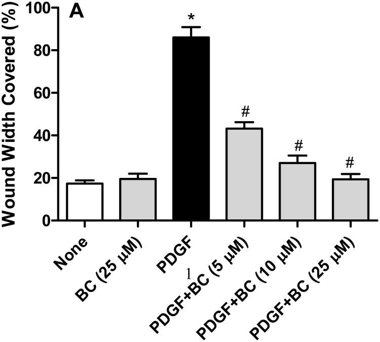
Scope: A key event in the development of plaque in the arteries is the migration and proliferation of smooth muscle cells (SMCs) from the media to the intima of the blood vessel. This study was conducted to evaluate the effects of bisdemethoxycurcumin (BC), a naturally occurring structural analog of curcumin (CC), on platelet-derived growth factor (PDGF)-stimulated migration and proliferation of SMCs.
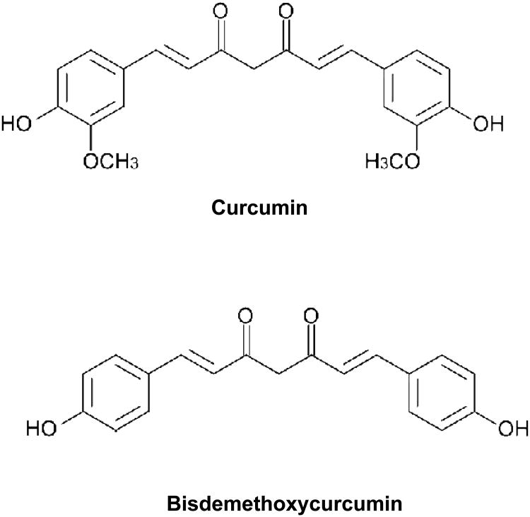
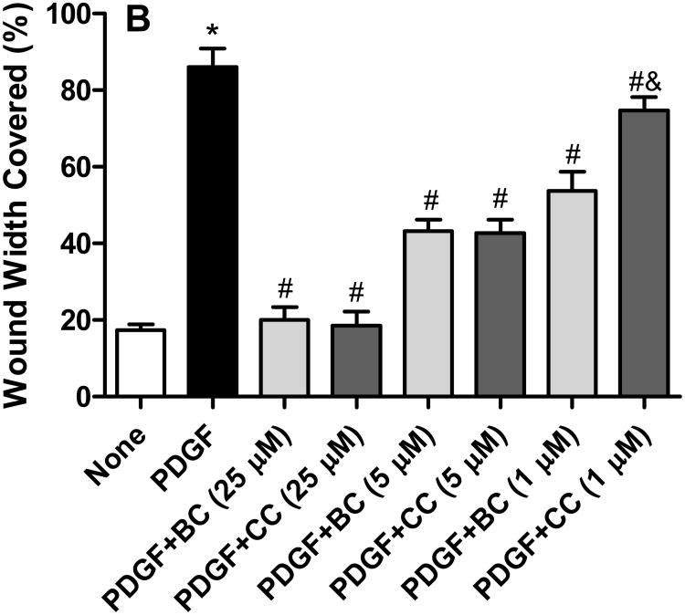
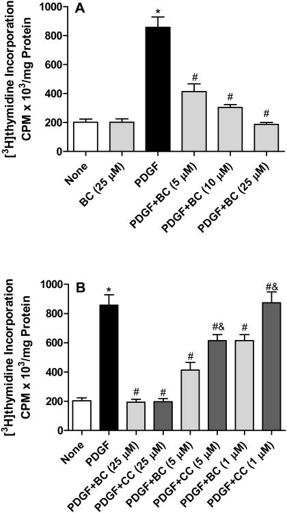
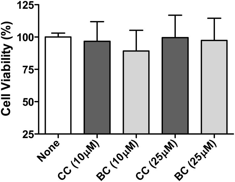
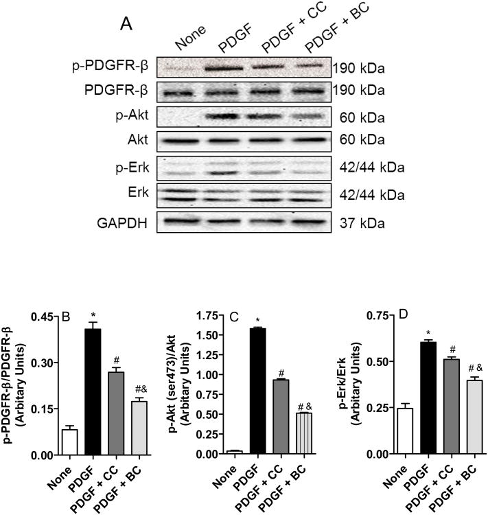
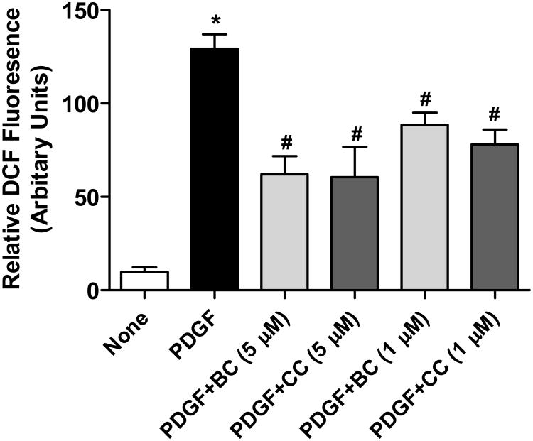
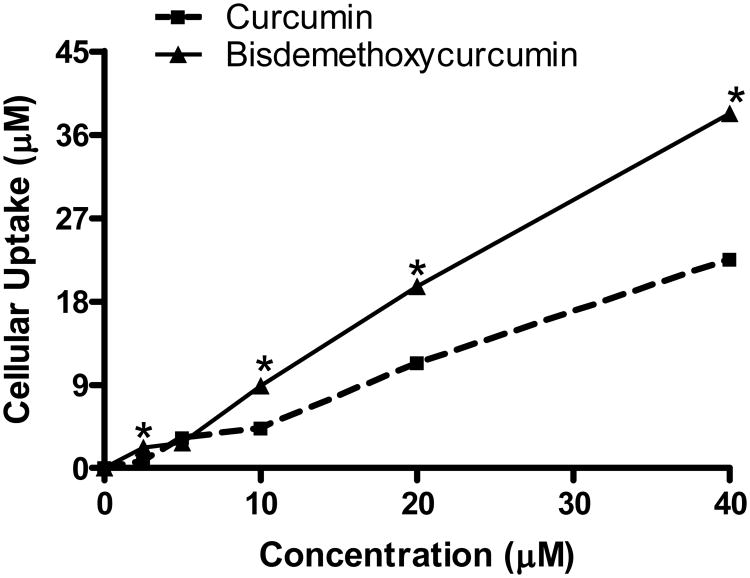
Methods and results: CC and BC were synthesized by condensing acetyl acetone with vanillin and 4-hydroxybenzaldehyde, respectively. SMCs isolated from adult rat aorta were stimulated with PDGF in the presence or absence of CC or BC following which, cell migration and proliferation were assessed by monolayer wound healing assay and [(3) H]-thymidine incorporation respectively. PDGF-stimulated phosphorylation of PDGF receptor-β and its downstream effectors Akt and ERK were assessed by Western blotting. Intracellular reactive oxygen species was assessed using the fluorescent dye 5-(6)-chloromethyl-2',7'-dichlorodihydrofluorescein diacetate. BC elicited a concentration-dependent inhibition of PDGF-stimulated phosphorylation of PDGF receptor-β, Akt and Erk as well as the PDGF-stimulated SMC migration and proliferation. BC was more potent than CC in inhibiting migration and proliferation and suppressing PDGF-signaling in SMCs. Both compounds were equipotent in inhibiting PDGF-stimulated generation of intracellular reactive oxygen species.
Conclusion: BC may be of potential use in the prevention or treatment of vascular disease.
Keywords: Curcumin; Migration; Platelet-derived growth factor; Proliferation; Vascular smooth muscle cell.
© 2013 WILEY-VCH Verlag GmbH & Co. KGaA, Weinheim.
Figures
Similar articles
-
Inhibitory effect of dehydrozingerone on vascular smooth muscle cell function.J Cardiovasc Pharmacol. 2008 Nov;52(5):422-9. doi: 10.1097/FJC.0b013e31818aed93. J Cardiovasc Pharmacol. 2008. PMID: 19033821
-
Gambogic acid induces G0/G1 cell cycle arrest and cell migration inhibition via suppressing PDGF receptor β tyrosine phosphorylation and Rac1 activity in rat aortic smooth muscle cells.J Atheroscler Thromb. 2010 Sep 30;17(9):901-13. doi: 10.5551/jat.3491. Epub 2010 Jun 11. J Atheroscler Thromb. 2010. PMID: 20543524
-
Curcumin inhibits platelet-derived growth factor-stimulated vascular smooth muscle cell function and injury-induced neointima formation.Arterioscler Thromb Vasc Biol. 2006 Jan;26(1):85-90. doi: 10.1161/01.ATV.0000191635.00744.b6. Epub 2005 Oct 20. Arterioscler Thromb Vasc Biol. 2006. PMID: 16239599
-
Empagliflozin Attenuates Neointima Formation After Arterial Injury and Inhibits Smooth Muscle Cell Proliferation and Migration by Suppressing Platelet-Derived Growth Factor-Related Signaling.J Am Heart Assoc. 2024 Nov 19;13(22):e035044. doi: 10.1161/JAHA.124.035044. Epub 2024 Nov 7. J Am Heart Assoc. 2024. PMID: 39508166 Free PMC article.
-
Emerging roles of PDGF-D signaling pathway in tumor development and progression.Biochim Biophys Acta. 2010 Aug;1806(1):122-30. doi: 10.1016/j.bbcan.2010.04.003. Epub 2010 Apr 28. Biochim Biophys Acta. 2010. PMID: 20434526 Free PMC article. Review.
Cited by
-
Curcumin induced oxidative stress attenuation by N-acetylcysteine co-treatment: a fibroblast and epithelial cell in-vitro study in idiopathic pulmonary fibrosis.Mol Med. 2019 Jun 13;25(1):27. doi: 10.1186/s10020-019-0096-z. Mol Med. 2019. PMID: 31195971 Free PMC article.
-
Preparation of poly(lactide-co-glycolide) microspheres and evaluation of pharmacokinetics and tissue distribution of BDMC-PLGA-MS in rats.Asian J Pharm Sci. 2018 Jan;13(1):82-90. doi: 10.1016/j.ajps.2017.09.002. Epub 2017 Oct 10. Asian J Pharm Sci. 2018. PMID: 32104381 Free PMC article.
-
A novel PDGF receptor inhibitor-eluting stent attenuates in-stent neointima formation in a rabbit carotid model.Mol Med Rep. 2017 Jan;15(1):21-28. doi: 10.3892/mmr.2016.5986. Epub 2016 Dec 5. Mol Med Rep. 2017. PMID: 27922693 Free PMC article.
-
Effects of curcumin on vascular smooth muscle cells: implications for health and disease.Pharmacol Rep. 2025 Jun 5. doi: 10.1007/s43440-025-00744-3. Online ahead of print. Pharmacol Rep. 2025. PMID: 40471441 Review.
-
Assessment of Safety Profile of Activated Curcumin C3 Complex (AC3®), Enriched Extract of Bisdemethoxycurcumin from the Rhizomes of Curcuma longa.J Toxicol. 2023 Oct 31;2023:3729399. doi: 10.1155/2023/3729399. eCollection 2023. J Toxicol. 2023. PMID: 37941801 Free PMC article.
References
-
- Newby AC, Zaltsman AB. Molecular mechanisms in intimal hyperplasia. J Pathol. 2000;190:300–309. - PubMed
-
- Willis AI, Pierre-Paul D, Sumpio BE, Gahtan V. Vascular smooth muscle cell migration: current research and clinical implications. Vasc Endovascular Surg. 2004;38:11–23. - PubMed
-
- Aggarwal BB, Sung B. Pharmacological basis for the role of curcumin in chronic diseases: an age-old spice with modern targets. Trends Pharmacol Sci. 2009;30:85–94. - PubMed
-
- Epstein J, Sanderson IR, Macdonald TT. Curcumin as a therapeutic agent: the evidence from in vitro, animal and human studies. Br J Nutr. 2010;103:1545–1557. - PubMed
-
- Lin JK. Molecular targets of curcumin. Adv Exp Med Biol. 2007;595:227–243. - PubMed
Publication types
MeSH terms
Substances
Grants and funding
LinkOut - more resources
Full Text Sources
Other Literature Sources
Miscellaneous
