. 2013 Apr;5(4):608-25.
doi: 10.1002/emmm.201202215.

Amyloid precursor protein controls cholesterol turnover needed for neuronal activity

Affiliations

Amyloid precursor protein controls cholesterol turnover needed for neuronal activity

Nathalie Pierrot et al. EMBO Mol Med. 2013 Apr.

Abstract

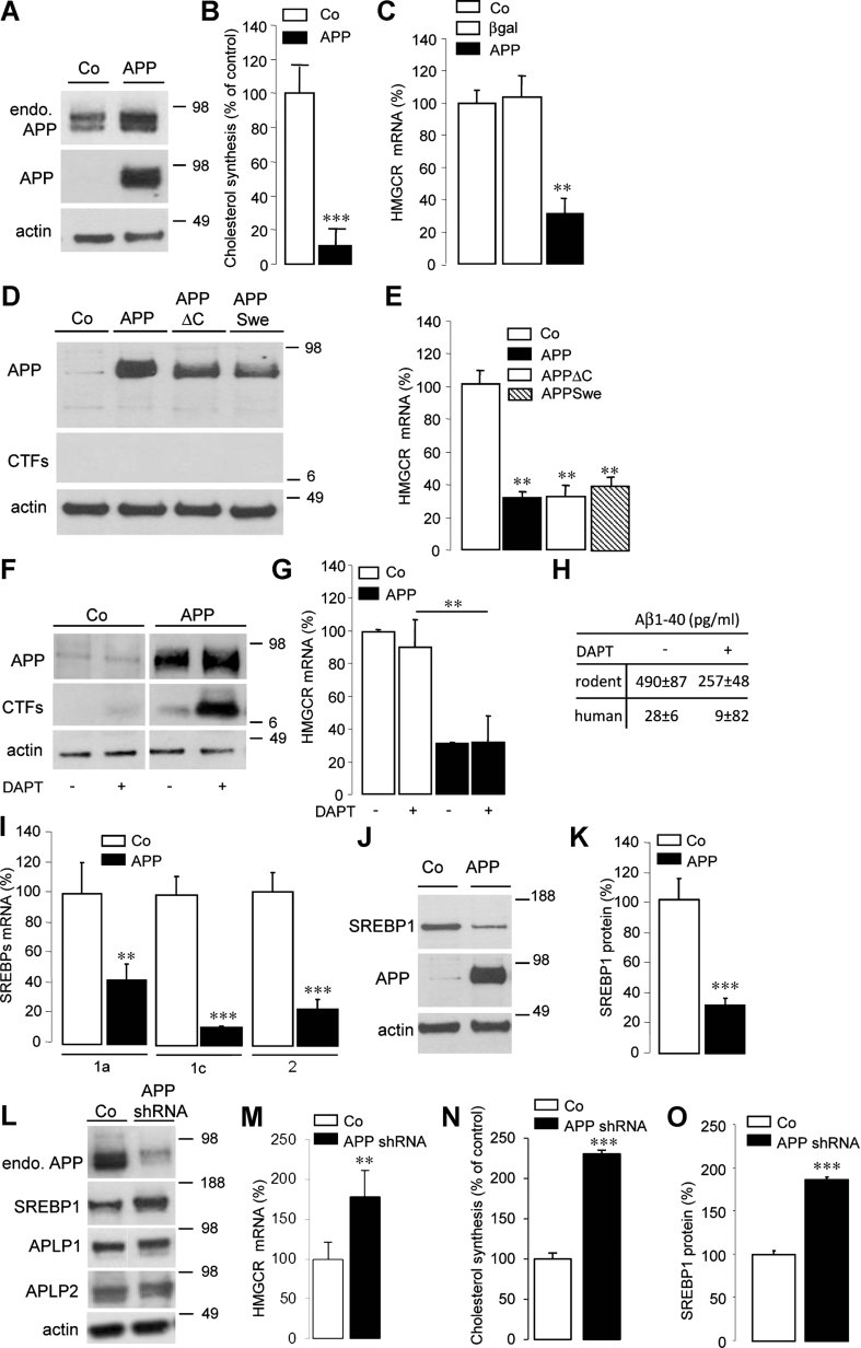
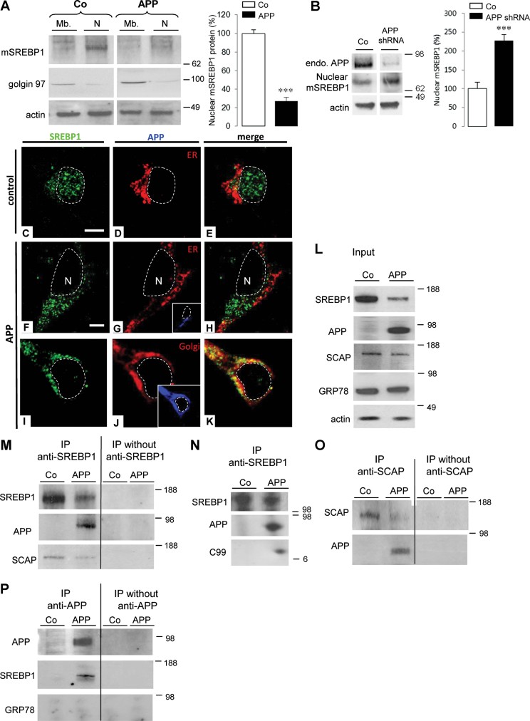
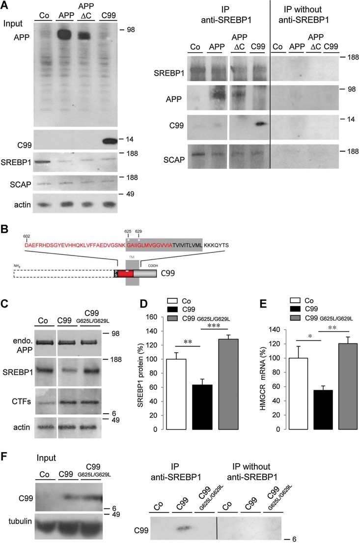
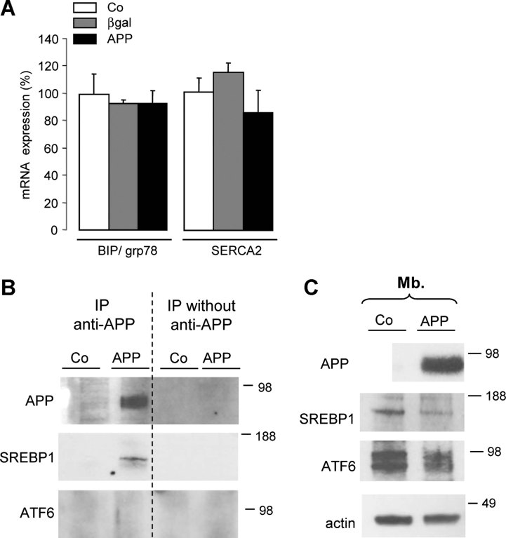
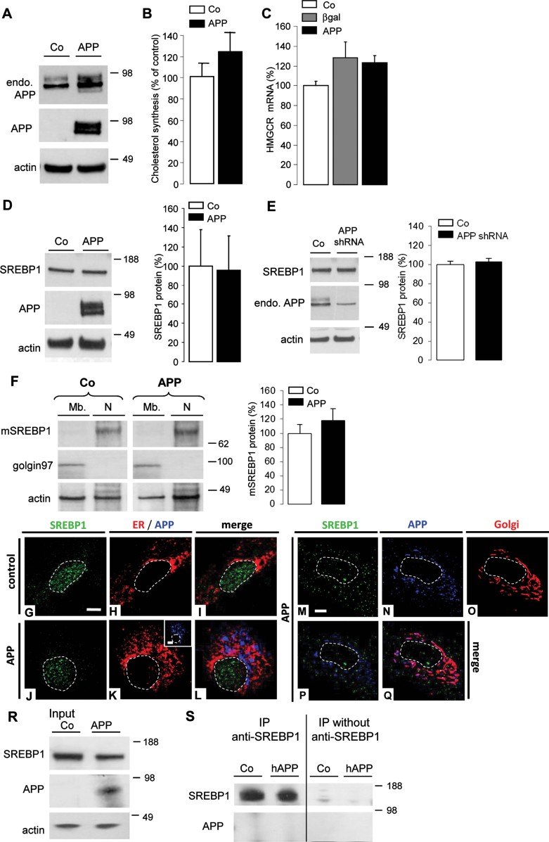
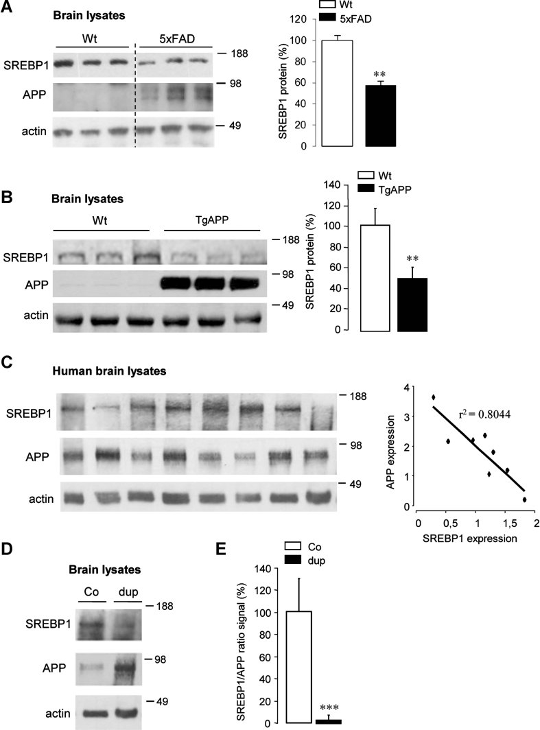
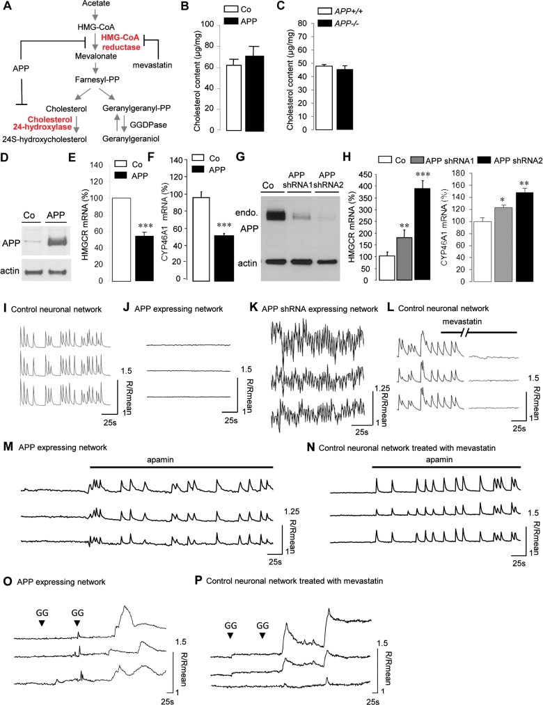
Perturbation of lipid metabolism favours progression of Alzheimer disease, in which processing of Amyloid Precursor Protein (APP) has important implications. APP cleavage is tightly regulated by cholesterol and APP fragments regulate lipid homeostasis. Here, we investigated whether up or down regulation of full-length APP expression affected neuronal lipid metabolism. Expression of APP decreased HMG-CoA reductase (HMGCR)-mediated cholesterol biosynthesis and SREBP mRNA levels, while its down regulation had opposite effects. APP and SREBP1 co-immunoprecipitated and co-localized in the Golgi. This interaction prevented Site-2 protease-mediated processing of SREBP1, leading to inhibition of transcription of its target genes. A GXXXG motif in APP sequence was critical for regulation of HMGCR expression. In astrocytes, APP and SREBP1 did not interact nor did APP affect cholesterol biosynthesis. Neuronal expression of APP decreased both HMGCR and cholesterol 24-hydroxylase mRNA levels and consequently cholesterol turnover, leading to inhibition of neuronal activity, which was rescued by geranylgeraniol, generated in the mevalonate pathway, in both APP expressing and mevastatin treated neurons. We conclude that APP controls cholesterol turnover needed for neuronal activity.


Figures

Figure 1
Figure 1
APP controls neuronal cholesterol synthesis via the SREBP pathway. Source data is available for this figure in the Supporting Information. A–O. Western blots of cell lysates from primary cultures of rat cortical neurons. Neurons were infected or not (A,D,F,J, control, Co) by AdAPP deleted or not from its C-terminal domain (APP or APPΔC, respectively, D), by AdAPPSwe carrying the Swedish mutation (APPSwe, D) and by APPshRNA1 recombinant lentivirus (APPshRNA, L). Expression of endogenous APP (endo. APP) and APP were monitored with anti-APP C-terminal and WO2 antibodies respectively (A). Cells were treated (+) or not (−) with 250 nM DAPT for 8 h and APP C-terminal fragments (CTFs) were analysed with the APP C-terminal antibody (F). Blots were further probed for the N-terminus of the SREBP1, APLP1/2 and actin (L). Cholesterol synthesis was measured by 14C acetate incorporation in neurons expressing APP (B) or APPshRNA (N) compared to Co (n = 4). Comparative qRT-PCR analyses of (C,E,G,M) HMGCR (n = 6) and (I) SREBP2 (n = 4), SREBP1a (n = 4) or SREBP1c (n = 4) mRNA levels in Co (C, E, G, I, M), APP (C, E, G, I, M), APPΔC (E), APPSwe (E), APPshRNA (M), β-galactosidase (βgal, C), APPΔC expressing neurons treated (+) or not (−) with 250 nM DAPT for 8 h (G, n = 6). Results were normalized by GAPDH mRNA and expressed as percentage of Co. (H) Effect of DAPT treatment on rodent and human extracellular Aβ40 release (p < 0.05, n = 6). Quantification of SREBP1/actin ratios; results were expressed as percentage of Co neurons (K, n = 12; O, n = 4), (**p < 0.01; ***p < 0.001).
Figure 2
Figure 2
Neuronal APP and SREBP1 interaction in the Golgi prevents the release of mature SREBP1. Source data is available for this figure in the Supporting Information. A,B. Membranes (Mb) and nuclear (N) fractions from primary cultures of rat cortical neurons infected or not (control, Co) by AdAPP (APP, A) or APPshRNA1 recombinant lentivirus (APPshRNA, B) was analysed by Western blotting for the N-terminus of SREBP1 to detect mature SREBP1 (mSREBP1), stripped and reprobed for golgin 97 (Golgi), anti-APP C-terminal to detect endogenous APP (endo. APP) and actin. APP causes depletion of mSREBP1 in the nuclear fraction. C–K. Subcellular localization of SREBP1 in control (C–E) and APP-expressing neurons (F–K) were analysed by confocal microscopy with anti-N-terminus SREBP1 antibody (green). Inserts show APP expression (blue) detected with the specific WO-2 antibody (G,J). The endoplasmic reticulum (ER, red) is detected with an antibody raised against membrane-associated KDEL-bearing proteins (D,E,G,H). In APP-positive neurons, the Golgi complex (red) was labelled with an anti-TGN46 antibody (J,K), a trans-Golgi marker. (E,H,K), Merged images. Scale bar: 5 µm. L. Cell lysates from rat control neurons (Co) or expressing APP (APP) were analysed by Western blotting (input) using anti-N-terminus SREBP1, specific anti-APP, anti-SCAP, a cargo protein of SREBP1, anti-GRP78, an endoplasmic reticulum chaperon protein, and with anti-actin antibodies. M–P. Cell lysates were immunoprecipitated (IP) with or without the anti-N-terminus SREBP1 (M,N), with the anti-SCAP (O) or the specific anti-APP (P) antibodies and further analysed in Western blotting using anti-APP, -SREBP1, -SCAP or -GRP78 antibodies.
Figure 3
Figure 3
Interaction occurs between the juxta-/transmembrane domain of APP and SREBP1. Source data is available for this figure in the Supporting Information. A. Cell lysates from control neurons (Co) and neurons expressing APP or APP deleted from its C- or N- terminal domain (APPΔC or C99, respectively) were analysed in Western blotting using anti-N-terminus SREBP1, anti-APP, anti-SCAP, and anti-actin antibodies (input). Cell lysates were immunoprecipitated (IP) with or without the anti-N-terminus SREBP1 antibody and further analysed in Western blotting using anti-APP, -SCAP or -SREBP1 antibodies. B. Schematic representation of the transmembrane (TM) and juxtamembrane domains of C99 (APP 695 numbering). For the C99 mutant (G625L/G629L C99 mutant, APP 695 numbering) the two positions (625 and 629) of the amino acid substitution (Gly (G) to Leu (L)), in the first GXXXG motif appear in white stars. Red boxes correspond to the Aβ sequence and SP, signal peptide. C. Western blots of cell lysates from primary cultures of rat cortical neurons expressing or not (control, Co) C99 mutated or not (C99 or C99 G625L/G629L). Expression of endogenous APP (endo. APP) and C-terminal fragments (CTFs) were monitored with anti-APP C-terminal. Blots were further probed for N-terminus SREBP1 and actin. D. Quantification of SREBP1/actin ratios. Results were expressed as percentage of Co neurons (n = 4), (**p < 0.01; ***p < 0.001). E. Comparative qRT-PCR analyses of HMGCR (n = 3) mRNA levels in Co, C99 and C99 G625L/G629L expressing neurons. Results were normalized by GAPDH mRNA and expressed as percentage of Co (*p < 0.1; **p < 0.01). F. Cell lysates from neurons expressing or not (control, Co) C99 mutated or not (C99 or C99 G625L/G629L) were analysed by Western blotting (input, left panel) using WO2 for the detection of C99 and anti-tubulin antibodies. Cell lysates were immunoprecipitated (IP) with or without the anti-N-terminus SREBP1 and further analyzed in Western blotting using WO2 antibody (right panel).
Figure 4
Figure 4
APP inhibits the cleavage of SREBP1 without affecting S2P proteolytic activity. Source data is available for this figure in the Supporting Information. A. Comparative qRT-PCR (n = 3) analysis of BIP/grp78 and SERCA2 mRNA in control (Co), APP and βgal expressing neurons. Results (mean ± SE) are normalized by GAPDH mRNA and expressed as percentage of Co. B. Cellular extracts from control (Co) and APP expressing neurons were immunoprecipitated with the WO-2 antibody. The presence of APP, ATF6 and SREBP1 in the immunoprecipitate was tested by Western blotting with WO-2, MBTPS2 and H160 antibodies, respectively. C. ATF6 expression in membrane (Mb.) protein fractions from control (Co) and APP expressing neurons was analysed by Western blotting with anti-ATF6 antibody. glycosylated (upper band) and non-glycosylated (lower band) forms of ATF6 were detected in both membrane fractions of Co and APP expressing neurons.
Figure 5
Figure 5
APP does not affect SREBP-mediated cholesterol synthesis in astrocytes. Source data is available for this figure in the Supporting Information. A–E. Western blots of cell lysates from primary cultures of rat cortical astrocytes infected or not (control, Co) by AdAPP (A,D, APP) or APPshRNA1 recombinant lentivirus (E, APPshRNA) were probed with antibodies to APP C-terminus (A and E), specific anti-APP (A and D), SREBP1 N-terminus (D and E) and actin (A, D and E). (B) Cholesterol synthesis measured by 14C acetate incorporation in APP expressing astrocytes compared to Co (n = 4). (C) Comparative qRT-PCR analyses of HMGCR (n = 3) mRNA levels in Co, APP and β-galactosidase (βgal) expressing astrocytes. Results were normalized by GAPDH mRNA and expressed as percentage of Co. (D,E) SREBP1/actin ratios were quantified and expressed as percentage of Co (n = 5) (right panels). F. Membrane (Mb.) and nuclear (N) fractions of Co and APP expressing astrocytes were analysed by Western blotting using an antibody raised against the N-terminus of SREBP1 to detect mature SREBP1 (mSREBP1). Blots were then probed with anti-golgin 97 and anti-actin antibodies. G–Q. Cellular localization of SREBP1 in control (G–I) and APP-expressing astrocytes (J–Q) was analysed by confocal multiplex fluorescence with anti-N-terminus SREBP1 antibody (green). Inserts show APP expression (blue) detected with the WO-2 antibody (K). The endoplasmic reticulum (ER, red) is marked for KDEL-bearing proteins (H,I,K,L). (M–Q) In APP expressing astrocytes (blue), cytoplasmic SREBP1 was labelled with an anti-Cterminus C20 (green, M,P) and Golgi apparatus with GM130 (red, O,Q) antibodies. (I,L,P,Q) Merged images. Scale bar: 5 µm. R. Cell lysates from Co and APP expressing astrocytes were analysed by Western blotting using anti-Nterminus SREBP1, -APP and -actin antibodies. S. Cell lysates were immunoprecipitated (IP) with or without the anti-N-terminus SREBP1 antibody and further analysed in Western blotting using anti-APP and -SREBP1 antibodies.
Figure 6
Figure 6
APP inversely correlates with SREBP1 in mice and man. Source data is available for this figure in the Supporting Information. A,B. Western blots analysis of brain tissue from wild type (Wt), 5×FAD mice at 15 months of age (A) and APP transgenic mice (TgAPP) at 5 months of age (B). The expression of APP was detected with anti-APP specific antibody, SREBP1 was detected with the anti-N-terminus SREBP1 antibody and protein loading was controlled using anti-actin antibody (left panels). SREBP1/actin ratios were quantified (n = 6, A and n = 3, B) and expressed as percentage of Wt controls (right panels). C. Western blot analysis of postmortem human brain homogenates with anti-APP specific antibody, SREBP1 and actin antibodies (left panel). Quantification of densitometry arbitrary units indicating an inverse correlation between APP and SREBP1 levels (right panel). D. Western blot analysis of postmortem human brain homogenates from control brain (Co) and an Alzheimer patient carrying a microduplication of the APP locus (dup) (left panel). The SREBP1/APP ratios were quantified on several Western blots from Co and dup brains and expressed as percentage of Co (n = 4) (right panel); (**p < 0.01, ***p < 0.0001).
Figure 7
Figure 7
Inhibition of mevalonate pathway by APP impairs neuronal activity. Source data is available for this figure in the Supporting Information. A. Diagram of the mevalonate pathway. HMG-CoA reductase (HMGCR), the rate-limiting enzyme in the biosynthesis of cholesterol and cholesterol 24-hydroxylase, converting cholesterol into 24S-hydroxycholesterol, are involved in cholesterol turnover. HMGCR is inhibited by statins drugs such as mevastatin but also after APP expression. Farnesyl diphosphate (farnesyl-PP), geranylgeranyl diphosphate (geranylgeranyl-PP) and geranylgeraniol are polyisoprenoid end products of the mevalonate pathway. GGDPase (geranylgeranyl diphosphatase). A,C. Cholesterol content in cell membranes from control (Co), APP expressing neurons (APP, B) and from primary cultures of mouse cortical neurons prepared from wild type (APP+/+) and APP knockout mice (APP−/−, C), (n = 6). D–H. Western blots analysis of cell lysates from rat cortical neurons infected or not (control, Co) with AdAPP (G, APP) or APPshRNA1 and two recombinant lentiviruses (G). The expression of APP and endogenous APP (endo. APP) were detected with anti-APP specific and anti-APPC-terminal antibodies, respectively; and protein loading was controlled using anti-actin antibody. Comparative qRT-PCR analysis of HMGCR mRNA in Co, APP (E) and APPshRNA1 and 2 neurons (H, left panel) respectively. Results (n = 6) are normalized by GAPDH mRNA and expressed as percentage of Co. Comparative qRT-PCR analysis of CYP46A1 mRNA in Co, APP (F) and APP shRNA1 and 2 neurons (H, right panel), respectively. Results (n = 6) are normalized by GAPDH mRNA and expressed as percentage of Co. I–P. Traces of free cytosolic calcium concentration in three neurons of a coverslip, measured with Fura-2 AM at 13 days in vitro in control (I), APP (J) and APPshRNA neurons (K), or in control neurons before and 30 min (//) after addition of mevastatin (12.5 µM) (L). Simultaneous traces of [Ca2+] in three cells of APP expressing neurons (M), and in control neurons pretreated with mevastatin (12.5 µM, for 12 h) (N) network show that cells recover synchronous calcium oscillations after bath application of 200 nM apamin, an SK channel inhibitor. In APP expressing neurons (O), and in control neurons pretreated with mevastatin (12.5 µM) for 12 h (P), a two round of geranylgeraniol treatment (GG, 2 mM) is needed to rescue neuronal activity. Traces are expressed as R/Rmean, were R is the ratio of F340/F380 and Rmean is the mean ratio value after recording.

Similar articles

Cited by

References

    1. Aydin D, Weyer SW, Muller UC. Functions of the APP gene family in the nervous system: insights from mouse models. Exp Brain Res. 2012;217:423–434. - PubMed
    1. Barrett PJ, Song Y, Van Horn WD, Hustedt EJ, Schafer JM, Hadziselimovic A, Beel AJ, Sanders CR. The amyloid precursor protein has a flexible transmembrane domain and binds cholesterol. Science. 2012;336:1168–1171. - PMC - PubMed
    1. Beel AJ, Mobley CK, Kim HJ, Tian F, Hadziselimovic A, Jap B, Prestegard JH, Sanders CR. Structural studies of the transmembrane C-terminal domain of the amyloid precursor protein (APP): does APP function as a cholesterol sensor. Biochemistry. 2008;47:9428–9446. - PMC - PubMed
    1. Bengoechea-Alonso MT, Ericsson J. SREBP in signal transduction: cholesterol metabolism and beyond. Curr Opin Cell Biol. 2007;19:215–222. - PubMed
    1. Bligh EG, Dyer WJ. A rapid method of total lipid extraction and purification. Can J Biochem Physiol. 1959;37:911–917. - PubMed

Publication types

MeSH terms

Substances