

Mobile digital fluorescence microscopy for diagnosis of tuberculosis
- PMID: 23554191
- PMCID: PMC3716054
- DOI: 10.1128/JCM.03432-12
Mobile digital fluorescence microscopy for diagnosis of tuberculosis
Abstract
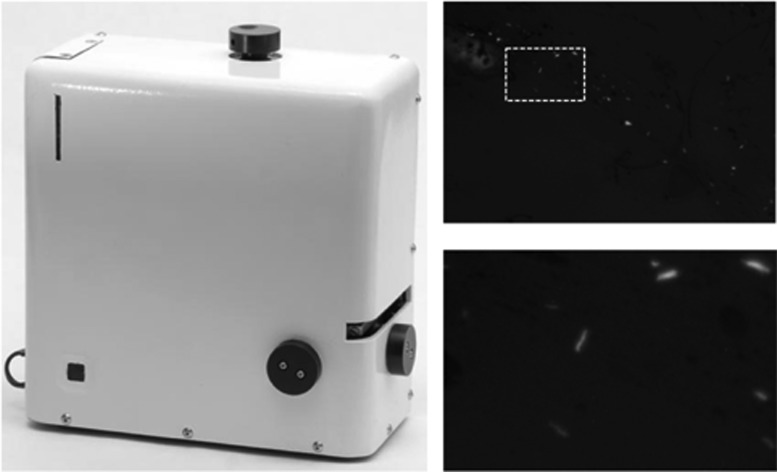
Access to sputum smear microscopy in high-tuberculosis (TB)-burden regions is limited by a scarcity of microscopes and experienced technicians. We evaluated the accuracy of CellScope, a novel digital fluorescence microscope that may expand access to microscopy. The study utilized smear microscopy slides prepared from sputum specimens submitted by consecutive adults with ≥ 2 weeks of cough who were admitted to Mulago Hospital (Kampala, Uganda). Conventional light-emitting diode (LED) fluorescence microscopy (FM) and mycobacterial culture were performed by experienced technicians. Two U.S.-based postgraduate researchers without prior microscopy experience restained, imaged, and interpreted the slides using CellScope. We assessed whether sensitivity and specificity of CellScope-based LED FM was noninferior to conventional LED FM by using a preselected margin of inferiority of 15%. Of 525 patients included, 72% were HIV seropositive and 39% had culture-confirmed TB. The proportions of positive results were similar with CellScope and conventional LED FM (34% versus 32%, respectively; P = 0.32), and agreement was substantial. CellScope accuracy was within the noninferiority margin for both sensitivity (63% versus 70%; difference, -7%; 95% confidence interval [CI], -13% to -1%) and specificity (85% versus 92%; difference, -7%; 95% CI, -12% to -3%). A subanalysis of 43 slides evaluated by each CellScope reader found substantial interreader reliability (custom-weighted kappa, 0.65) and variable intrareader reliability (custom-weighted kappa, 0.11 versus 0.48). CellScope offers promise for expanding microscopy services. Future studies should evaluate the device when operated by health workers in low-resource settings, the feasibility of image transmission and analysis by experienced microscopists, and the accuracy of automated image analysis algorithms.
Figures
References
-
- Lawn SD, Zumla AI. 2011. Tuberculosis. Lancet 378:57–72 - PubMed
-
- World Health Organization 2012. Global tuberculosis report 2012. World Health Organization, Geneva, Switzerland
-
- Keeler E, Perkins MD, Small P, Hanson C, Reed S, Cunningham J, Aledort JE, Hillborne L, Rafael ME, Girosi F, Dye C. 2006. Reducing the global burden of tuberculosis: the contribution of improved diagnostics. Nature 444(Suppl 1):49–57 - PubMed
-
- Dowdy DW, Chaisson RE, Moulton LH, Dorman SE. 2006. The potential impact of enhanced diagnostic techniques for tuberculosis driven by HIV: a mathematical model. AIDS 20:751–762 - PubMed
-
- Boehme CC, Nabeta P, Hillemann D, Nicol MP, Shenai S, Krapp F, Allen J, Tahirli R, Blakemore R, Rustomjee R, Milovic A, Jones M, O'Brien SM, Persing DH, Ruesch-Gerdes S, Gotuzzo E, Rodrigues C, Alland D, Perkins MD. 2010. Rapid molecular detection of tuberculosis and rifampin resistance. N. Engl. J. Med. 363:1005–1015 - PMC - PubMed
Publication types
MeSH terms
Grants and funding
LinkOut - more resources
Full Text Sources
Other Literature Sources
Medical
