Review
. 2013 Apr;8(4):434-47.
doi: 10.1002/biot.201200199.

Perivascular cells in blood vessel regeneration

Affiliations
Review

Perivascular cells in blood vessel regeneration

Maureen Wanjare et al. Biotechnol J. 2013 Apr.

Abstract

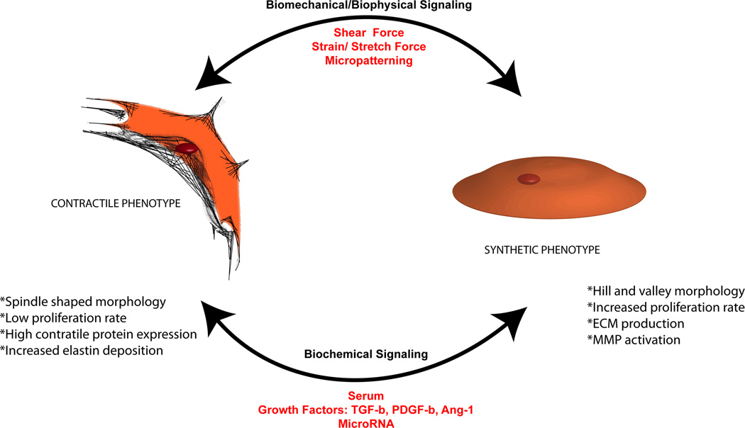
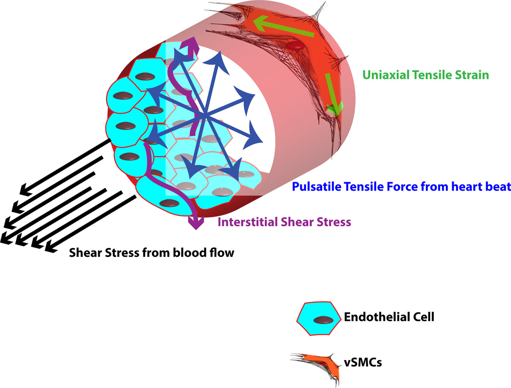
Vascular engineering seeks to design and construct functional blood vessels comprising endothelial cells (ECs) and perivascular cells (PCs), with the ultimate goal of clinical translation. While EC behavior has been extensively investigated, PCs play an equally significant role in the development of novel regenerative strategies, providing functionality and stability to vessels. The two major classes of PCs are vascular smooth muscle cells (vSMCs) and pericytes; vSMCs can be further sub-classified as either contractile or synthetic. The inclusion of these cell types is crucial for successful regeneration of blood vessels. Furthermore, understanding distinctions between vSMCs and pericytes will enable improved therapeutics in a tissue-specific manner. Here we focus on the approaches and challenges facing the use of PCs in vascular regeneration, including their characteristics, stem cell sources, and interactions with ECs. Finally, we discuss biochemical and microRNA (miR) regulators of PC behavior and engineering approaches that mimic various cues affecting PC function.


Conflict of interest statement

The authors declare no conflict of interest.

Figures

Figure 1
Figure 1
Phenotypic plasticity of vSMCs. Characteristics of the synthetic and contractile phenotypes – including morphology, proliferation, ECM and contractile protein expression, and phenotypic switch – are regulated by various biochemical and biomechanical cues.
Figure 2
Figure 2
Functionality of hPSC derivatives. Synthetic-vSMCs and contractile-vSMCs derived from hPSCs, transplanted subcutaneously, were shown to: (A) migrate to the host vasculature and locate in the outer layers of the mouse blood vessels that penetrated into the Matrigel plug. (B) On some occasions, the human contractile vSMCs were found to wrap the smaller mouse vasculature circumferentially. Human cells in red, mouse vasculature in green, and nuclei in blue. Some human cells are indicated with white arrows. For details about method please refer to ref. [75].
Figure 3
Figure 3
Comparison of hESC-derived SMLCs and control vSMCs. Comparative immunofluorescence analysis demonstrates the expression of specific SMC markers such as SMA, calponin, SM22, and SM-MHC. Scale bar is 100 µm. Reproduced with kind permission from Springer Science and Business Media [51].
Figure 4
Figure 4
Network stabilization by SMLCs. hESC-derived SMLCs contribute to formation and stabilization of endothelial progenitor cell (EPC) networks. Fluorescent microscopy images of viable cord like structures formed on Matrigel following seeding with ratios of 100:0, 60:40, 40:60, 20:80, and 0:100 (EPCs:SMLCs). Reproduced with kind permission from Springer Science and Business Media [51].
Figure 5
Figure 5
Biomechanical forces affecting vSMCs. Various forces in the blood vessel act on the vSMCs, including shear stress, interstitial shear stress, pulsatile tensile force, and uniaxial tensile strain.

Similar articles

Cited by

References

    1. Discher DE, Mooney DJ, Zandstra PW. Growth factors, matrices, and forces combine and control stem cells. Science. 2009;324:1673–1677. - PMC - PubMed
    1. Mancini ML, Terzic A, Conley BA, Oxburgh LH, et al. Endoglin plays distinct roles in vascular smooth muscle cell recruitment and regulation of arteriovenous identity during angiogenesis. Dev. Dynam. 2009;238:2479–2493. - PMC - PubMed
    1. Lindahl P, Johansson BR, Levéen P, Betsholtz C. Pericyte loss and microaneurysm formation in PDGF-B-deficient mice. Science. 1997;277:242–245. - PubMed
    1. Alberts B, Johnson A, Lewis J, Raff M, Roberts K, Walter P. Molecular Biology of the Cell. New York: Garland Science; 2002.
    1. Armulik A, Genove G, Mae M, Nisancioglu MH, et al. Pericytes regulate the blood-brain barrier. Nature. 2010;468:557–561. - PubMed

Publication types