. 2013 Jul;31(7):1363-70.
doi: 10.1002/stem.1385.

Uterine vasculature remodeling in human pregnancy involves functional macrochimerism by endothelial colony forming cells of fetal origin

Affiliations
Free PMC article

Uterine vasculature remodeling in human pregnancy involves functional macrochimerism by endothelial colony forming cells of fetal origin

Peter I Sipos et al. Stem Cells. 2013 Jul.
Free PMC article

Abstract

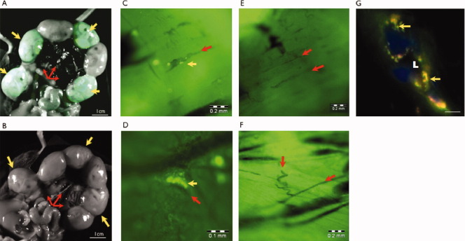
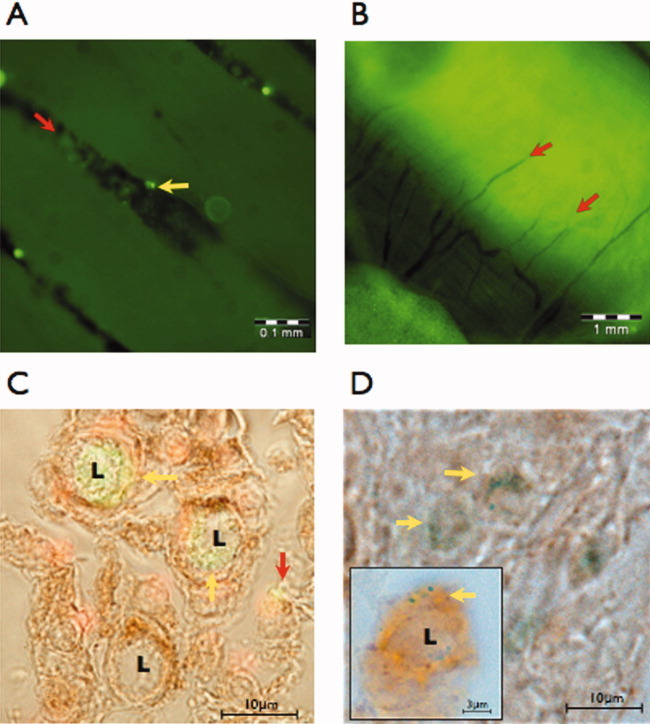
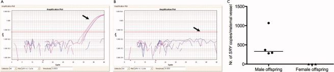
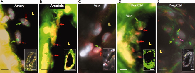
The potency of adult-derived circulating progenitor endothelial colony forming cells (ECFCs) is drastically surpassed by their fetal counterparts. Human pregnancy is associated with robust intensification of blood flow and vascular expansion in the uterus, crucial for placental perfusion and fetal supply. Here, we investigate whether fetal ECFCs transmigrate to maternal bloodstream and home to locations of maternal vasculogenesis, primarily the pregnant uterus. In the first instance, endothelial-like cells, originating from mouse fetuses expressing paternal eGFP, were identified within uterine endothelia. Subsequently, LacZ or enhanced green fluorescent protein (eGFP)-labeled human fetal ECFCs, transplanted into immunodeficient (NOD/SCID) fetuses on D15.5 pregnancy, showed similar integration into the mouse uterus by term. Mature endothelial controls (human umbilical vein endothelial cells), similarly introduced, were unequivocally absent. In humans, SRY was detected in 6 of 12 myometrial microvessels obtained from women delivering male babies. The copy number was calculated at 175 [IQR 149-471] fetal cells per millimeter square endothelium, constituting 12.5% of maternal vessel lumina. Cross-sections of similar human vessels, hybridized for Y-chromosome, positively identified endothelial-associated fetal cells. It appears that through ECFC donation, fetuses assist maternal uterine vascular expansion in pregnancy, potentiating placental perfusion and consequently their own fetal supply. In addition to fetal growth, this cellular mechanism holds implications for materno-fetal immune interactions and long-term maternal vascular health.

Keywords: Chimerism; Circulation; Maternal-Fetal Exchange; Neovascularization; Physiologic; Uteroplacenta.


Figures

Figure 1
Figure 1
Fetal cells of endothelial characteristics cross the mouse placenta and colonize the uterine microvasculature in a transgenic murine model of enhanced green fluorescent protein (eGFP)-expressing offspring. Images taken with small animal imager demonstrated eGFP-positive offspring (yellow arrows) in dissected intact uterine horns of eGFP-negative mothers, supplied by vessels of broad ligaments (red arrows, A). Pups (yellow arrows) did not express eGFP in the event of control matings (B). Fluorescent cells of fetal origin were detected by small animal imager at D18.5 within maternal uteri (C) and broad ligaments (D). These cells (yellow arrows) were aligned along lumina of arterial and venous branches (red arrows), and frequently formed obvious vascular colonies, while eGFP signals were undetected in uteri (E) and broad ligaments (F) of controls. With crossmating, immunofluorescence of uterine cross-sections (G) showed coexpression (amber, yellow arrows) of fetus-associated eGFP (green) and endothelial Von Willebrand Factor (red) in cells surrounding the vessel lumina (L), confirming fetal origin and endothelial character. Blue = Nuclei. Scale bars (unless indicated) = 10 μm.
Figure 2
Figure 2
Transplanted fetal-derived human endothelial colony forming cells (ECFCs) traverse the mouse placenta and home to the pregnant uterus. On D15.5, enhanced green fluorescent protein (eGFP) and LacZ expressing culture-propagated human cord blood ECFCs were transplanted by ultrasound guided intracardiac injection into Non-Obese Diabetes/Severe Combined Immunodeficient (NOD/SCID) mouse fetuses. Using a small animal imager, eGFP-fluorescent cells (yellow arrows) were subsequently located along the vascular lumina (red arrows) within the pregnant mouse uterus (A). Although detectable in the blood of mouse placentas [8], fluorescent-tracked mature endothelial cells (human umbilical vein endothelial cells) could not be defined in the maternal uterine microvasculature (B). Combined fluorescence and light microscopy of uterine tissues (red nuclei) showed transplanted eGFP-ECFCs (yellow arrows) undertaking de novo lumen (L) formation. Positive staining for claudin-5, a tight junction marker (brown stain), confirms their integration, while nonintegrated ECFCs fail to express tight junctions (red arrow). (C) Transplanted human LacZ-ECFCs (punctate blue stain, yellow arrows) similarly demonstrated vasculogenic activities, including vacuole formation (L, insert), and expressed the gap junction marker connexin 40 (brown), confirming full vascular integration (D).
Figure 3
Figure 3
The presence of fetal-derived male cells within maternal human uterine vessels. Reverse transcription quantitative polymerase chain reaction (PCR) amplification plots of the male-specific SRY gene in microvessels from biopsies of the upper uterine segments showed gene presence in 6 of 12 cases of mothers with male offspring (A, arrow points at positive SRY-amplification, fluorescence higher than threshold [red line]). Gene copies were absent in women with female babies (B, n = 3, arrow points at intact detection threshold). Horizontal axes: PCR cycle numbers, vertical axes: ΔRn (arbitrary units (log)). In 4 of 6 positive cases the copies of SRY were successfully quantified (C).
Figure 4
Figure 4
Endothelial location of fetal-derived cells within human uterine vessels. Y-chromosome-specific FISH in uterine tissue cross-sections detected male cells in the endothelium in 8 of 11 mothers with male babies. Examples given for prominent Y-signals in detached endothelial cells of a larger arterial branch (A), a colony of fetal cells in the intact endothelial lining of an arteriole (B) and fetal cells in a venous endothelium (C). Male placentas were used as positive controls (D, n = 4), and uterine tissue of mothers with female offspring as negatives (E, n = 2, magenta arrow = artifact resembling Y-signal but outside nuclear margin). Somatic chromosomes were hybridized for internal control (red color/arrows: Cy3-conjugated Y chromosomes, green color/arrows: Fluorescein isothiocyanate (FITC)-conjugated chromosomes 20 or 15, gray: 4’,6-diamidino-2-phenylindole (DAPI), L: lumen, Scale bars = 5 μm, 20 μm (insets), 50 μm (inset A)).

Similar articles

Cited by

References

    1. Berg CJ, Mackay AP, Qin C, et al. Overview of maternal morbidity during hospitalization for labor and delivery in the United States: 1993–1997 and 2001–2005. Obstet Gynecol. 2009;113:1075–1081. - PubMed
    1. Mikolajczyk RT, Zhang J, Betran AP, et al. A global reference for fetal-weight and birthweight percentiles. Lancet. 2011;377:1855–1861. - PubMed
    1. Romo A, Carceller R, Tobajas J. Intrauterine growth retardation (IUGR): Epidemiology and etiology. Pediatr Endocrinol Rev. 2009;6(suppl 3):332–336. - PubMed
    1. Barker DJ, Osmond C, Forsen TJ, et al. Trajectories of growth among children who have coronary events as adults. N Engl J Med. 2005;353:1802–1809. - PubMed
    1. Fraser A, Nelson SM, Macdonald-Wallis C, et al. Associations of pregnancy complications with calculated CVD risk and cardiovascular risk factors in middle age: The Avon Longitudinal Study of Parents and Children. Circulation. 2012;15:1367–1380. - PMC - PubMed

Publication types

MeSH terms