. 2013 May 1;190(9):4830-5.
doi: 10.4049/jimmunol.1202884. Epub 2013 Apr 3.

Connexin43 is dispensable for phagocytosis

Affiliations

Connexin43 is dispensable for phagocytosis

Aaron M Glass et al. J Immunol. .

Abstract

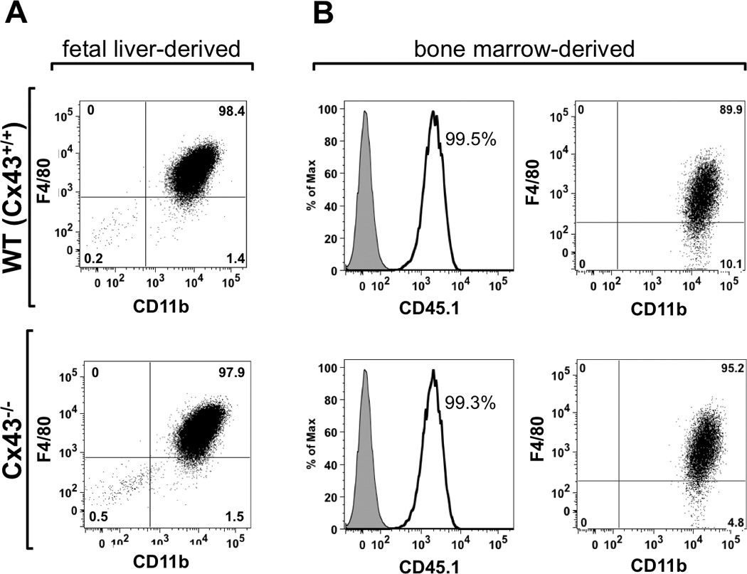
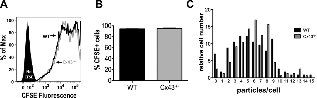
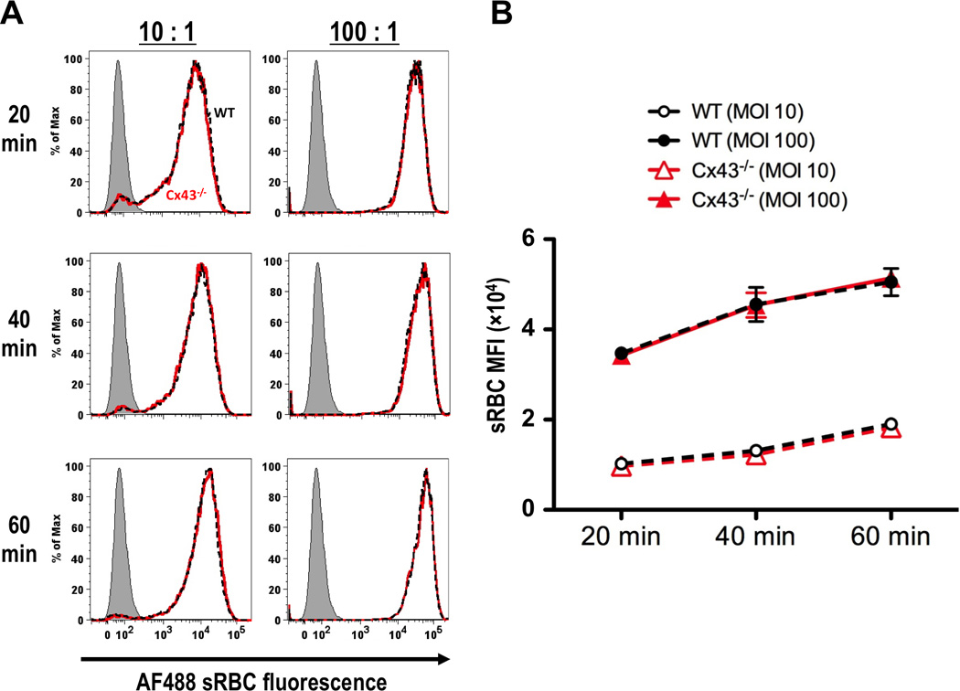
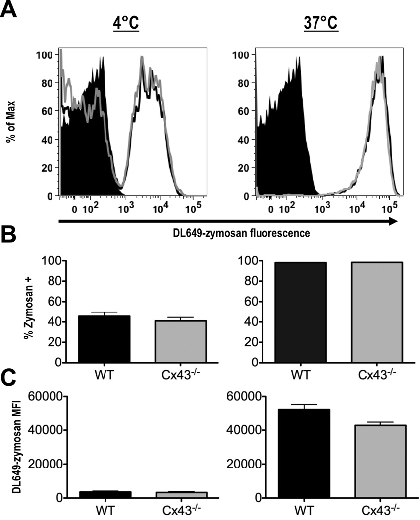
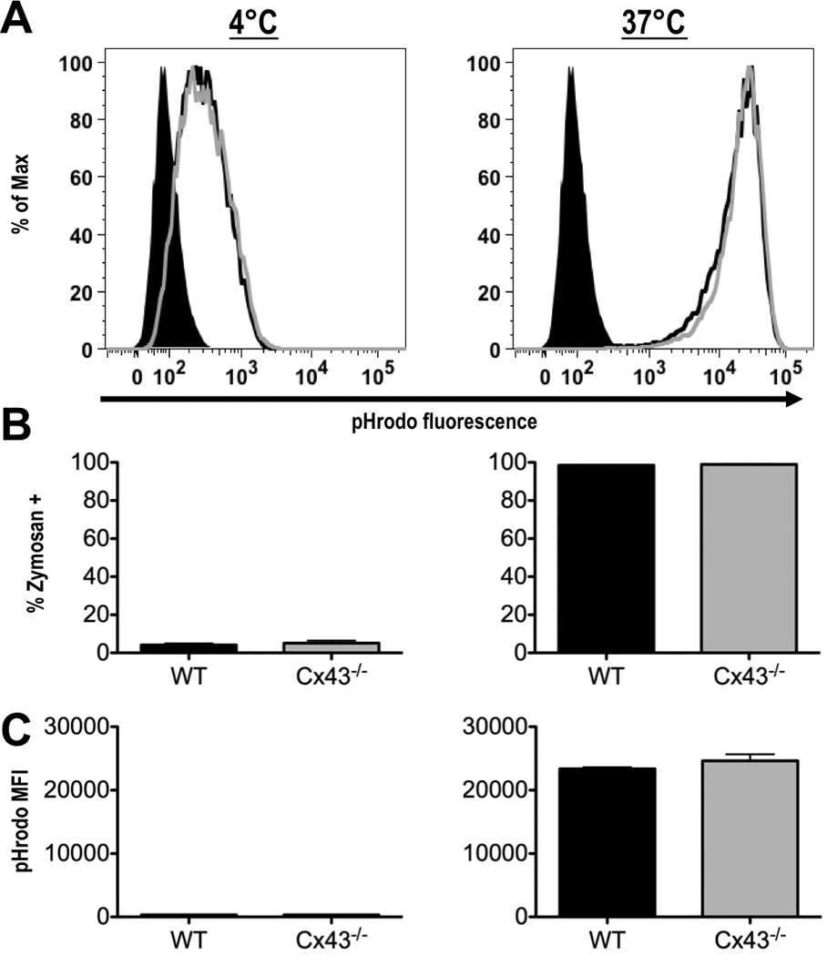
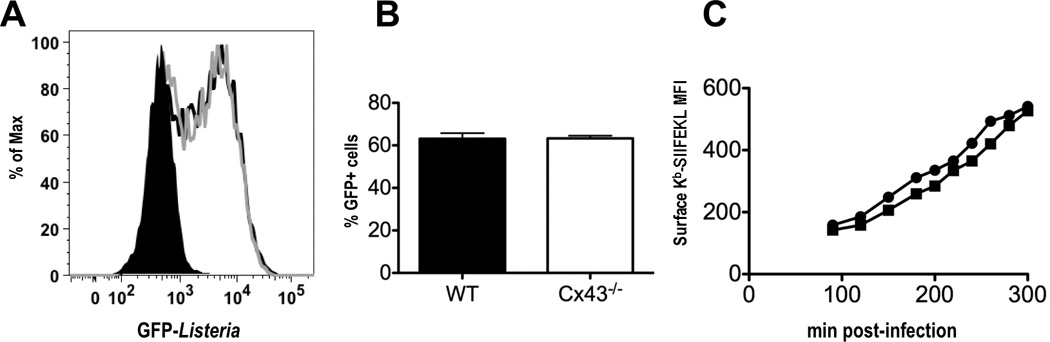
Macrophages that lack connexin43 (Cx43), a gap junction protein, have been reported to exhibit dramatic deficiencies in phagocytosis. In this study, we revisit these findings using well-characterized macrophage populations. Cx43 knockout (Cx43(-/-)) mice die soon after birth, making the harvest of macrophages from adult Cx43(-/-) mice problematic. To overcome this obstacle, we used several strategies: mice heterozygous for the deletion of Cx43 were crossed to produce Cx43(+/+) (wild type [WT]) and Cx43(-/-) fetuses. Cells isolated from 12- to 14-d fetal livers were used to reconstitute irradiated recipient animals. After reconstitution, thioglycollate-elicited macrophages were collected by peritoneal lavage and bone marrow was harvested. Bone marrow cells and, alternatively, fetal liver cells were cultured in media containing M-CSF for 7-10 d, resulting in populations of cells that were >95% macrophages based on flow cytometry. Phagocytic uptake was detected using flow cytometric and microscopic techniques. Quantification of phagocytic uptake of IgG-opsonized sheep erythrocytes, zymosan particles, and Listeria monocytogenes failed to show any significant difference between WT and Cx43(-/-) macrophages. Furthermore, the use of particles labeled with pH-sensitive dyes showed equivalent acidification of phagosomes in both WT and Cx43(-/-) macrophages. Our findings suggest that modulation of Cx43 levels in cultured macrophages does not have a significant impact on phagocytosis.


Figures

FIGURE 1
FIGURE 1
Characterization of the macrophages populations used in this study. (A) Wild type (WT, top panel) and Cx43−/− (bottom panel) macrophages derived from fetal liver cells were stained for the macrophage markers F4/80 and CD11b. In both cases, cultures contained a high proportion of double-positive cells, indicating that these cultures were predominantly comprised of macrophages. (B) WT (top panel) and Cx43−/− (bottom panel) bone marrow-derived macrophages from chimeric mice were stained for the donor marker, CD45.1, as well as CD11b and F4/80. Nearly all of the cultured macrophages were donor derived, and of these a high proportion were positive for macrophage markers, suggesting that WT and Cx43−/− populations were essentially pure and faithful to their respective genotypes.
FIGURE 2
FIGURE 2
WT and Cx43−/− macrophages are equally capable of phagocytosing opsonized sRBCs. (A) Wild type (black line) and Cx43−/− (gray line) macrophages were incubated for 20 min with IgG-opsonized CFSE-labeled sRBCs and analyzed by flow cytometry (shaded black peak represents macrophages incubated with unlabeled sRBCs). (B) Percentage of cells positive for CFSE fluorescence after 20 minutes of incubation with opsonized sRBCs (average and S.E.M. of 8 experiments). (C) Histogram of phagocytosis by WT (black bars) and Cx43−/− (grey bars) fetal liver-derived macrophages after 20 min of incubation with sRBCs. Histograms were normalized for cell counts by dividing the number of cells in each bin by the total number of cells counted. At least 100 cells were counted for each genotype.
FIGURE 3
FIGURE 3
Bone marrow-derived macrophages from radiation chimeric mice reconstituted with fetal liver cells from WT and Cx43−/− mice are equally capable of phagocytosis of sRBCs. (A) Fluorescence histograms comparing sRBC uptake of WT (black dashed lines) and Cx43−/− (red lines) bone marrow-derived macrophages after 20, 40 and 60 min co-incubation. Macrophages were treated with particles at 10:1 (left panels) and 100:1 (right panels) particle-to-macrophage ratios. Grey shaded peaks represent non-fluorescent sRBCs. (B) Kinetic plot of phagocytosis by bone marrow-derived macrophages from radiation chimeric animals reconstituted with WT (black dashed lines) and Cx43−/− (red lines) fetal liver cells. Macrophages were incubated with 100:1 (closed symbols) and 10:1 (open symbols) sRBC to macrophage ratios for 20, 40 and 60 min time points before analysis by flow cytometry (average and S.E.M. for 3 experiments).
FIGURE 4
FIGURE 4
Fetal liver-derived macrophages phagocytose zymosan, independently of Cx43 genotype. (A) Wild type (black line) and Cx43−/− (gray line) macrophages were incubated with DL649-labeled zymosan particles for 60 min at 4°C (left panel), or at 37°C (right panel), (black peak represents macrophages without zymosan). (B) Percentage of cells positive for labeled zymosan after incubation on at 4°C (left panel), or at 37°C (right panel). (C) Mean fluorescence intensity (MFI) of DL649 signal in macrophages incubated at 4°C (left panel), or at 37°C (right panel), (averages from 3 experiments, +/− S.E.M).
FIGURE 5
FIGURE 5
Fetal liver-derived macrophages from wild type and Cx43−/− mice can phagocytose zymosan, and are capable of phagosome acidification. (A) Wild type (black line) and Cx43−/− (gray line) macrophages were incubated with zymosan particles labeled with pHrodo for 60 min at 4°C (left panel), or at 37°C (right panel), (shaded black peak represents macrophages not treated with zymosan). (B) Percentage of cells positive for labeled zymosan after incubation at 4°C (left panel), or at 37°C (right panel). (C) Mean fluorescence intensity (MFI) of pHrodo signal in macrophages incubated at 4°C (left panel), or at 37°C (right panel), (averages from 3 experiments, error bars depict S.E.M.).
FIGURE 6
FIGURE 6
Cx43 deficient macrophages are capable of phagocytosis of Listeria monocytogenes and presentation of a foreign antigen. (A) Flow cytometry histogram of gated live cells demonstrating engulfment of GFP-positive Listeria at 90 min post-infection; black line represents Listeria uptake by WT macrophages, gray line represents uptake by Cx43−/− cells. (B) Percentage of WT and Cx43−/− macrophages positive for GFP fluorescence at 90 min post-infection (graph representative of three independent experiments. (C) Kinetics of surface Kb-SIINFEKL production in WT (circle) and Cx43−/− (square). Analysis of Kb-SIINFEKL was limited to live, GFP-positive cells only. One representative experiment of three is shown.

References

    1. Mendoza-Naranjo A, Bouma G, Pereda C, Ramirez M, Webb KF, Tittarelli A, Lopez MN, Kalergis AM, Thrasher AJ, Becker DL, Salazar-Onfray F. Functional gap junctions accumulate at the immunological synapse and contribute to T cell activation. J Immunol. 2011;187:3121–3132. - PMC - PubMed
    1. Pang B, Neijssen J, Qiao X, Janssen L, Janssen H, Lippuner C, Neefjes J. Direct antigen presentation and gap junction mediated cross-presentation during apoptosis. J Immunol. 2009;183:1083–1090. - PubMed
    1. Neijssen J, Pang B, Neefjes J. Gap junction-mediated intercellular communication in the immune system. Prog Biophys Mol Biol. 2007;94:207–218. - PubMed
    1. Nguyen TD, Taffet SM. A model system to study Connexin 43 in the immune system. Mol Immunol. 2009;46:2938–2946. - PubMed
    1. Anand RJ, Dai S, Gribar SC, Richardson W, Kohler JW, Hoffman RA, Branca MF, Li J, Shi XH, Sodhi CP, Hackam DJ. A role for connexin43 in macrophage phagocytosis and host survival after bacterial peritoneal infection. J Immunol. 2008;181:8534–8543. - PMC - PubMed

Publication types

MeSH terms