. 2013 Apr 3;33(14):6143-53.
doi: 10.1523/JNEUROSCI.5399-12.2013.

The p38α MAPK regulates microglial responsiveness to diffuse traumatic brain injury

Affiliations

The p38α MAPK regulates microglial responsiveness to diffuse traumatic brain injury

Adam D Bachstetter et al. J Neurosci. .

Abstract

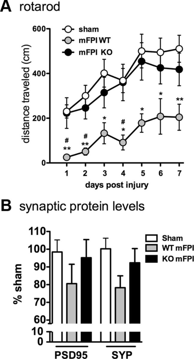
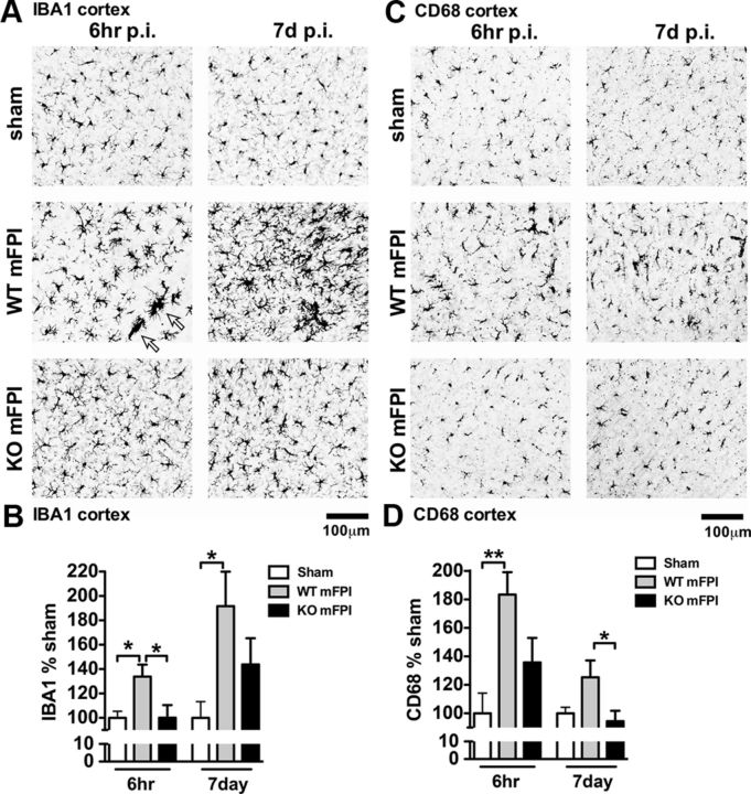
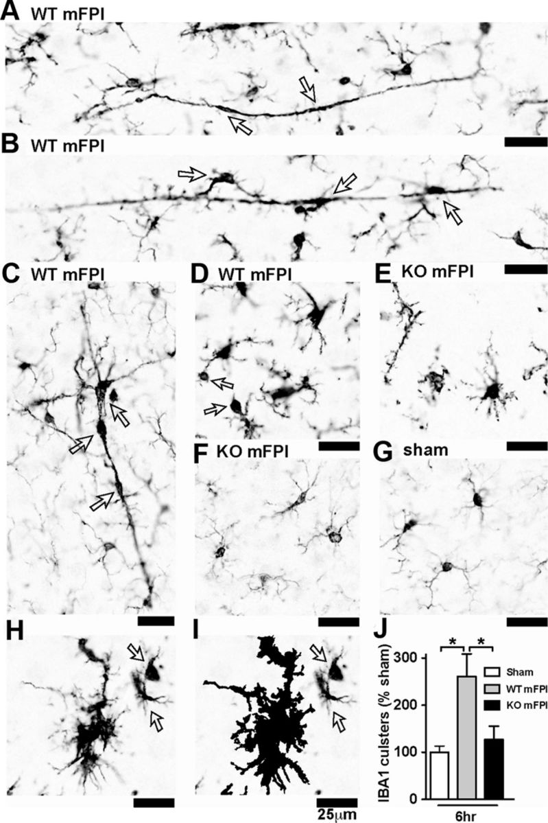
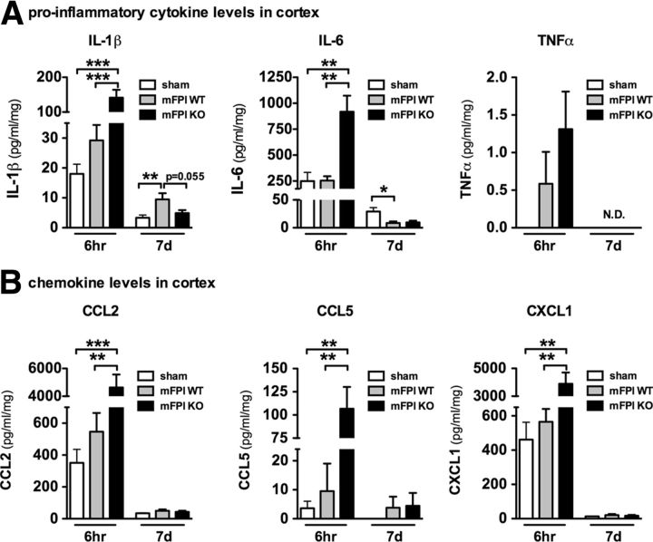
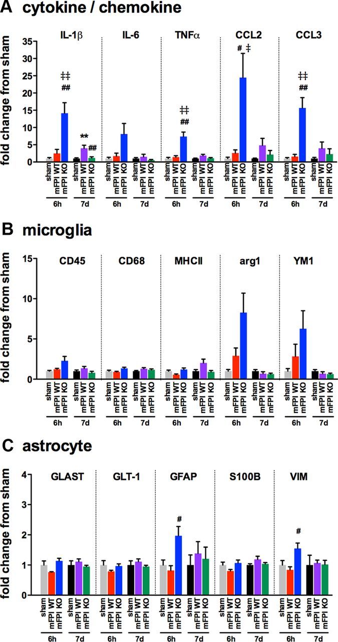
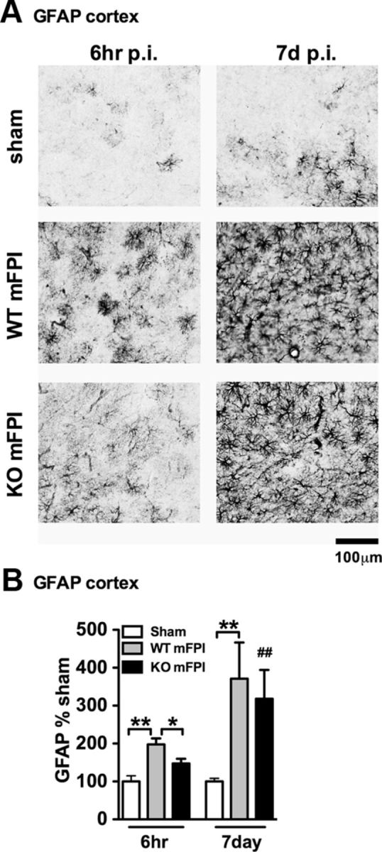
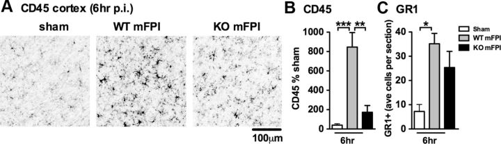
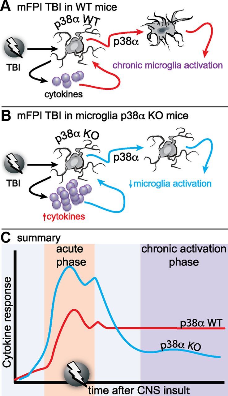
Neuropathology after traumatic brain injury (TBI) is the result of both the immediate impact injury and secondary injury mechanisms. Unresolved post-traumatic glial activation is a secondary injury mechanism that contributes to a chronic state of neuroinflammation in both animal models of TBI and human head injury patients. We recently demonstrated, using in vitro models, that p38α MAPK signaling in microglia is a key event in promoting cytokine production in response to diverse disease-relevant stressors and subsequent inflammatory neuronal dysfunction. From these findings, we hypothesized that the p38α signaling pathway in microglia could be contributing to the secondary neuropathologic sequelae after a diffuse TBI. Mice where microglia were p38α-deficient (p38α KO) were protected against TBI-induced motor deficits and synaptic protein loss. In wild-type (WT) mice, diffuse TBI produced microglia morphological activation that lasted for at least 7 d; however, p38α KO mice failed to activate this response. Unexpectedly, we found that the peak of the early, acute phase cytokine and chemokine levels was increased in injured p38α KO mice compared with injured WT mice. The increased cytokine levels in the p38α KO mice could not be accounted for by more infiltration of macrophages or neutrophils, or increased astrogliosis. By 7 d after injury, the cytokine and chemokine levels remained elevated in injured WT mice but not in p38α KO mice. Together, these data suggest that p38α balances the inflammatory response by acutely attenuating the early proinflammatory cytokine surge while perpetuating the chronic microglia activation after TBI.


Figures

Figure 1.
Figure 1.
Myeloid-specific deletion of p38α protects mice from diffuse brain injury-induced vestibulomotor impairments and synaptic protein loss. A, Injured WT mice (mFPI WT) demonstrated significant impairments in rotarod performance compared with sham-injured mice and mFPI p38α KO mice. The injured KO mice were not significantly different from the sham mice. B, Although the differences did not reach statistical significance, at 7 d after injury, levels of the synaptic proteins PSD95 and synaptophysin (SYP) were decreased in the WT mice compared with sham, whereas the injury-induced decrease was not seen in the KO mice; n = 8–14 per group. *p < 0.05, sham versus mFPI WT. **p < 0.01, sham versus mFPI WT. #p < 0.05, mFPI WT versus mFPI KO.
Figure 2.
Figure 2.
Changes in gene expression associated with glial activation after diffuse brain injury. Temporal changes in cytokine/chemokine (A), microglia (B), and astrocyte markers (C) were measured in the cortex of male C57BL/6J mice subjected to sham conditions (gray), or at 3 h (red), 9 h (green), 24 h (blue), or 7 d (black) after mFPI; n = 3 per time point.
Figure 3.
Figure 3.
The increase in p-p38 after diffuse brain injury is associated with myeloid cells. A, p-p38 IHC was seen in the WT mFPI mice at 6 h and 7 d post-injury (p.i.) but was nearly absent in the sham-injured mice and the p38α KO mFPI mice. B, Quantification shows a significant increase in the number of p-p38+ cells in the injured WT mice at 6 h, which persists to 7 d after injury. Injured p38α KO mice show little to no p-p38 immunoreactivity and are not different from sham-injured mice at either time point (n = 6–12 per group). *p < 0.05. **p < 0.01.
Figure 4.
Figure 4.
p38α KO microglia fail to respond morphologically to a diffuse brain injury. A, IBA1 IHC shows a strong increase in staining and morphological alterations in the WT mFPI mice post-injury (p.i.), which were absent in the KO mFPI mice 6 h after injury and remained diminished in the KO mice 7 d after injury. Arrows indicate clusters of microglia around what appears to be a blood vessel. B, Quantification shows a significant increase in IBA1 staining in the injured WT mice at 6 h, which persists to 7 d after injury. Microglia in the KO mice demonstrated a failure to activate during the acute (6 h) or chronic phase (7 d) after the injury. C, CD68 IHC further supports the lack of a microglia morphological response to the diffuse brain injury in the absence of p38α, both at acute time points and during the chronic phase after the injury. D, Quantification shows a significant increase in CD68 staining in the WT-injured mice at 6 h that persisted to 7 d after injury, with minimal response in the KO mice. Data are presented as percentage of sham; n = 6–8 per group. *p < 0.05. **p < 0.01.
Figure 5.
Figure 5.
Microglia clustering is dependent on p38α KO after a diffuse TBI. A–D, IBA1 IHC shows the spectrum of microglia activation seen in the WT mice 6 h after the diffuse brain injury. Note the long, rod-shaped microglia (A), trains of multiple microglia in plane with the axonal columns in the cortex (B), and multiple microglia in the shape of a layer-V pyramidal cell (C), as well as IBA1+ cells with retracted processes with large swellings (D). In contrast, the p38α KO mice exhibited thin ramified microglia processes (E) or looked morphologically similar (F) to sham-injured mice (G). A cluster of microglia from a WT mFPI mouse shows the IBA1 staining (H), and the Aperio nuclear algorithm generated markup shows the reliable detection of the microglia clusters (demonstrated here as the black markup) and not individual cells (arrows) (I). Quantification shows a significant increase in IBA1 clusters in the WT-injured mice at 6 h after injury (J). Data are presented as percentage of sham; n = 6–8 per group. *p < 0.05.
Figure 6.
Figure 6.
Cytokine and chemokine levels are acutely enhanced in the p38α KO mice after a diffuse brain injury. The protein levels of a panel of inflammatory cytokines (A) and chemokines (B) were measured in the neocortex by MSD multiplex immunoassay. The injured p38α KO mice were found to have significantly enhanced cytokine response at 6 h; but by 7 d after injury, the cytokine response in the KO mice was below (IL-1β) or equal to (IL-6) the response of the WT-injured mice. The chemokines were also significantly increased at 6 h in the p38α KO mice; and by 7 d, the levels were near baseline; n = 6–12 per group. *p < 0.05. **p < 0.01. ***p < 0.001. N.D., Not detected.
Figure 7.
Figure 7.
Enhanced cytokine and chemokine gene expression is seen in the p38α KO mice at 6 h after diffuse brain injury. Temporal changes in cytokine/chemokine (A), microglia (B), and astrocyte markers (C) were measured from the cortex harvested from sham mice (gray represents 6 h; black, 7 d), injured WT mice (red, 6 h; purple, 7 d), or injured p38α KO mice (blue, 6 h; green, 7 d); n = 4–10 per group. **p < 0.01, sham versus mFPI WT. #p < 0.05, mFPI WT versus mFPI KO. ##p < 0.01, mFPI WT versus mFPI KO. p < 0.05, sham versus mFPI KO. ‡‡p < 0.01, sham versus mFPI KO.
Figure 8.
Figure 8.
There is intact astrogliosis in myeloid-deficient p38α KO mice after injury. A, GFAP IHC shows a strong increase in staining and morphological alterations in the WT mFPI mice 6 h after injury, which was not as prominent in the p38α KO mFPI mice. By 7 d after injury, WT and KO mice showed similar GFAP staining. B, Quantification shows a significant increase in GFAP at 6 h after injury in WT mice but not KO mice. At 7 d, there was no difference in astrogliosis between the WT and KO injured mice. Data are presented as percentage of sham (n = 6–12 per group). *p < 0.05. **p < 0.01. ##p < 0.01, sham versus mFPI KO (t test).
Figure 9.
Figure 9.
Myeloid-deficient p38α KO mice have a limited increase in CD45, but similar changes in GR1+ monocytes/neutrophils, compared with WT mice. A, Representative photomicrographs of CD45+ IHC in the cortex 6 h post-injury (p.i.). B, A significant increase in CD45+ staining was found in the cortex of WT mFPI mice compared with sham-injured mice, whereas p38α KO mice showed very little increase in CD45+ staining. C, The number of GR1+ cells were counted in the cortex and presented as the average number of GR1+ cells per coronal tissue section. WT and KO mice showed similar numbers of GR1+ cells after injury (n = 6–12 per group). *p < 0.05. **p < 0.01. ***p < 0.001.
Figure 10.
Figure 10.
Proposed model of the role of p38α in the microglia response to diffuse brain injury. A, In WT mice, a mild to moderate diffuse brain injury induces a reactive glia response associated with elevated inflammatory cytokines and chemokines, morphological hypertrophy, and loss of microglia spatial domains as early as 6 h after injury. The reactive glia response, which is exemplified by an “M1” proinflammatory phenotype, persists for at least 7 d after the injury. B, In the p38α KO mice, the acute phase cytokine response is elevated compared with the WT mice. However, despite the higher cytokine response, there is a failure of the microglia deficient in p38α to exhibit an acute or chronic phase morphological activation response. C, Our results suggest that p38α is a critical kinase in the detrimental inflammatory feedforward loop responsible for neuromotor deficits.

References

    1. Bachstetter AD, Van Eldik LJ. The p38 MAP kinase family as regulators of proinflammatory cytokine production in degenerative diseases of the CNS. Aging Dis. 2010;1:199–211. - PMC - PubMed
    1. Bachstetter AD, Xing B, de Almeida L, Dimayuga ER, Watterson DM, Van Eldik LJ. Microglial p38alpha MAPK is a key regulator of proinflammatory cytokine upregulation induced by toll-like receptor (TLR) ligands or beta-amyloid (Abeta) J Neuroinflammation. 2011;8:79. - PMC - PubMed
    1. Bachstetter AD, Norris CM, Sompol P, Wilcock DM, Goulding D, Neltner JH, St Clair D, Watterson DM, Van Eldik LJ. Early stage drug treatment that normalizes proinflammatory cytokine production attenuates synaptic dysfunction in a mouse model that exhibits age-dependent progression of Alzheimer's disease-related pathology. J Neurosci. 2012;32:10201–10210. - PMC - PubMed
    1. Barone FC, Irving EA, Ray AM, Lee JC, Kassis S, Kumar S, Badger AM, White RF, McVey MJ, Legos JJ, Erhardt JA, Nelson AH, Ohlstein EH, Hunter AJ, Ward K, Smith BR, Adams JL, Parsons AA. SB 239063, a second-generation p38 mitogen-activated protein kinase inhibitor, reduces brain injury and neurological deficits in cerebral focal ischemia. J Pharmacol Exp Ther. 2001;296:312–321. - PubMed
    1. Cao T, Thomas TC, Ziebell JM, Pauly JR, Lifshitz J. Morphological and genetic activation of microglia after diffuse traumatic brain injury in the rat. Neuroscience. 2012;225:65–75. - PMC - PubMed

Publication types

MeSH terms