Preliminary feasibility and hemodynamic performance of a newly-developed self-expanding bioprosthesis and 16-F delivery system in transcatheter aortic valve implantation in sheep
- PMID: 23554751
- PMCID: PMC3596071
- DOI: 10.7555/JBR.26.20120011
Preliminary feasibility and hemodynamic performance of a newly-developed self-expanding bioprosthesis and 16-F delivery system in transcatheter aortic valve implantation in sheep
Abstract
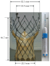
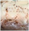
We sought to evaluate the feasibility and hemodynamic performance of a new self-expanding bioprosthesis and 16-F delivery system in sheep. A 23-mm new self-expanding aortic bioprosthesis was implanted in sheep (n = 10) with a 16-F catheter via the right common carotid artery. Each sheep underwent angiography and coronary angiography before intervention, immediately and 1 h after stent implantation. Electrocardiographic monitoring was carried out during and 2 h after the procedure. Transthoracic echocardiography was employed to detect hemodynamic performance before intervention, immediately and 1 and 2 h after stent implantation. All sheep were euthanized 2 h after successful implantation for macroscopic inspection. In all cases, the new self-expanding aortic bioprosthesis was successfully delivered to the aortic root and released with a 16-F catheter. Successful implantation was achieved in 8 of 10 sheep. Hemodynamic performance and device position of successful implantation were stable 2 h after device deployment. Atrioventricular block was not observed. We conclude that it is feasible to implant the new self-expanding aortic valve with a 16-F delivery system into sheep hearts via the retrograde route.
Keywords: aortic valve; percutaneous; self-expanding bioprothesis; sheep.
Conflict of interest statement
The authors reported no conflict of interest.
Figures


References
-
- Nkomo VT, Gardin JM, Skelton TN, Gottdiener JS, Scott CG, Enriquez-Sarano M. Burden of valvular heart diseases: a populationbased study. Lancet. 2006;368:1005–11. - PubMed
-
- Iung B, Cachier A, Baron G, Messika-Zeitoun D, Delahaye F, Tornos P, et al. Decision-making in elderly patients with severe aortic stenosis: why are so many denied surgery? Eur Heart J. 2005;26:2714–20. - PubMed
-
- Dal-Bianco JP, Khandheria BK, Mookadam F, Gentile F, Sengupta PP. Management of asymptomatic severe aortic stenosis. J Am Coll Cardiol. 2008;52:1279–92. - PubMed
-
- Routledge HC, Lefevre T, Morice MC, DeMarco F, Salmi L, Cormier B. Percutaneous aortic valve replacement: New hope for inoperable and high-risk patients. J Invasive Cardiol. 2007;19:478–83. - PubMed
-
- Cribier A, Savin T, Berland J, Rocha P, Mechmeche R, Saoudi N, et al. Percutaneous transluminal balloon valvuloplasty of adult aortic stenosis: report of 92 cases. J Am Coll Cardiol. 1987;9:381–6. - PubMed
LinkOut - more resources
Full Text Sources
Other Literature Sources

