

Serum IL-10 from systemic lupus erythematosus patients suppresses the differentiation and function of monocyte-derived dendritic cells
- PMID: 23554785
- PMCID: PMC3597043
- DOI: 10.7555/JBR.26.20120115
Serum IL-10 from systemic lupus erythematosus patients suppresses the differentiation and function of monocyte-derived dendritic cells
Abstract
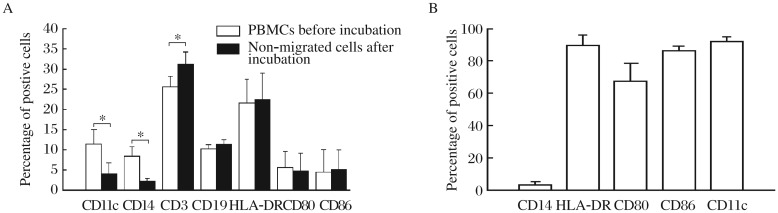
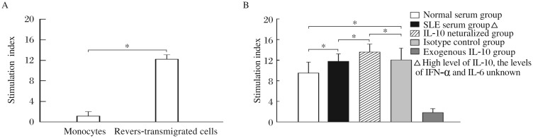
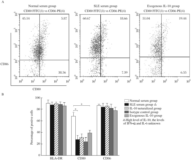
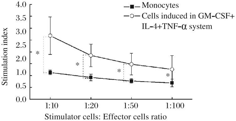
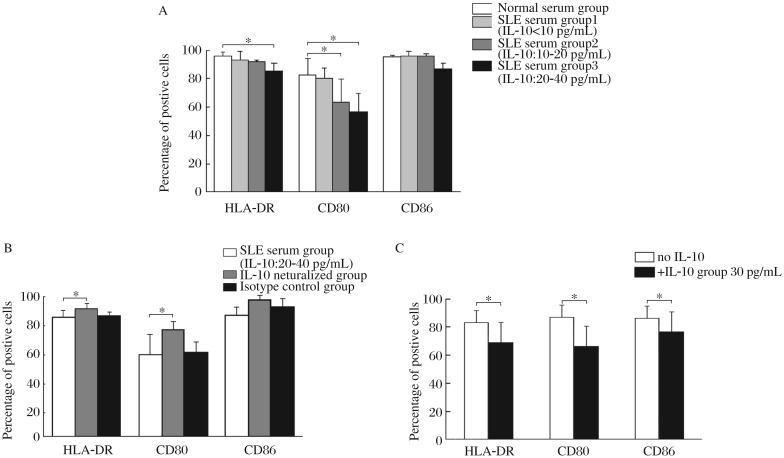
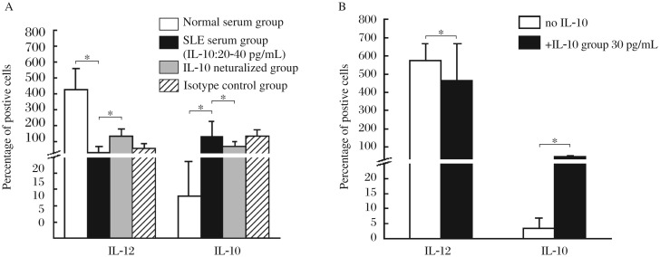
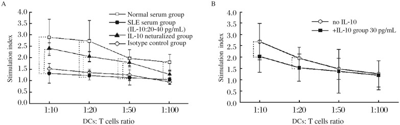
The role played by cytokines, other than interferon (IFN)-α, in the differentiation and function of dendritic cells (DCs) in systemic lupus erythematosus (SLE), remains unclear. Serum interleukin-10 (IL-10) levels are generally elevated in SLE patients, which might modulate the differentiation of DCs. In this study, DCs were induced from monocytes either by transendothelial trafficking or by culture with granulocyte-macrophage colony-stimulating factor (GM-CSF) + IL-4 + tumor necrosis factor (TNF)-α. Both systems were used to investigate the effects of elevated serum IL-10 level on DC differentiation in SLE patients. The results showed that monocyte-derived DCs induced by either SLE serum or exogenous IL-10 reduced the expression of human leukocyte antigen (HLA)-DR and CD80, decreased IL-12p40 level, and increased IL-10 level, and exhibited an impaired capacity to stimulate allogenic T-cell proliferation. These results indicate that serum IL-10 may be involved in the pathogenesis of SLE by modulating the differentiation and function of DCs.
Keywords: dendritic cells (DCs); differentiation; interleukin-10 (IL-10); lupus erythematosus systemic (SLE).
Conflict of interest statement
The authors declared no financial conflict of interests.
Figures
Similar articles
-
Interferon-alpha and interleukin-6 in SLE serum induce the differentiation and maturation of dendritic cells derived from CD34+ hematopoietic precursor cells.Cytokine. 2010 May;50(2):195-203. doi: 10.1016/j.cyto.2010.02.017. Epub 2010 Mar 19. Cytokine. 2010. PMID: 20303780
-
Macrophage colony-stimulating factor could evaluate both disease activity and renal involvement in systemic lupus erythematosus.Ann Palliat Med. 2021 Feb;10(2):2098-2107. doi: 10.21037/apm-20-2607. Epub 2021 Feb 3. Ann Palliat Med. 2021. PMID: 33549023
-
Interferon-induced protein IFIT4 is associated with systemic lupus erythematosus and promotes differentiation of monocytes into dendritic cell-like cells.Arthritis Res Ther. 2008;10(4):R91. doi: 10.1186/ar2475. Epub 2008 Aug 15. Arthritis Res Ther. 2008. PMID: 18706081 Free PMC article.
-
Interferon-α in the generation of monocyte-derived dendritic cells: recent advances and implications for dermatology.Br J Dermatol. 2011 Aug;165(2):247-54. doi: 10.1111/j.1365-2133.2011.10301.x. Epub 2011 Jun 2. Br J Dermatol. 2011. PMID: 21410666 Review.
-
Cytokines in the generation and maturation of dendritic cells: recent advances.Eur Cytokine Netw. 2002 Apr-Jun;13(2):186-99. Eur Cytokine Netw. 2002. PMID: 12101074 Review.
Cited by
-
Dendritic Cells in Systemic Lupus Erythematosus: From Pathogenic Players to Therapeutic Tools.Mediators Inflamm. 2016;2016:5045248. doi: 10.1155/2016/5045248. Epub 2016 Mar 30. Mediators Inflamm. 2016. PMID: 27122656 Free PMC article. Review.
-
Cytokines and MicroRNAs as Candidate Biomarkers for Systemic Lupus Erythematosus.Int J Mol Sci. 2015 Oct 13;16(10):24194-218. doi: 10.3390/ijms161024194. Int J Mol Sci. 2015. PMID: 26473848 Free PMC article. Review.
-
High Interleukin 21 Levels in Patients with Systemic Lupus Erythematosus: Association with Clinical Variables and rs2221903 Polymorphism.J Clin Med. 2024 Aug 2;13(15):4512. doi: 10.3390/jcm13154512. J Clin Med. 2024. PMID: 39124778 Free PMC article.
-
The Expansion of CD25 high IL-10 high FoxP3 high B Regulatory Cells Is in Association with SLE Disease Activity.J Immunol Res. 2015;2015:254245. doi: 10.1155/2015/254245. Epub 2015 Oct 4. J Immunol Res. 2015. PMID: 26504851 Free PMC article.
-
Dual Role of Interleukin-10 in Murine NZB/W F1 Lupus.Int J Mol Sci. 2021 Jan 29;22(3):1347. doi: 10.3390/ijms22031347. Int J Mol Sci. 2021. PMID: 33572870 Free PMC article.
References
-
- Figdor CG, de Vries IJ, Lesterhuis WJ, Melief CJ. Dendritic cell immunotherapy: mapping the way. Nat Med. 2004;10:475–80. - PubMed
-
- Scheinecker C, Zwölfer B, Köller M, Männer G, Smolen JS. Alterations of dendritic cells in systemic lupus erythematosus: phenotypic and functional deficiencies. Arthritis Rheum. 2001;44:856–65. - PubMed
-
- Blanco P, Palucka AK, Gill M, Pascual V, Banchereau J. Induction of dendritic cell differentiation by IFN-alpha in systemic lupus erythematosus. Science. 2001;294:1540–3. - PubMed
-
- Pascual V, Farkas L, Banchereau J. Systemic lupus erythematosus: all roads lead to type I interferons. Curr Opin Immunol. 2006;18:676–82. - PubMed
LinkOut - more resources
Full Text Sources
Research Materials