

Low dose bisphenol A impairs spermatogenesis by suppressing reproductive hormone production and promoting germ cell apoptosis in adult rats
- PMID: 23554804
- PMCID: PMC3602871
- DOI: 10.7555/JBR.27.20120076
Low dose bisphenol A impairs spermatogenesis by suppressing reproductive hormone production and promoting germ cell apoptosis in adult rats
Abstract
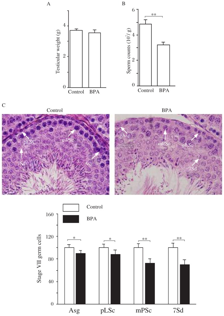
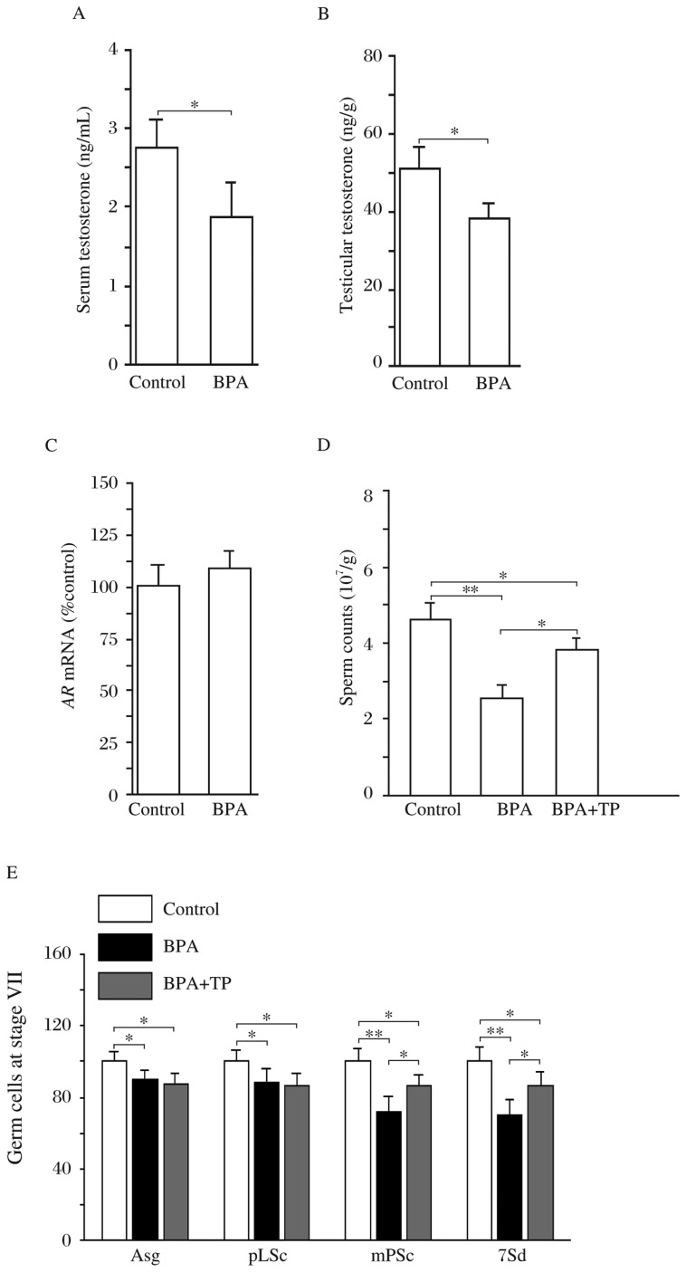
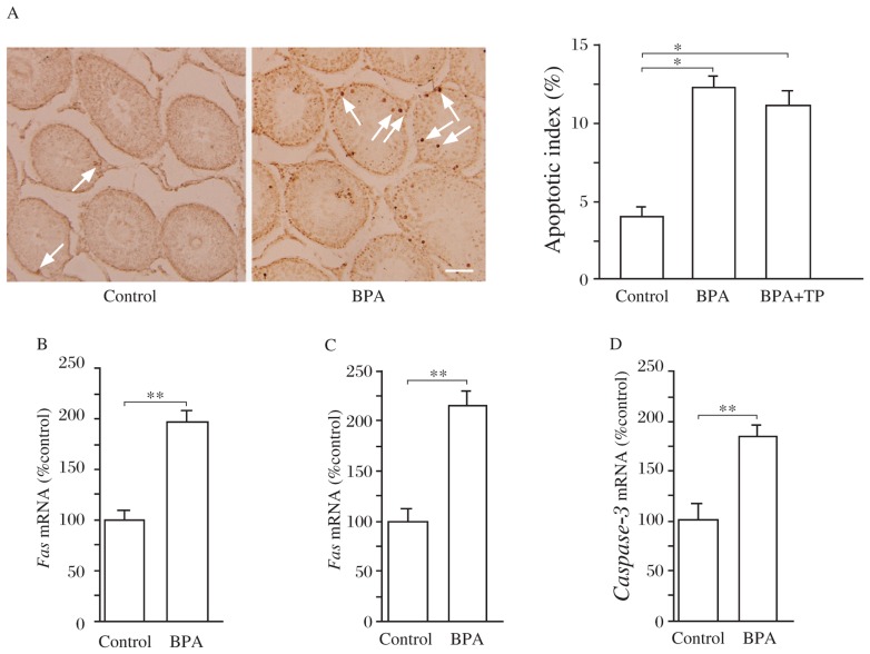
Bisphenol A (BPA), an estrogenic chemical, has been shown to reduce sperm count; however, the underlying mechanisms remain unknown. Herein, we show that oral administration of BPA (2 µg/kg) for consecutive 14 days in adult rats (BPA rats) significantly reduced the sperm count and the number of germ cells compared to controls. The serum levels of testosterone and follicle-stimulating hormone (FSH), as well as the level of GnRH mRNA in BPA rats were lower than those of control rats. Testosterone treatment could partially rescue the reduction of germ cells in BPA rats. Notably, the number of apoptotic germ cells was significantly increased in BPA rats, which was insensitive to testosterone. Furthermore, the levels of Fas, FasL and caspase-3 mRNA in the testicle of BPA rats were increased in comparison with controls. These results indicate that exposure to a low dose of BPA impairs spermatogenesis through decreasing reproductive hormones and activating the Fas/FasL signaling pathway.
Keywords: apoptosis; bisphenol A (BPA); spermatogenesis; testosterone.
Conflict of interest statement
The authors declare no conflict of interests.
Figures

Similar articles
-
Adult exposure to bisphenol A (BPA) in Wistar rats reduces sperm quality with disruption of the hypothalamic-pituitary-testicular axis.Toxicology. 2015 Mar 2;329:1-9. doi: 10.1016/j.tox.2015.01.002. Epub 2015 Jan 6. Toxicology. 2015. PMID: 25575453
-
Permanent effects of neonatal estrogen exposure in rats on reproductive hormone levels, Sertoli cell number, and the efficiency of spermatogenesis in adulthood.Endocrinology. 1999 Nov;140(11):5364-73. doi: 10.1210/endo.140.11.7108. Endocrinology. 1999. PMID: 10537168
-
Bisphenol A exposure induces apoptosis and upregulation of Fas/FasL and caspase-3 expression in the testes of mice.Toxicol Sci. 2009 Apr;108(2):427-36. doi: 10.1093/toxsci/kfp024. Epub 2009 Feb 4. Toxicol Sci. 2009. PMID: 19193734
-
Effects of chronic testosterone administration in normal men: safety and efficacy of high dosage testosterone and parallel dose-dependent suppression of luteinizing hormone, follicle-stimulating hormone, and sperm production.J Clin Endocrinol Metab. 1990 Jan;70(1):282-7. doi: 10.1210/jcem-70-1-282. J Clin Endocrinol Metab. 1990. PMID: 2104626 Clinical Trial.
-
Neonatal exposure of male rats to Bisphenol A impairs fertility and expression of sertoli cell junctional proteins in the testis.Toxicology. 2009 Nov 9;265(1-2):56-67. doi: 10.1016/j.tox.2009.09.012. Epub 2009 Sep 25. Toxicology. 2009. PMID: 19782717
Cited by
-
Exposure to environmental bisphenol A inhibits HTR-8/SVneo cell migration and invasion.J Biomed Res. 2020 Jun 30;34(5):369-378. doi: 10.7555/JBR.34.20200013. J Biomed Res. 2020. PMID: 32981897 Free PMC article.
-
The Impact of Endocrine-Disrupting Chemicals in Male Fertility: Focus on the Action of Obesogens.J Xenobiot. 2021 Nov 29;11(4):163-196. doi: 10.3390/jox11040012. J Xenobiot. 2021. PMID: 34940512 Free PMC article. Review.
-
Characterization of Estrogenic Activity and Site-Specific Accumulation of Bisphenol-A in Epididymal Fat Pad: Interfering Effects on the Endocannabinoid System and Temporal Progression of Germ Cells.Int J Mol Sci. 2021 Mar 3;22(5):2540. doi: 10.3390/ijms22052540. Int J Mol Sci. 2021. PMID: 33802611 Free PMC article.
-
BPA and Nutraceuticals, Simultaneous Effects on Endocrine Functions.Endocr Metab Immune Disord Drug Targets. 2019;19(5):594-604. doi: 10.2174/1871530319666190101120119. Endocr Metab Immune Disord Drug Targets. 2019. PMID: 30621569 Free PMC article. Review.
-
The Methylcytosine Dioxygenase Ten-Eleven Translocase-2 (tet2) Enables Elevated GnRH Gene Expression and Maintenance of Male Reproductive Function.Endocrinology. 2016 Sep;157(9):3588-603. doi: 10.1210/en.2016-1087. Epub 2016 Jul 6. Endocrinology. 2016. PMID: 27384303 Free PMC article.
References
LinkOut - more resources
Full Text Sources
Other Literature Sources
Research Materials
Miscellaneous