

Correlation of immunoglobulin G expression and histological subtype and stage in breast cancer
- PMID: 23554916
- PMCID: PMC3595271
- DOI: 10.1371/journal.pone.0058706
Correlation of immunoglobulin G expression and histological subtype and stage in breast cancer
Abstract
Introduction: Recently, growing evidence indicates that immunoglobulins (Igs) are not only produced by mature B lymphocytes or plasma cells, but also by various normal cells types at immune privileged sites and neoplasm, including breast cancer. However, the association of breast cancer derived IgG with genesis and development of the disease has not yet been established.
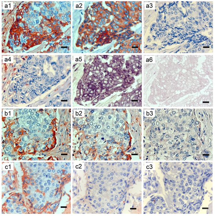
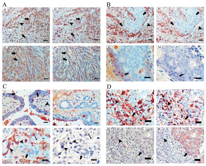
Methods: In this study we examined the expression of IgG in 186 breast cancers, 20 benign breast lesions and 30 normal breast tissues. Both immunohistochemistry with antibodies to Igκ (immunoglobulin G κ light chain) and Igγ (immunoglobulin G heavy chain) and in situ hybridization with an antisense probe to IgG1 heavy chain constant region gene were performed. Various clinicopathological features were also analyzed.
Results: We found that IgG is specifically expressed in human breast cancer cells. Both infiltrating ductal carcinoma and infiltrating lobular carcinoma had significantly greater numbers of Igκ and Igγ positive cancer cells as compared with medullary carcinoma, carcinoma in situ, and benign lesions (all p<0.05). In addition, IgG expression was correlated with breast cancer histological subtypes (p<0.01) and AJCC stages (p<0.05), with more abundance of IgG expression in more malignant histological subtypes or in more advanced stage of the disease.
Conclusions: IgG expression in breast cancer cells is correlated with malignancy and AJCC stages of the cancers. This suggests that breast cancer derived IgG may be associated with genesis, development and prognosis of the cancer.
Conflict of interest statement
Figures
Similar articles
-
Non B Cell-Derived Immunoglobulins in Lung Epithelial Cells and Lung Cancer.Adv Exp Med Biol. 2024;1445:157-168. doi: 10.1007/978-981-97-0511-5_13. Adv Exp Med Biol. 2024. PMID: 38967758 Review.
-
Immunoglobulin G expression and its colocalization with complement proteins in papillary thyroid cancer.Mod Pathol. 2012 Jan;25(1):36-45. doi: 10.1038/modpathol.2011.139. Epub 2011 Sep 9. Mod Pathol. 2012. PMID: 21909078
-
Immunoglobulin G is present in a wide variety of soft tissue tumors and correlates well with proliferation markers and tumor grades.Cancer. 2010 Apr 15;116(8):1953-63. doi: 10.1002/cncr.24892. Cancer. 2010. PMID: 20186824
-
Immunoglobulin G locus events in soft tissue sarcoma cell lines.PLoS One. 2011;6(6):e21276. doi: 10.1371/journal.pone.0021276. Epub 2011 Jun 23. PLoS One. 2011. PMID: 21731691 Free PMC article.
-
Expression and Function of Mammary Epithelial Cell-Derived Immunoglobulins.Adv Exp Med Biol. 2024;1445:169-177. doi: 10.1007/978-981-97-0511-5_14. Adv Exp Med Biol. 2024. PMID: 38967759 Review.
Cited by
-
Clinical and biological heterogeneities in triple-negative breast cancer reveals a non-negligible role of HER2-low.Breast Cancer Res. 2023 Mar 30;25(1):34. doi: 10.1186/s13058-023-01639-y. Breast Cancer Res. 2023. PMID: 36998014 Free PMC article.
-
Six novel immunoglobulin genes as biomarkers for better prognosis in triple-negative breast cancer by gene co-expression network analysis.Sci Rep. 2019 Mar 14;9(1):4484. doi: 10.1038/s41598-019-40826-w. Sci Rep. 2019. PMID: 30872752 Free PMC article.
-
Immunoglobulin free light chains are biomarkers of poor prognosis in basal-like breast cancer and are potential targets in tumor-associated inflammation.Oncotarget. 2014 May 30;5(10):3159-67. doi: 10.18632/oncotarget.1868. Oncotarget. 2014. PMID: 24931643 Free PMC article.
-
A Comparative Analysis of the Immunoglobulin Repertoire in Leukemia Cells and B Cells in Chinese Acute Myeloid Leukemia by High-Throughput Sequencing.Biology (Basel). 2024 Aug 13;13(8):613. doi: 10.3390/biology13080613. Biology (Basel). 2024. PMID: 39194551 Free PMC article.
-
Non B Cell-Derived Immunoglobulins in Lung Epithelial Cells and Lung Cancer.Adv Exp Med Biol. 2024;1445:157-168. doi: 10.1007/978-981-97-0511-5_13. Adv Exp Med Biol. 2024. PMID: 38967758 Review.
References
-
- Siegel R, Naishadham D, Jemal A (2012) Cancer statistics, 2012. CA Cancer J Clin 62: 10–29. - PubMed
-
- Forouzanfar MH, Foreman KJ, Delossantos AM, Lozano R, Lopez AD, et al. (2011) Breast and cervical cancer in 187 countries between 1980 and 2010: a systematic analysis. Lancet 378: 1461–1484. - PubMed
-
- Tirona MT, Sehgal R, Ballester O (2010) Prevention of breast cancer (part I): epidemiology, risk factors, and risk assessment tools. Cancer Invest 28: 743–750. - PubMed
-
- Chen Z, Gu J (2007) Immunoglobulin G expression in carcinomas and cancer cell lines. FASEB J 21: 2931–2938. - PubMed
Publication types
MeSH terms
Substances
LinkOut - more resources
Full Text Sources
Other Literature Sources
Medical
