

Atrophy rates in asymptomatic amyloidosis: implications for Alzheimer prevention trials
- PMID: 23554933
- PMCID: PMC3599038
- DOI: 10.1371/journal.pone.0058816
Atrophy rates in asymptomatic amyloidosis: implications for Alzheimer prevention trials
Abstract
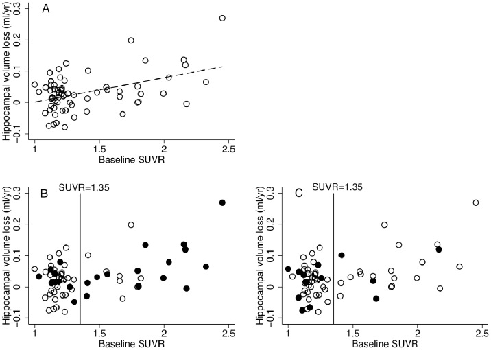
There is considerable interest in designing therapeutic studies of individuals at risk of Alzheimer disease (AD) to prevent the onset of symptoms. Cortical β-amyloid plaques, the first stage of AD pathology, can be detected in vivo using positron emission tomography (PET), and several studies have shown that ~1/3 of healthy elderly have significant β-amyloid deposition. Here we assessed whether asymptomatic amyloid-PET-positive controls have increased rates of brain atrophy, which could be harnessed as an outcome measure for AD prevention trials. We assessed 66 control subjects (age = 73.5±7.3 yrs; MMSE = 29±1.3) from the Australian Imaging Biomarkers & Lifestyle study who had a baseline Pittsburgh Compound B (PiB) PET scan and two 3T MRI scans ~18-months apart. We calculated PET standard uptake value ratios (SUVR), and classified individuals as amyloid-positive/negative. Baseline and 18-month MRI scans were registered, and brain, hippocampal, and ventricular volumes and annualized volume changes calculated. Increasing baseline PiB-PET measures of β-amyloid load correlated with hippocampal atrophy rate independent of age (p = 0.014). Twenty-two (1/3) were PiB-positive (SUVR>1.40), the remaining 44 PiB-negative (SUVR≤1.31). Compared to PiB-negatives, PiB-positive individuals were older (76.8±7.5 vs. 71.7±7.5, p<0.05) and more were APOE4 positive (63.6% vs. 19.2%, p<0.01) but there were no differences in baseline brain, ventricle or hippocampal volumes, either with or without correction for total intracranial volume, once age and gender were accounted for. The PiB-positive group had greater total hippocampal loss (0.06±0.08 vs. 0.02±0.05 ml/yr, p = 0.02), independent of age and gender, with non-significantly higher rates of whole brain (7.1±9.4 vs. 4.7±5.5 ml/yr) and ventricular (2.0±3.0 vs. 1.1±1.0 ml/yr) change. Based on the observed effect size, recruiting 384 (95%CI 195-1080) amyloid-positive subjects/arm will provide 80% power to detect 25% absolute slowing of hippocampal atrophy rate in an 18-month treatment trial. We conclude that hippocampal atrophy may be a feasible outcome measure for secondary prevention studies in asymptomatic amyloidosis.
Conflict of interest statement
Figures
References
-
- Reiman EM, Langbaum JBS, Fleisher AS, Caselli RJ, Chen K, et al. (2011) Alzheimer’s Prevention Initiative: a plan to accelerate the evaluation of presymptomatic treatments. J Alzheimers Dis 26 Suppl 3321–329 doi:10.3233/JAD-2011-0059. - DOI - PMC - PubMed
-
- Herholz K, Ebmeier K (2011) Clinical amyloid imaging in Alzheimer’s disease. Lancet Neurol 10: 667–670 doi:10.1016/S1474-4422(11)70123-5. - DOI - PubMed
-
- Klunk WE, Engler H, Nordberg A, Wang Y, Blomqvist G, et al. (2004) Imaging brain amyloid in Alzheimer’s disease with Pittsburgh Compound-B. Ann Neurol 55: 306–319 doi:10.1002/ana.20009. - DOI - PubMed
-
- Mattsson N, Zetterberg H, Hansson O, Andreasen N, Parnetti L, et al. (2009) CSF biomarkers and incipient Alzheimer disease in patients with mild cognitive impairment. JAMA 302: 385–393 doi:10.1001/jama.2009.1064. - DOI - PubMed
-
- Fox NC, Schott JM (2004) Imaging cerebral atrophy: normal ageing to Alzheimer’s disease. Lancet 363: 392–394 doi:10.1016/S0140-6736(04)15441-X. - DOI - PubMed
Publication types
MeSH terms
Substances
Grants and funding
LinkOut - more resources
Full Text Sources
Other Literature Sources
Medical
