. 2013;8(3):e58857.
doi: 10.1371/journal.pone.0058857. Epub 2013 Mar 15.

Zebrafish hoxd4a acts upstream of meis1.1 to direct vasculogenesis, angiogenesis and hematopoiesis

Affiliations

Zebrafish hoxd4a acts upstream of meis1.1 to direct vasculogenesis, angiogenesis and hematopoiesis

Aseervatham Anusha Amali et al. PLoS One. 2013.

Abstract

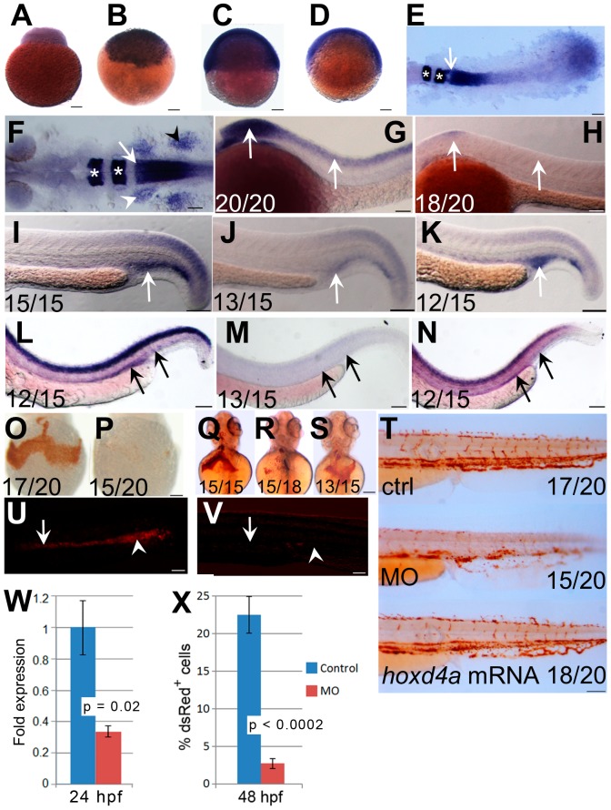
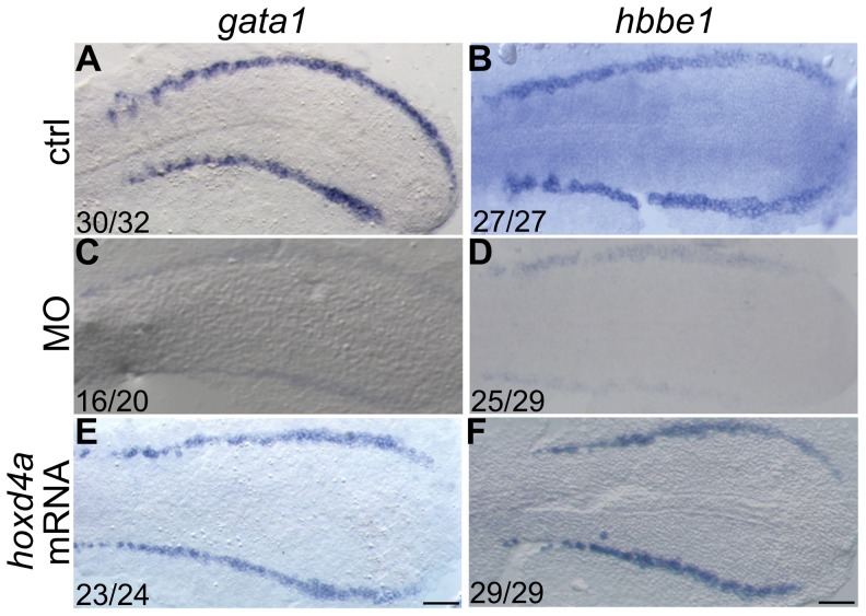
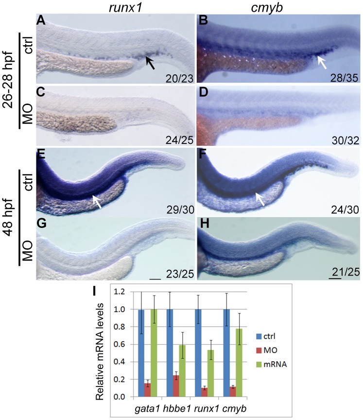
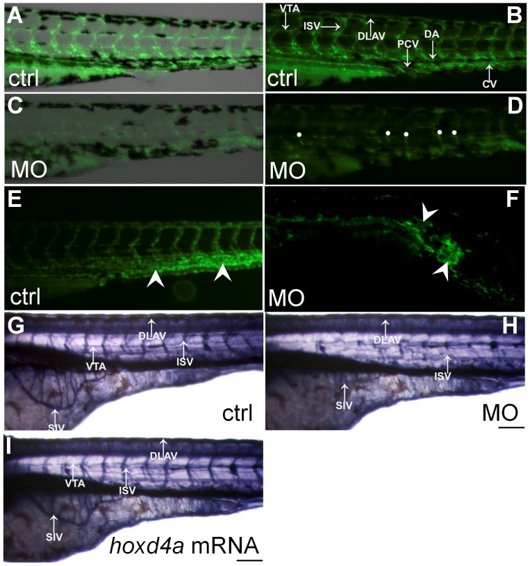
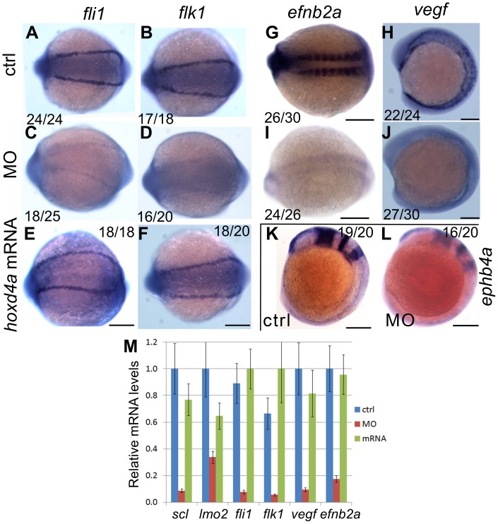
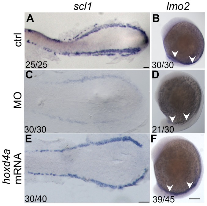
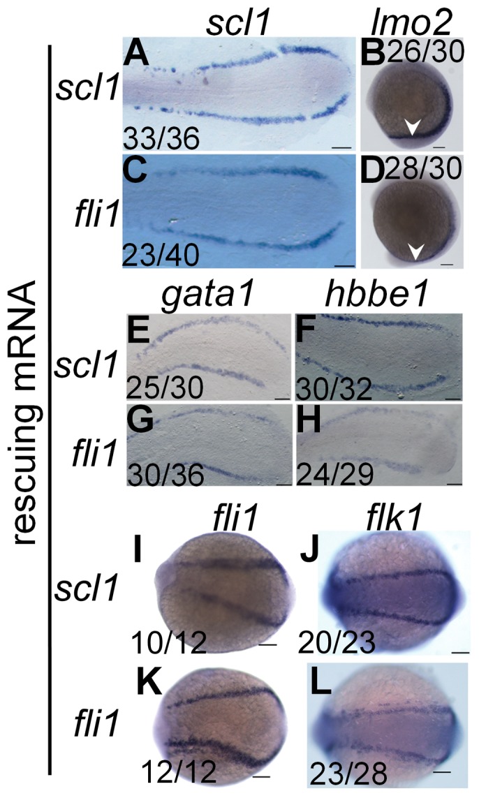
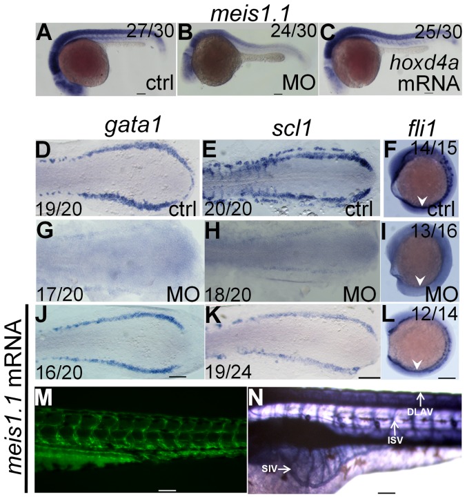
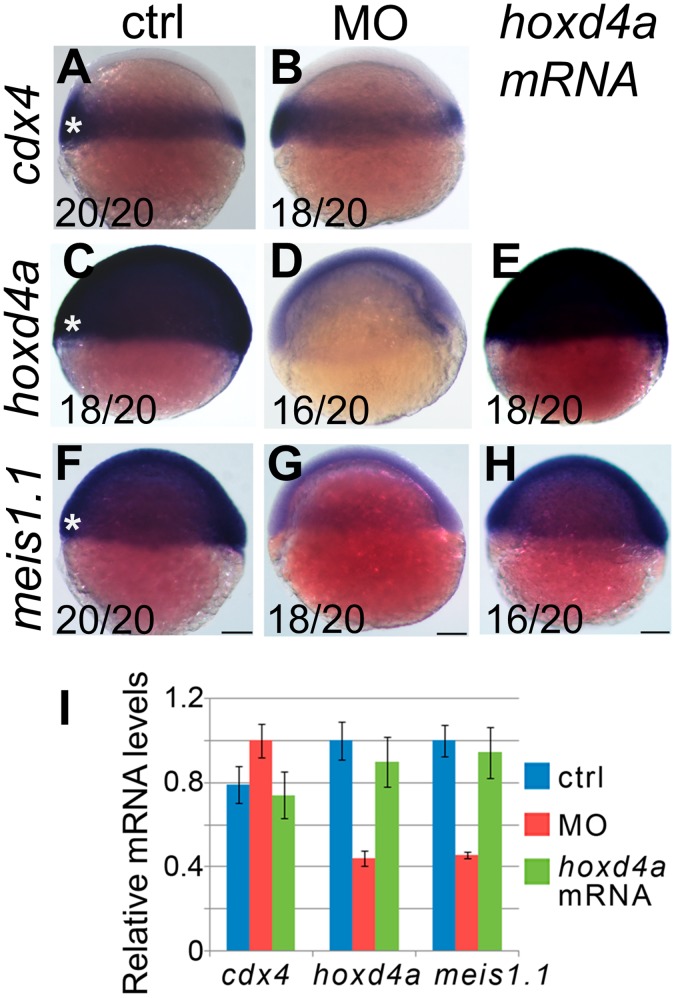
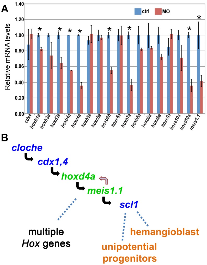
Mice lacking the 4th-group paralog Hoxd4 display malformations of the anterior vertebral column, but are viable and fertile. Here, we report that zebrafish embryos having decreased function of the orthologous hoxd4a gene manifest striking perturbations in vasculogenesis, angiogenesis and primitive and definitive hematopoiesis. These defects are preceded by reduced expression of the hemangioblast markers scl1, lmo2 and fli1 within the posterior lateral plate mesoderm (PLM) at 13 hours post fertilization (hpf). Epistasis analysis revealed that hoxd4a acts upstream of meis1.1 but downstream of cdx4 as early as the shield stage in ventral-most mesoderm fated to give rise to hemangioblasts, leading us to propose that loss of hoxd4a function disrupts hemangioblast specification. These findings place hoxd4a high in a genetic hierarchy directing hemangioblast formation downstream of cdx1/cdx4 and upstream of meis1.1. An additional consequence of impaired hoxd4a and meis1.1 expression is the deregulation of multiple Hox genes implicated in vasculogenesis and hematopoiesis which may further contribute to the defects described here. Our results add to evidence implicating key roles for Hox genes in their initial phase of expression early in gastrulation.


Conflict of interest statement

Competing Interests: The authors have declared that no competing interests exist.

Figures

Figure 1
Figure 1. Expression pattern of hoxd4a and phenotype of hoxd4a morphants.
(A–D) Detection of hoxd4a transcripts in early zebrafish embryos. Animal pole is to the top. (A) 1 cell. (B) 3 hpf. (C) 50% epiboly. (D) 75% epiboly. (E) Dorsal view of flat-mounted embryo at 12 hpf (5–6 somites) doubly stained for expression of hoxd4a and krox20a. The arrow indicates the anterior expression border of hoxd4a, while asterisks denote expression of krox20a in r3 and r5. Anterior is to the left. The rostral-most portion of the embryo is not captured in the image. (F) Dorsal view of a flat-mounted embryo at 26–28 hpf (>26 somites) showing hoxd4a expression in hindbrain, branchial arches (white arrowhead, left side only) and pectoral fin field (black arrowhead, right side only). The white arrow marks the hoxd4a anterior expression border in the hindbrain at the boundary between r6 and r7. Asterisks denote krox20a expression in r3 and r5. (G–H) Lateral views of 26–28 hpf embryos showing hoxd4a expression in the central nervous system (arrows) of control embryos (G) and reduced expression in hoxd4a morphants (H). (I–K) hoxd4a expression in the caudal half at 26–28 hpf as shown in lateral views, anterior to the left. hoxd4a expression seen in the PBI of control embryos (I, white arrow) is greatly reduced in hoxd4a morphants (J). Expression is rescued following co-injection with capped hoxd4a mRNA (K). (L–N) Results of In situ hybridization for hoxd4a at 48 hpf showing expression in the AGM and caudal vein plexus (site of future CHT) of control injected embryos (L, black arrows), greatly reduced expression in hoxd4a morphants (M), and rescued expression in embryos simultaneously injected with capped mRNA for hoxd4a (N). (O–T) Hemoglobin within RBCs revealed by o-dianisidine staining. Ventral views at 48 hpf (O,P) and 72 hpf (Q,R) show greatly reduced levels of hemoglobin in the ducts of Cuvier in hoxd4a morphants (P and R) vs controls (O and Q). Co-injection with capped mRNA for hoxd4a results in rescued RBC production (S). (T) O-dianisidine staining of the caudal half of a control larva (upper panel) and hoxd4a morphant (middle panel) showing overall reduction in hemoglobin levels at 72 hpf in morphants, and rescue by co-injection of capped hoxd4a mRNA (lower panel). (U,V) Lateral views of trunk regions of Tg(gata1:dsRed) embryos at 26 hpf, anterior to the left. The expression of dsRed within proerythroblasts is readily detected in the ICM (arrow) and PBI (arrowhead) of control-injected embryos (U) but not hoxd4a morphants (V). Scale bars = 100 µm. Ratios indicate the number of embryos showing the presented phenotype. (W) qRT-PCR shows an overall 3-fold reduction of hoxd4a expression in morphants at 26–28 hpf. Error bars = standard error. p = 0.02. (X) Quantitation by flow cytometry of dsRed-positive cells in Tg(gata1:dsRed) embryos at 48 hpf showing that morphants display an 88% reduction in RBCs relative to control-injected embryos. Error bars = standard deviation. p<0.0002.
Figure 2
Figure 2. hoxd4a knockdown disrupts primitive hematopoiesis and is highly specific.
In situ hybridization revealing expression at 13 hpf of erythroid lineage markers gata1 (A,C,E) and β embryonic globin 1 (hbbe1) (B,D,F). (A,B) Normal expression of gata1 and hbbe1 in the LPM. (C,D) Expression of gata1 and hbbe1 is strongly down-regulated in hoxd4a morphants, but rescued by co-injection of capped mRNA for hoxd4a (E,F). All embryos have been flat-mounted and are shown in dorsal view. Anterior is to the left. Ratios in the bottom left corner of all panels indicate the number of embryos showing the presented phenotype. ctrl, embryos injected with a non-specific morpholino. MO, embryos injected with the anti-hoxd4a morpholino. hoxd4a mRNA, embryos simultaneously injected with the anti-hoxd4a MO plus capped mRNA for hoxd4a. Scale bars equal 100 µm. All images are at the same magnification.
Figure 3
Figure 3. hoxd4a expression is required for transient and definitive hematopoiesis.
In situ hybridization on 28 hpf (A–D) and 48 hpf (E–H) embryos showing expression of runx1 (A,C,E,G) and cmyb (B,D,F,H) in presumptive HSCs arising in the PBI (arrows in A and B) and AGM (arrows in E and F). Expression of both genes was severely reduced in hoxd4a morphants (C,D and G,H). Ratios in the bottom right corner of images indicate the fraction of embryos showing the presented phenotype. ctrl, embryos injected with a non-specific morpholino. MO, embryos injected with the anti-hoxd4a morpholino. hoxd4a mRNA, embryos simultaneously injected with the anti-hoxd4a MO plus capped mRNA for hoxd4a. Scale bars equal 100 µm. All images are at the same magnification. (I) qRT-PCR confirms the strong depletion of hematopoietic gene expression in hoxd4a morphants at 26–28 hpf, and restored expression following co-injection with capped mRNA for hoxd4a. Samples were normalized to β-actin. Error bars indicate standard error. By comparison to controls and rescuants, the gene expression levels of all morphants were statistically different to p≤0.02 except for gata1 control vs morphant (p = 0.04) and hbbe1 rescuant vs morphant (p = 0.09).
Figure 4
Figure 4. Loss of hoxd4a function impairs development of the vasculature.
(A–D) Fluorescent images of the trunk and tail regions of Tg(fli1:EGFP) embryos at 48 hpf. The panels present merged bright field and fluorescent images (A,C) or fluorescent images only (B,D) The normal pattern of the vasculature (A,B) is severely disrupted in hoxd4a morphants (C,D) Dorsal extremities of ISV sprouts that fail to contact the DLAV are marked by white dots (D). The caudal vein plexus of control embryos (E, arrowheads) is replaced by a disorganized mass of endothelial tissue in hoxd4a morphants (F, arrowheads). (G–I) Alkaline phosphatase staining at 72 hpf revealing the vasculature in (G) control-injected larvae, (H) hoxd4a morphants, and (I) rescued larvae co-injected with capped mRNA for hoxd4a. Dorsal aorta (DA), posterior cardinal vein (PCV), inter-segmental vessels (ISV), caudal artery (CA), dorsal longitudinal anastomotic vessel (DLAV), caudal vein (CV) and vertebral artery (VTA). All images show lateral views, with anterior to the left and dorsal on top. Scale bars equal 100 µm.
Figure 5
Figure 5. Knockdown of hoxd4a disrupts the endothelial programme in zebrafish embryos.
In situ hybridization at 13 hpf revealing expression of pan-endothelial markers fli1 and flk1 in control-injected embryos (A,B), hoxd4a morphants (C,D), and rescued embryos co-injected with anti-hoxd4a MO and capped mRNA for hoxd4a (E,F). (G,I) Expression of the marker of arterial identity efnb2a in controls (G) and hoxd4a morphants (I). (H,J) Expression of the endothelial inducer vegf in controls (H) and hoxd4a morphants (J). (K,L) Expression of the venous marker ephb4a in controls (K) and hoxd4a morphants (L). Ratios indicate the fraction of embryos showing the presented phenotype. All images show dorsal views with anterior to the left except H, J, K and L which are lateral views with anterior to the left. Scale bars equal 100 µm. (M) qRT-PCR results showing depletion at 26–28 hpf of angioblast and vascular gene expression in hoxd4a morphants and rescue by co-injection of capped mRNA for hoxd4a. Samples were normalized to β-actin. Error bars indicate standard error. By comparison to controls and rescuants, the gene expression levels of all morphants were statistically different to p≤0.02 except for lmo2 control vs morphant (p = 0.04) and lmo2 rescuant vs morphant (p = 0.05).
Figure 6
Figure 6. hoxd4a is required for hemangioblast formation.
(A,B) Normal expression at 13 hpf of posterior hemangioblast markers scl1 (A) and lmo2 (B) in the PLM. (C,D) Expression of these markers is greatly reduced in hoxd4a morphants, but is rescued by co-injection of capped mRNA for hoxd4a (E,F). Ratios indicate the fraction of embryos showing the presented phenotype. Anterior is to the left. A, C and E are dorsal views of flat-mounted specimens while B, D and F are lateral views. Scale bars equal 100 µm. A is at a lower magnification than C and E.
Figure 7
Figure 7. scl1 and lmo2 act downstream of hoxd4a to direct formation of the hemangioblast.
All images are of hoxd4a morphants at 13 hpf. In situ hybridization was performed to detect expression of scl1 and lmo2 (A–D), gata1 and hbbe1 (E–H) and fli1 and flk1 (I–L). To test for rescue of gene expression, embryos were co-injected with capped mRNA for either scl1 or fli1 as indicated on the left. Ratios indicate the fraction of embryos showing the presented phenotype. Scale bars equal 100 µm.
Figure 8
Figure 8. meis1.1 is down regulated in hoxd4a morphants and meis1.1 mRNA rescues hematopoietic and vasculogenic gene expression in hoxd4a morphants.
Expression of meis1.1 in control (A), morphant (B) and hoxd4a-rescuant (C) embryos at 26–28 hpf. (D–I) At 13 hpf, normal expression of gata1, scl1 and fli1 (D–F) is reduced in hoxd4a morphants (G–I), but rescued upon co-injection with capped mRNA for meis1.1 (J–L). (M,N) Rescue of vascular patterning in hoxd4a morphants by co-injection with capped mRNA for meis1.1 as visualized in Tg(fli1:EGFP) transgenics at 48 hpf (M) and by alkaline phosphatase staining at 72 hpf (N). Scale bars equal 100 µm.
Figure 9
Figure 9. Expression of meis1.1, but not cdx4, is impaired in hoxd4a morphants at the shield stage.
Expression of cdx4 (A,B), hoxd4a (C–E) and meis1.1 (F–H) in control (A,C,F), hoxd4a morphants (B,D,G) and rescuants injected with hoxd4a mRNA (E,H) observed at the shield stage. Asterisks denote the ventral-most mesoderm fated to give rise to hemangioblast in addition to unipotential hematopoietic and angiogenic progenitors. Ratios indicate the fraction of embryos showing the presented phenotype. Scale bars equal 100 µm. (I) qRT-PCR was used to quantitate relative mRNA levels in controls, morphants and rescuants as indicated. Error bars present the standard error. Decreased expression of hoxd4a and meis1.1 in hoxd4a morphants is significantly lower than both controls and rescuants to p≤0.02.
Figure 10
Figure 10. cdx4, meis1.1 and Hox gene expression in hoxd4a morphants, and a genetic pathway for specification of hemangioblasts and unipotential stem cells.
(A) qRT-PCR showing decreased expression of meis1.1 and many, but not all, hox genes in hoxd4a morphants at 26–28 hpf. By contrast, cdx4 levels are unchanged. Samples were normalized to β-actin. Error bars indicate standard error. All pairs marked with an asterisk meet statistical significance (p≤0.02). (B) Based on the results presented here and those in the literature, we propose a pathway in which hoxd4a and meis1.1 occupy sequential steps downstream of cloche and cdx1/4 in a genetic programme leading to the specification of hemangioblasts and unipotential angiogenic and hematopoietic stem cells. The effects of hoxd4a knockdown may be magnified through positive cross-regulatory interactions with meis1.1. The observed effects on the expression of multiple Hox genes could be due to the direct action of hoxd4a and meis1.1. Non-exclusively, cdx1 and cdx4 may act in conjunction with hoxd4a and meis1.1 in a feed-forward type of mechanism to regulate one or more of these same Hox genes with widespread consequences for vasculogenesis, angiogenesis and hematopoiesis at all levels.

Similar articles

Cited by

References

    1. Paik EJ, Zon LI (2010) Hematopoietic development in the zebrafish. Int J Dev Biol 54: 1127–1137. - PubMed
    1. Ellertsdottir E, Lenard A, Blum Y, Krudewig A, Herwig L, et al. (2010) Vascular morphogenesis in the zebrafish embryo. Dev Biol 341: 56–65. - PubMed
    1. Ciau-Uitz A, Liu F, Patient R (2010) Genetic control of hematopoietic development in Xenopus and zebrafish. Int J Dev Biol 54: 1139–1149. - PubMed
    1. Chen AT, Zon LI (2009) Zebrafish blood stem cells. J Cell Biochem 108: 35–42. - PubMed
    1. Vogeli KM, Jin SW, Martin GR, Stainier DY (2006) A common progenitor for haematopoietic and endothelial lineages in the zebrafish gastrula. Nature 443: 337–339. - PubMed

Publication types

MeSH terms

Substances

LinkOut - more resources