. 2013:2013:215064.
doi: 10.1155/2013/215064. Epub 2013 Mar 11.

Benign hepatocellular tumors in children: focal nodular hyperplasia and hepatocellular adenoma

Affiliations

Benign hepatocellular tumors in children: focal nodular hyperplasia and hepatocellular adenoma

Stéphanie Franchi-Abella et al. Int J Hepatol. 2013.

Abstract

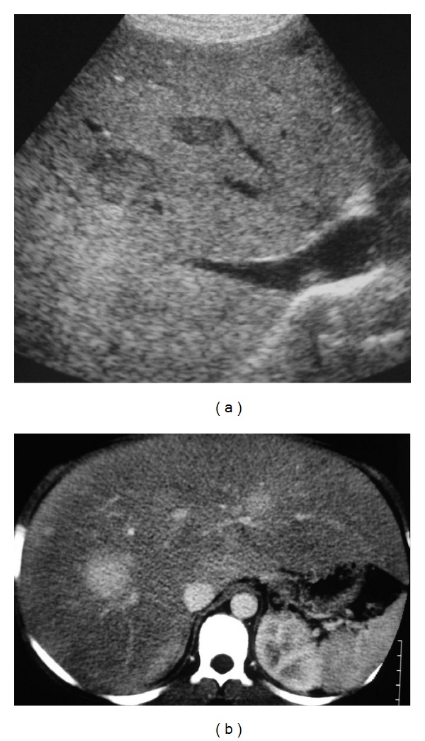
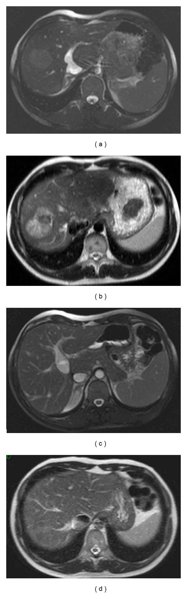
Benign liver tumors are very rare in children. Most focal nodular hyperplasia (FNH) remain sporadic, but predisposing factors exist, as follows: long-term cancer survivor (with an increasing incidence), portal deprivation in congenital or surgical portosystemic shunt. The aspect is atypical on imaging in two-thirds of cases. Biopsy of the tumor and the nontumoral liver is then required. Surgical resection will be discussed in the case of large tumors with or without symptoms. In the case of associated vascular disorder with portal deprivation, restoration of the portal flow will be discussed in the hope of seeing the involution of FNH. HepatoCellular Adenoma (HCA) is frequently associated with predisposing factors such as GSD type I and III, Fanconi anemia especially if androgen therapy is administered, CPSS, and SPSS. Adenomatosis has been reported in germline mutation of HNF1- α . Management will depend on the presence of a predisposing factor and may include metabolic control, androgen therapy withdrawn, or closure of the shunt when appropriate. Surgery is usually performed on large lesions. In the case of adenomatosis or multiple lesions, surgery will be adapted. Close followup is required in all cases.


Figures

Figure 1
Figure 1
Nine-year-old girl with a history of metastatic right nephroblastoma treated with chemo- and radiotherapy. Liver MRI with T1-weighted images without (a) and with contrast injection at the arterial (b) and portal phases (c), performed six years after the end of treatment, displays multiple hepatic nodules, from 1 to 3 cm large, that enhance strongly after contrast injection at the arterial phase (b) and have almost the same signal as the surrounding liver before contrast injection (a) and at the portal phase (c). Simple clinical and imaging followup was performed (courtesy of Dr. H. Brisse, Institut Curie Paris, France).
Figure 2
Figure 2
Six-year-old boy with a congenital portohepatic shunt complicated by a biopsy-proven FNH measuring 7 cm diameter (white star). (a) Contrast-enhanced CT scan at diagnosis shows an abnormal and large communication between the right portal branch (RPB) and the right hepatic vein (RHV). (b) Phlebography with opacification of the shunt between the RPB and the RHV. Closure of the shunt was performed by interventional radiology. (c) MRI performed seven years later shows the disappearance of FNH on the T2-weighted images. No enhancement was present at the arterial phase after gadolinium injection (not shown). (d) Note the normal aspect of the portal bifurcation (RPB and left portal branch (LPB)) on coronal MIP reconstruction of the T2-balanced sequence.
Figure 3
Figure 3
Spontaneous evolution of fortuitously discovered FNH in two children. (a) and (b): three-year-old girl with sickle-cell disease. CT scan after contrast injection at the portal phase at diagnosis (a). And three years later (b) shows growth of the tumor from 5 to 12 cm diameter. (c) and (d): eight-year-old girl. CT scan after contrast injection at diagnosis (c) and 12 years later (d) shows the spontaneous disappearance of the 5 cm diameter tumor with capsular retraction.
Figure 4
Figure 4
Adenomatosis related to HNF1-α germline mutation aspect on MRI in a 14-year-old girl: (a) and (b): T1 WI with chemical shift shows two lesions in the left lobe of the liver with drop of the signal of the largest lesion on the out-phase sequence that reveals the presence of fat in the tumor. (c) and (d): T1 WI after contrast injection shows early arterial enhancement of the largest lesion with washout in the late portal phase.
Figure 5
Figure 5
Fourteen-year-old boy with glycogen storage disease type I and multiple HCA measuring from 1 to 3 cm diameter. (a) US shows an enlarged hyperechoic liver (steatotic) with well-delimited hypoechoic nodules. (b) CT performed at the arterial phase after contrast injection shows enlarged steatotic liver with multiple nodules that are strongly enhanced.
Figure 6
Figure 6
Adenoma associated with CPSS. Aspect on MRI at diagnosis and 10 months after surgical closure of the shunt: (a) T2 WI-balanced sequence shows the CPSS consisting of a patent ductus venosus. Note that the tumor is not easily visible in this sequence. (b) The 4 cm diameter HCA lies in segment 8, and it is better seen on T2 WI and appears heterogeneous and mainly hyperintense compared to the surrounding liver. MRI performed 10 months after surgical closure of the shunt (c) shows the complete disappearance of the tumor (d).

References

    1. Fukukura Y, Nakashima O, Kusaba A, Kage M, Kojiro M. Angioarchitecture and blood circulation in focal nodular hyperplasia of the liver. Journal of Hepatology. 1998;29(3):470–475. - PubMed
    1. Wanless IR, Mawdsley C, Adams R. On the pathogenesis of focal nodular hyperplasia of the liver. Hepatology. 1985;5(6):1194–1200. - PubMed
    1. Rebouissou S, Bioulac-Sage P, Zucman-Rossi J. Molecular pathogenesis of focal nodular hyperplasia and hepatocellular adenoma. Journal of Hepatology. 2008;48(1):163–170. - PubMed
    1. Nguyen BN, Fléjou JF, Terris B, Belghiti J, Degott C. Focal nodular hyperplasia of the liver: a comprehensive pathologic study of 305 lesions and recognition of new histologic forms. American Journal of Surgical Pathology. 1999;23(12):1441–1454. - PubMed
    1. Kumagai H, Masuda T, Oikawa H, Endo K, Endo M, Takano T. Focal nodular hyperplasia of the liver: direct evidence of circulatory disturbances. Journal of Gastroenterology and Hepatology. 2000;15(11):1344–1347. - PubMed

LinkOut - more resources