. 2013 Apr;15(4):421-34.
doi: 10.1593/neo.122098.

γ-Catenin at adherens junctions: mechanism and biologic implications in hepatocellular cancer after β-catenin knockdown

Affiliations

γ-Catenin at adherens junctions: mechanism and biologic implications in hepatocellular cancer after β-catenin knockdown

Emily Diane Wickline et al. Neoplasia. 2013 Apr.

Abstract

β-Catenin is important in liver homeostasis as a part of Wnt signaling and adherens junctions (AJs), while its aberrant activation is observed in hepatocellular carcinoma (HCC). We have reported hepatocyte-specific β-catenin knockout (KO) mice to lack adhesive defects as γ-catenin compensated at AJ. Because γ-catenin is a desmosomal protein, we asked if its increase in KO might deregulate desmosomes. No changes in desmosomal proteins or ultrastructure other than increased plakophilin-3 were observed. To further elucidate the role and regulation of γ-catenin, we contemplate an in vitro model and show γ-catenin increase in HCC cells upon β-catenin knockdown (KD). Here, γ-catenin is unable to rescue β-catenin/T cell factor (TCF) reporter activity; however, it sufficiently compensates at AJs as assessed by scratch wound assay, centrifugal assay for cell adhesion (CAFCA), and hanging drop assays. γ-Catenin increase is observed only after β-catenin protein decrease and not after blockade of its transactivation. γ-Catenin increase is associated with enhanced serine/threonine phosphorylation and abrogated by protein kinase A (PKA) inhibition. In fact, several PKA-binding sites were detected in γ-catenin by in silico analysis. Intriguingly γ-catenin KD led to increased β-catenin levels and transactivation. Thus, γ-catenin compensates for β-catenin loss at AJ without affecting desmosomes but is unable to fulfill functions in Wnt signaling. γ-Catenin stabilization after β-catenin loss is brought about by PKA. Catenin-sensing mechanism may depend on absolute β-catenin levels and not its activity. Anti-β-catenin therapies for HCC affecting total β-catenin may target aberrant Wnt signaling without negatively impacting intercellular adhesion, provided mechanisms leading to γ-catenin stabilization are spared.


Figures
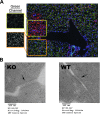
Figure 1
Figure 1
Assessment of AJs and desmosomes in β-catenin KO and WT livers. (A) Membranous γ-catenin (green) and β-catenin (red) are mutually exclusive at the cell membranes/AJ in β-catenin of hepatocytes in β-catenin KO livers that become repopulated with β-catenin-positive hepatocytes at elderly ages as shown by representative double IF images of fixed liver sections. Boxes show the staining pattern of γ-catenin (green channel) in β-catenin-positive and β-catenin-negative sections of the KO liver. (B) TEM of β-catenin KO and WT livers shows the presence of desmosomes (arrows). The intercellular distance between hepatocytes connected by desmosomes is not significantly different (mean distances: KO = 27.4 nm, WT = 25.1 nm; P > .10).
Figure 2
Figure 2
β-Catenin KO mouse livers show no changes in desmosomal proteins, except Pkp3. (A) β-Catenin KO livers have no apparent changes in most desmosomal protein levels in Triton X-100-soluble (S) and Triton X-100-insoluble/cytoskeleton-associated lysates (I) through WBs, despite mRNA changes (Table 1): DPI (250 kDa), DPII (210 kDa), Dsg1 (150 kDa), Dsg2 (59–150 kDa), Dsg3 (55–130 kDa), Dsg4 (100–115 kDa), Dsc2 (110 kDa), and Pkp2 (100 kDa). However, Pkp3 (87 kDa) is slightly increased in KO livers in both soluble (S) and insoluble (I) CAL fractions over WT age- and sex-matched liver lysates. (B) β-Catenin KO livers show more soluble and insoluble γ-catenin (83 kDa) than WTs by WBs. WB for actin (42 kDa) verifies comparable loading. (C) Representative IPs of γ-catenin in insoluble CAL fraction show no apparent changes in desmosomal protein associations with γ-catenin in β-catenin KO and WT livers. (D) Pkp3 co-precipitates with γ-catenin (upper panel) and E-cadherin (lower panel) equally in the soluble CAL fractions of KO and WT livers. (E) Nuclear lysates show that Pkp3 protein levels are similar between KO and WT livers.
Figure 3
Figure 3
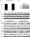
sKD of β-catenin protein accurately replicates conditions of β-catenin KO in vivo. (A) siRNA (siβ-catenin) induces efficient KD of β-catenin (92 kDa) in Hep3B cells and leads to an increase of γ-catenin (83 kDa) protein levels. (B) Whole-cell lysates from siβ-catenin and siNegative-treated Hep3B cells were immunoprecipitated with anti-E-cadherin or anti-γ-catenin antibodies. WB performed for γ-catenin (upper panel) and E-cadherin (120 kDa; lower panel) shows differential co-precipitation with sKD of β-catenin. (C) Hep3B and HepG2 cells treated with ASO to β-catenin show a decrease in β-catenin protein and subsequent increase in γ-catenin by WB. Both the WT and truncated forms of β-catenin in HepG2 cells show decrease with ASO. (D) KD of β-catenin activity using small molecule inhibitor ICG-001 does not change the protein levels of β- or γ-catenin, despite the decrease in β-catenin activity indicated by a decrease in its target cyclin D1 (37 kDa).
Figure 4
Figure 4
Mechanism of γ-catenin stabilization after sKD of β-catenin. (A) Real-time PCR for γ-catenin gene (JUP) and GAPDH reference gene shows insignificant difference in average mRNA expression (±SD) in siβ-catenin versus siNegative Hep3B-treated cells at 48-hour KD (n = 3). (B) Treatment of Hep3B cells for 3 hours with 10 or 25 nM OA, a known serine/threonine phosphatase inhibitor, results in increase in γ-catenin (83 kDa) as shown in a representative WB. (C) Treatment of Hep3B cells with various serine/threonine kinase inhibitors for 3 hours after 45 hours of transfection with control or β-catenin siRNA reveals a decrease in stabilization of γ-catenin after H-89 (PKA inhibitor). The numbers below the WB represent integrated optical density normalized to GAPDH (loading control) for the respective lane. (D) Treatment of Hep3B cells with H-89 at 150 nM and 20 µM for 3 hours after 21 or 45 hours of transfection with siβ-catenin or siNegative reveals a dose- and time-dependent decrease in extent of γ-catenin stabilization. The numbers below the WB represent integrated optical density normalized to GAPDH (loading control) for the respective lane.
Figure 5
Figure 5
sKD and dKD of β- and γ-catenins with siRNA in Hep3B, a human HCC cell line, and impact on Wnt signaling, DNA synthesis, and E-cadherin. (A) Representative WBs showing a time course of sKD and dKD of β- and γ-catenins using siRNA in Hep3B cells. KD begins at 24 hours and persists for 72 hours after transfection. (B) Whole-cell lysates from siγ-catenin and siNegative-treated Hep3B cells were immunoprecipitated with an anti-β-catenin antibody. WB shows no changes in β-catenin's association with AJ protein E-cadherin. (C) Wnt reporter (TOPflash) activity is significantly decreased after sKD of β-catenin and dKD; however, there are insignificant differences between these two groups. A significant increase of TOPflash activity is observed after sKD of γ-catenin at 48 hours (P < .01; ±SD; asterisk indicates significance compared to siNegative; NS, not significant). (D) Upper panel: Thymidine incorporation (counts per minute) for sKD and dKD shows a significant but comparable decrease in DNA synthesis with sKD of β-catenin and dKD compared to siNegative. No change in DNA synthesis with sKD of γ-catenin at 48-hour KD was observed. Lower panel: Thymidine incorporation decreased comparably and significantly for sKD of β-catenin and dKD at 72 hours as well. A significant increase in DNA synthesis was observed with sKD of γ-catenin at 72-hour KD (P < .01; ±SD; asterisk indicates significance compared to siNegative; NS, not significant). (E) Representative WBs from lysates of Hep3B cells show a modest decrease in E-cadherin after dKD at 48 hours, while comparably higher levels are observed in sKD and siNegative conditions.
Figure 6
Figure 6
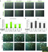
dKD of β- and γ-catenins affects cell migration and cell-cell adhesion in vitro. (A) Representative images of wound closure at 24 hours after initiation of the wound and 48 hours after transfection with various siRNAs. (B) Scratch wound assay for sKD and dKD Hep3B cells shows a significant increase in wound closure percentage for dKD of β- and γ-catenins only (P < .01; ±SD; asterisk indicates significance compared to siNegative). (C) CAFCA with sKD and dKD Hep3B cells shows a significant decrease in heterotypic cell-cell adhesion with dKD of β- and γ-catenins only (P < .01; ±SD; asterisk indicates significance compared to siNegative) as measured by increase in percentage of cells that de-adhere from the monolayer after centrifugation at 375g for 10 minutes. (D) Hanging drop assay shows that at 24 hours after drop formation (72-hour transfection) the colonies formed appear similar in size, though the dKD and sKD of β-catenin colonies appear to be less symmetric than the sKD γ-catenin and siNegative colonies. After pipetting, the dKD cells dissociate the most, indicating weaker homotypic cell-cell adhesions. Addition of EGTA, a calcium chelator, to siNegative-treated cells was used as a control to show that colonies formed were calcium-dependent or cadherin-based cell-cell adhesion.

References

    1. El-Serag HB. Hepatocellular carcinoma: recent trends in the United States. Gastroenterology. 2004;127:S27–S34. - PubMed
    1. Parkin DM, Bray F, Ferlay J, Pisani P. Global cancer statistics, 2002. CA Cancer J Clin. 2005;55:74–108. - PubMed
    1. Waly Raphael S, Yangde Z, Yuxiang C. Hepatocellular carcinoma: focus on different aspects of management. ISRN Oncol. 2012;2012:421673. - PMC - PubMed
    1. Kondo Y, Kanai Y, Sakamoto M, Genda T, Mizokami M, Ueda R, Hirohashi S. Beta-catenin accumulation and mutation of exon 3 of the beta-catenin gene in hepatocellular carcinoma. Jpn J Cancer Res. 1999;90:1301–1309. - PMC - PubMed
    1. Legoix P, Bluteau O, Bayer J, Perret C, Balabaud C, Belghiti J, Franco D, Thomas G, Laurent-Puig P, Zucman-Rossi J. Beta-catenin mutations in hepatocellular carcinoma correlate with a low rate of loss of heterozygosity. Oncogene. 1999;18:4044–4046. - PubMed

Publication types

MeSH terms