. 2013 Mar;9(3):e1003400.
doi: 10.1371/journal.pgen.1003400. Epub 2013 Mar 28.

Differential evolutionary fate of an ancestral primate endogenous retrovirus envelope gene, the EnvV syncytin, captured for a function in placentation

Affiliations

Differential evolutionary fate of an ancestral primate endogenous retrovirus envelope gene, the EnvV syncytin, captured for a function in placentation

Cécile Esnault et al. PLoS Genet. 2013 Mar.

Abstract

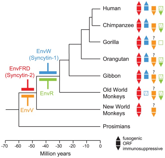
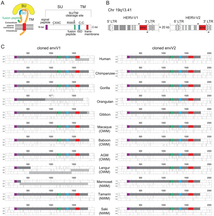
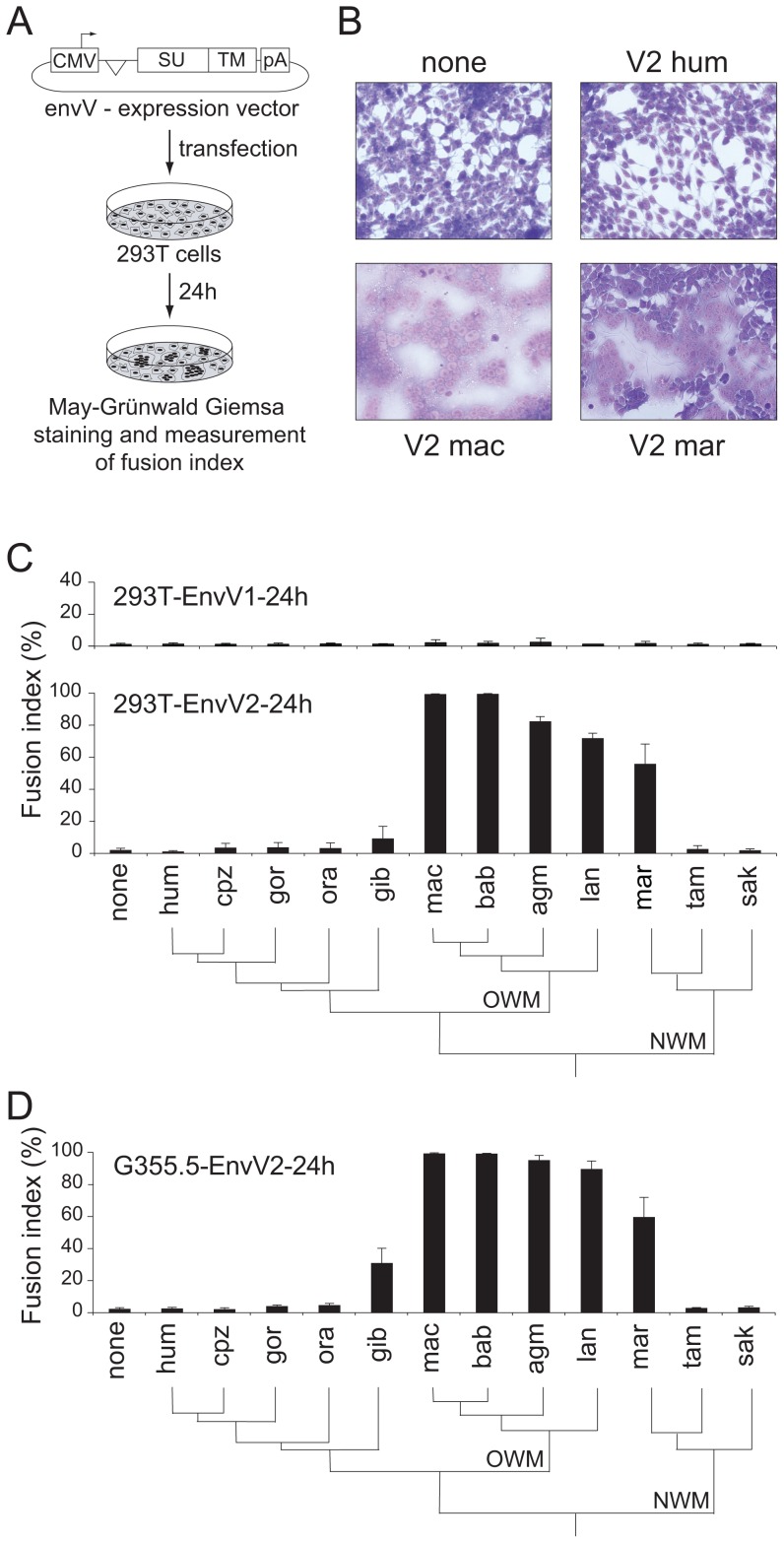
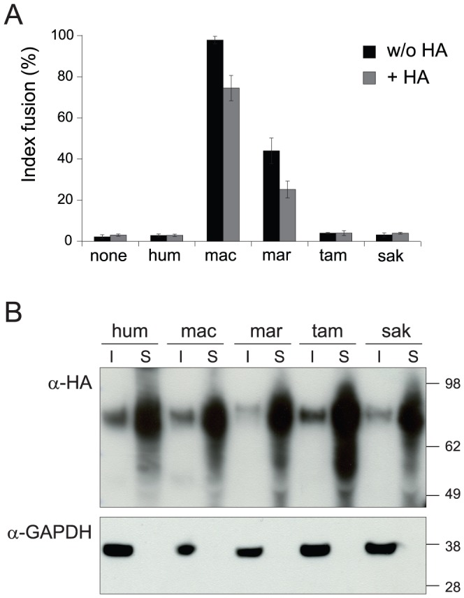
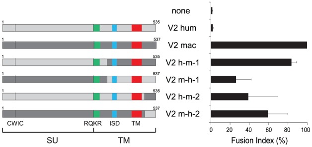
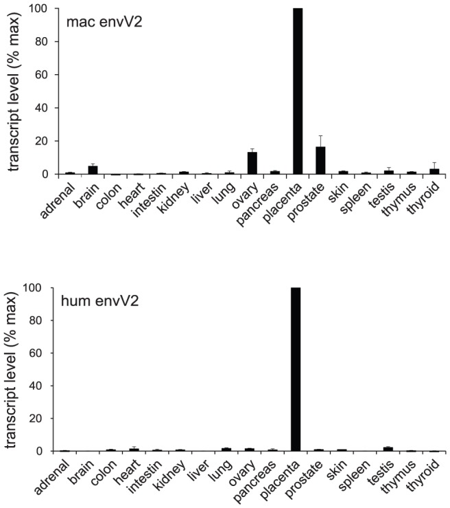
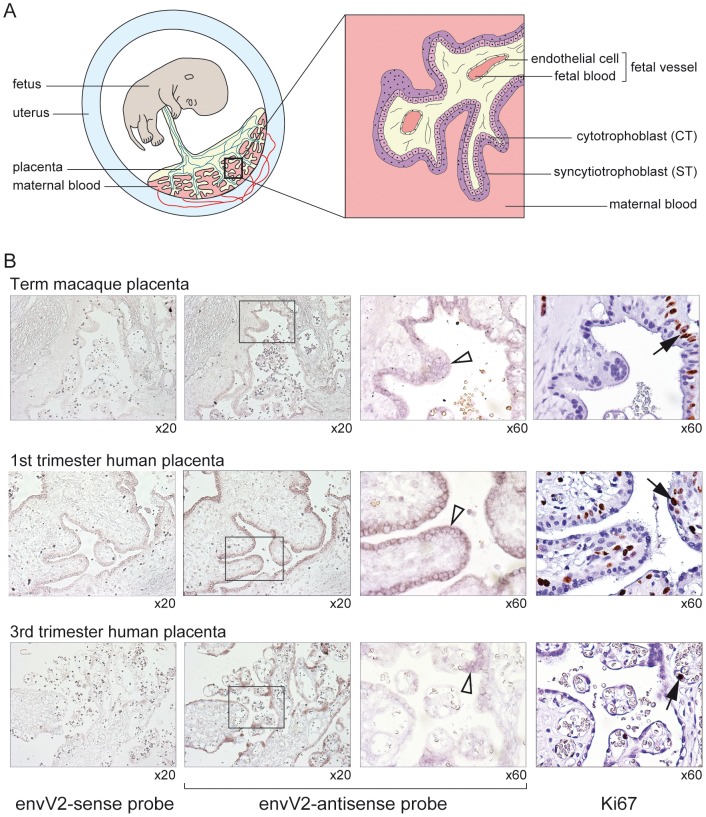
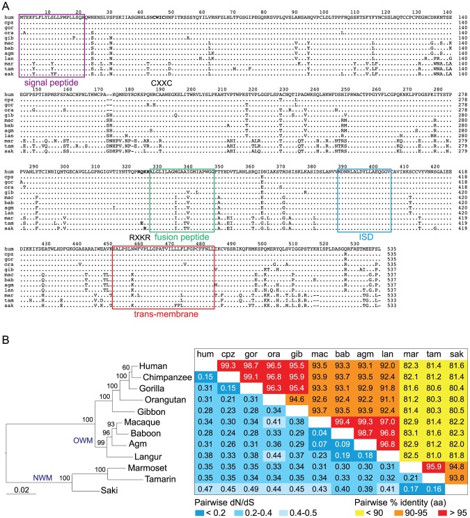
Syncytins are envelope genes of retroviral origin that have been co-opted for a role in placentation. They promote cell-cell fusion and are involved in the formation of a syncytium layer--the syncytiotrophoblast--at the materno-fetal interface. They were captured independently in eutherian mammals, and knockout mice demonstrated that they are absolutely required for placenta formation and embryo survival. Here we provide evidence that these "necessary" genes acquired "by chance" have a definite lifetime with diverse fates depending on the animal lineage, being both gained and lost in the course of evolution. Analysis of a retroviral envelope gene, the envV gene, present in primate genomes and belonging to the endogenous retrovirus type V (ERV-V) provirus, shows that this captured gene, which entered the primate lineage >45 million years ago, behaves as a syncytin in Old World monkeys, but lost its canonical fusogenic activity in other primate lineages, including humans. In the Old World monkeys, we show--by in situ analyses and ex vivo assays--that envV is both specifically expressed at the level of the placental syncytiotrophoblast and fusogenic, and that it further displays signs of purifying selection based on analysis of non-synonymous to synonymous substitution rates. We further show that purifying selection still operates in the primate lineages where the gene is no longer fusogenic, indicating that degeneracy of this ancestral syncytin is a slow, lineage-dependent, and multi-step process, in which the fusogenic activity would be the first canonical property of this retroviral envelope gene to be lost.


Conflict of interest statement

The authors have declared that no competing interests exist.

Figures

Figure 1
Figure 1. Phylogenetic tree of primates, and status of the captured endogenous retrovirus envelope genes with placental expression.
The date of insertion of the indicated retroviral envelope genes into the genome of primate ancestors is indicated for the syncytin genes (i.e. the EnvFRD- and EnvW-encoding genes, in red and blue , respectively, and for the EnvV- and EnvR-encoding genes, in orange and green , respectively). Their presence in the various primate lineages is indicated with filled squares when the gene still possesses a full-length ORF, with hatched squares when the coding sequence is prematurely interrupted, and an empty square when the gene is no longer present. In addition, fusogenic activities (as determined by ex vivo cell-cell fusion assays in ref [5], [8]) are schematized with an upper triangle when present, and immunosuppressive activities (as assayed in ref [19]) with lower triangles. Branch length is proportional to time (in million years).
Figure 2
Figure 2. Structure of a canonical retroviral envelope protein and characterization of the envV1 and envV2 genes in primates.
(A) Schematic representation of a retroviral Env protein, delineating the surface (SU) and transmembrane (TM) subunits. The furin cleavage site (consensus: R/K-X-R/K-R) between the two subunits, the C-X-X-C motif involved in SU-TM interaction, the hydrophobic signal peptide (purple), the fusion peptide (green), the transmembrane domain (red), and the putative immunosuppressive domain (ISD) (blue) along with the conserved C-C motif are indicated. (B) Genomic organization of the HERV-V1 and HERV-V2 proviruses. The retroviral env ORF (red open box) and long terminal repeats (LTRs; arrowed open boxes), and the Alu (light gray boxes) and MER50 (dark gray boxes) retroelements are indicated. Positions of the primers designed to amplify the env coding sequences are indicated. (C) ORF map of the cloned envV genes. The dark gray boxes delineate the envelope coding sequences and the light gray boxes represent the ORFs still present downstream of stop codons or frameshifts. The deletion in the orangutan envV1 gene is depicted by an open triangle.
Figure 3
Figure 3. Cell–cell fusion assay for the primate EnvV proteins.
(A) Env-expressing vectors and rationale of the assay. Each of the 24 cloned envV genes was introduced into the phCMV expression vector, between the beta-globin intron and polyadenylation (pA) sequences. Cells were transfected and stained with May-Grünwald-Giemsa 24 h after transfection. The fusion index is defined as [(N-S)/T]×100, where N is the number of nuclei in the syncytia, S is the number of syncytia, and T is the total number of nuclei counted. (B) 293T cells transfected with expression vectors for no protein (none), and for the human, macaque and marmoset EnvV2 (V2 hum, V2 mac and V2 mar, respectively), displaying large multinucleated syncytia 24 h later, only upon transfection with the latter two. (C) Histogram showing the fusion index of the indicated series of primate envV1 (upper) and envV2 (lower) genes in 293T cells transfected with the corresponding expression vectors. The primate phylogeny is illustrated below, with the Old World monkeys (OWM) and New World monkeys (NWM) indicated. None, no protein; hum, human; cpz, chimpanzee; gor, gorilla; gib, gibbon; mac, macaque; bab, baboon; agm, African green monkey; lan, langur; mar, marmoset; tam, tamarin; sak, saki. (D) Same as (C) but with feline G355.5 cells transfected with the envV2 expression vectors.
Figure 4
Figure 4. EnvV2 protein expression and cell surface localization.
(A) Cell-cell fusion assay for hemagglutinin (HA)-tagged EnvV2 proteins in G355.5 cells. Transient transfection of feline cells was assayed with a control plasmid (none) or plasmids expressing the EnvV2 protein, tagged (or not) with the HA epitope. May-Grünwald-Giemsa staining was carried out 24 h posttransfection and fusion index measured as in Figure 3. (B) Cell-surface expression of EnvV2 proteins. A23 cells were transfected with the indicated EnvV2-HA expression vectors. The cell surface-expressed proteins were biotinylated for further purification (see Methods). Detection of the EnvV2 proteins was performed by Western blotting of the two cellular fractions obtained –i.e. the intracellular cell lysates (I) and the cell surface biotinylated proteins (S)- with a monoclonal anti-HA antibody (upper). As a control for the fractioning, the blot was hybridized with a monoclonal antibody against the GAPDH soluble cellular protein (lower).
Figure 5
Figure 5. Cell–cell fusion assay for chimeric EnvV2 proteins.
Structure of the chimeric envelope proteins between the human (light gray) and macaque (dark gray) EnvV2 proteins, with the characteristic Env domains indicated (same color code as in Figure 2 for the fusion peptide, the immunosuppressive domain (ISD) and the transmembrane domain (TM), and the positions of the CWIC motif and the SU/TM cleavage site RQKR). 293T cells were transfected with the chimeric EnvV2-expressing plasmids, stained 48 h after transfection with May-Grünwald-Giemsa, and fusion indexes were measured as in Figure 3.
Figure 6
Figure 6. qRT–PCR analysis of envV2 transcripts in macaque and human tissues.
Real-time qRT-PCR analysis of the macaque and human envV2 transcripts in a panel of 17 macaque (upper) and human (lower) tissues, with transcript expressed as percent of maximum, after normalization with a control gene (PPIA) mRNA (see Methods).
Figure 7
Figure 7. In situ hybridization for envV2 expression in the macaque and human placenta.
(A) Schematic representation of the simian placenta, with an enlarged villus bathed in maternal blood and displaying –from the maternal to the fetal side- the syncytiotrophoblast (ST) layer, the mononucleated cytotrophoblasts (CT) and the fetal vessels. (B) Serial sections of macaque (upper row) and human (two lower rows) placenta. (columns 1–3) in situ hybridization with digoxigenin-labeled envV2 sense (negative control, column 1) and antisense (columns 2,3) riboprobes, revealed with an alkaline phosphatase-conjugated anti-digoxigenin antibody. (column 4) immunohistochemical staining of the Ki67 nuclear antigen. (columns 3,4) enlarged views with the empty arrowheads pointing to the syncytiotrophoblast positively stained for envV2 mRNA, and the filled arrows to Ki67-marked mononucleated cytotrophoblasts.
Figure 8
Figure 8. Sequence conservation and purifying selection of the envV2 gene in primates.
(A) Aligned amino acid sequences and characteristic structural features of the primate EnvV2 proteins. The SU (surface) and TM (transmembrane) subunits are delineated with the position of the putative proteolytic cleavage site (RQKR, in bold) between the two subunits, of the signal peptide (in purple) and of the CWIC motif (in bold) in the SU subunit, and the position of the fusion peptide (green), of the immunosuppressive domain (blue) and of the transmembrane domain (red) in the TM subunit. Dots indicate amino acid identity and hyphens codon deletions. Abbreviations are as in Figure 3. (B) EnvV2–based maximum likelihood phylogenetic tree was determined using nucleotide alignment of the env gene, inferred with the RAxML program. The horizontal branch length and scale indicate the percentage of nucleotide substitutions. Percent bootstrap values obtained from 1.000 replicates are indicated at the nodes. Double-entry table for the pairwise percentage of amino acid sequence identity (upper triangle) and the pairwise value of dN/dS (lower triangle) between the envV2 genes among primate species. A color code is provided for both series of values.
Figure 9
Figure 9. Branch-specific analysis of selection along the envV2-based phylogenetic tree.
Analysis was performed using the GA-branch method from the HyPhy Package on the webserver www.datamonkey.org, and the selected model was the one with the best Akaike Information Criterion (AIC). Branch-specific analysis allows the non-synonymous to synonymous mutation ratio (dN/dS) to vary between phylogenetic branches. Such an analysis on the envV2 gene reveals that all the branches are under strong purifying selection (dN/dS<1) with no significant difference in the strength of selection between New World monkeys (NWM), Old World monkeys (OWM) and hominoids. Left: maximum likelihood tree of envV2 (same as in Figure 8) is represented together with the branch numbers. The color code for each branch class is indicated on top. Right: estimated branch-specific dN/dS values are indicated ± standard deviation.

References

    1. Black SG, Arnaud F, Palmarini M, Spencer TE (2010) Endogenous retroviruses in trophoblast differentiation and placental development. Am J Reprod Immunol 64: 255–264 doi:10.1111/j.1600-0897.2010.00860.x. - DOI - PMC - PubMed
    1. Dupressoir A, Lavialle C, Heidmann T (2012) From ancestral infectious retroviruses to bona fide cellular genes: role of the captured syncytins in placentation. Placenta 33: 663–671 doi:10.1016/j.placenta.2012.05.005. - DOI - PubMed
    1. Mi S, Lee X, Li X, Veldman G, Finnerty H, et al. (2000) Syncytin is a captive retroviral envelope protein involved in human placental morphogenesis. Nature 17: 785–789 doi:10.1038/35001608. - DOI - PubMed
    1. Blond JL, Lavillette D, Cheynet V, Bouton O, Oriol G, et al. (2000) An envelope glycoprotein of the human endogenous retrovirus HERV-W is expressed in the human placenta and fuses cells expressing the type D mammalian retrovirus receptor. J Virol 74: 3321–3329 doi:10.1128/JVI.74.7.3321-3329.2000. - DOI - PMC - PubMed
    1. Mallet F, Bouton O, Prudhomme S, Cheynet V, Oriol G, et al. (2004) The endogenous retroviral locus ERVWE1 is a bona fide gene involved in hominoid placental physiology. Proc Natl Acad Sci U S A 101: 1731–1736 doi:10.1073/pnas.0305763101. - DOI - PMC - PubMed

Publication types

MeSH terms