

Three-dimensional CT Venography: A Diagnostic Modality for the Preoperative Assessment of Patients with Varicose Veins
- PMID: 23555458
- PMCID: PMC3595794
- DOI: 10.3400/avd.oa.11.00021
Three-dimensional CT Venography: A Diagnostic Modality for the Preoperative Assessment of Patients with Varicose Veins
Abstract
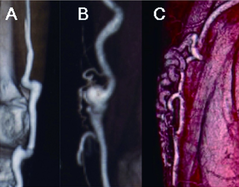
Objective: We preoperatively assessed varicose veins by means of computed tomography (CT) with contrast injection in the veins of the lower extremity (CT venography). This paper reports the procedures, results and implications of CT venography from the surgical aspect.
Methods: A total of 48 legs in 39 patients were examined. Contrast medium was diluted ten-fold and injected into the lower extremity veins, often using a dual route of injection. The images were reconstructed with the volume-rendering method.
Results: CT venography clearly visualized the veins with a small amount of contrast medium and facilitated the identification of anatomy that was not suitable for passing the stripper. In addition, CT venography helped identify unusual types of varicose veins or uncommon sites of inflow of small saphenous veins. Such information was helpful for avoiding unexpected vascular injury or for minimizing skin incision. Dual-route injection was beneficial to minimize the blind zones. Doppler ultrasound could be more focused on hemodynamic assessment and determination of incision sites.
Conclusions: CT Venography is feasible in all cases of varicose veins. When performed in conjunction with ultrasonography, it appears to facilitate the safe and efficient treatment of various types of varicose veins.
Keywords: computed tomography; varicose vein; venography.
Figures


References
-
- Kurdal AT, Cerrahoglu M, Iskesen I.Subfascial endoscopic perforator surgery ameliorates the symotoms of chronic venous ulcer (C6) patients. Int Angiol 2010; 29: 70-4 - PubMed
-
- Nadarajah ST, Demos D, Andrew IDM.Endovenous laser ablation: dose standard above-knee great saphenous vein ablation provide optimum results in patients with both above- and below-knee reflux? A randomized controlled trial. J Vasc Surg 2008; 48: 173-8 - PubMed
-
- Koyano K, Sakaguchi S. Selective stripping operation based on Doppler ultrasonic findings for primary varicose veins of the lower extremities. Surgery 1988; 103: 615-9 - PubMed
-
- Kistner RL, Ferris EB, Randhawa G.A method of performing descending venography. J Vasc Surg 1986; 4: 464-8 - PubMed
-
- Jutley RS, Cadle I, Cross KS. Preoperative assessment of primary varicose veins: a Duplex study of venous incompetence. Eur J Vasc Endovasc Surg 2001; 21: 370-3 - PubMed
LinkOut - more resources
Full Text Sources