. 2013;8(3):e58979.
doi: 10.1371/journal.pone.0058979. Epub 2013 Mar 26.

Platelets recognize brain-specific glycolipid structures, respond to neurovascular damage and promote neuroinflammation

Affiliations

Platelets recognize brain-specific glycolipid structures, respond to neurovascular damage and promote neuroinflammation

Ilya Sotnikov et al. PLoS One. 2013.

Erratum in

Abstract

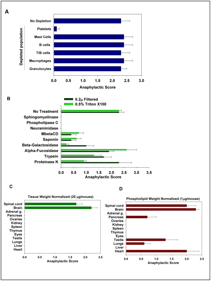
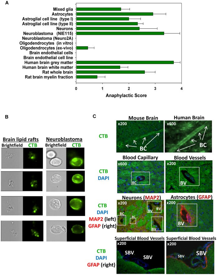
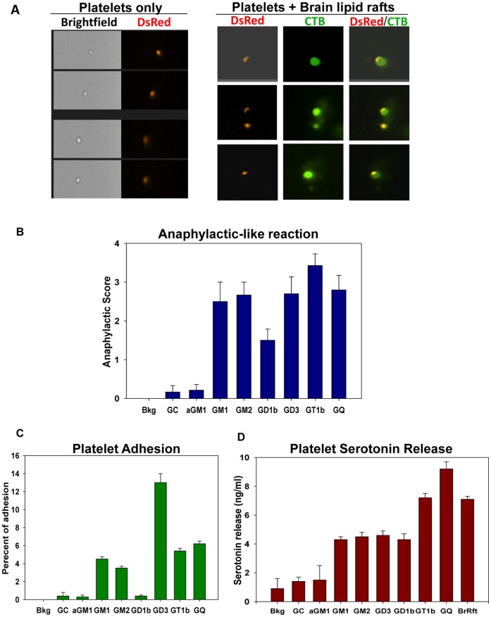
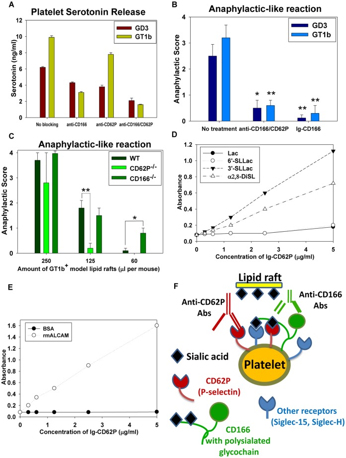
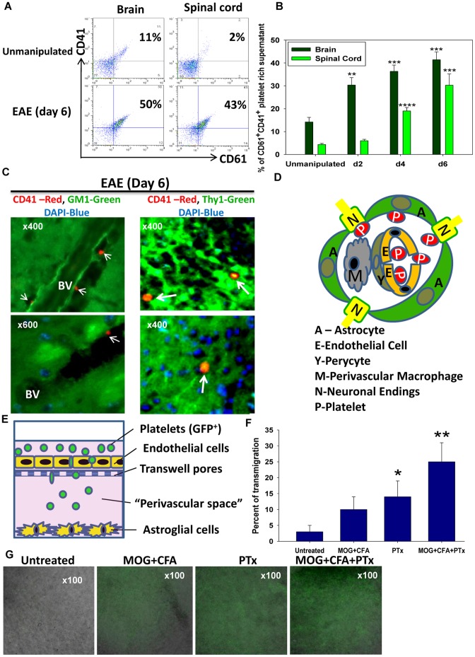
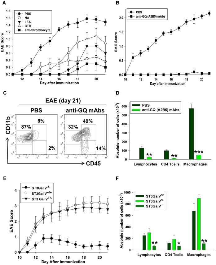
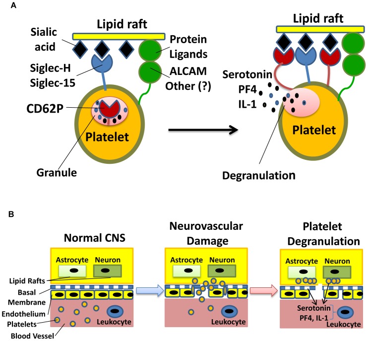
Platelets respond to vascular damage and contribute to inflammation, but their role in the neurodegenerative diseases is unknown. We found that the systemic administration of brain lipid rafts induced a massive platelet activation and degranulation resulting in a life-threatening anaphylactic-like response in mice. Platelets were engaged by the sialated glycosphingolipids (gangliosides) integrated in the rigid structures of astroglial and neuronal lipid rafts. The brain-abundant gangliosides GT1b and GQ1b were specifically recognized by the platelets and this recognition involved multiple receptors with P-selectin (CD62P) playing the central role. During the neuroinflammation, platelets accumulated in the central nervous system parenchyma, acquired an activated phenotype and secreted proinflammatory factors, thereby triggering immune response cascades. This study determines a new role of platelets which directly recognize a neuronal damage and communicate with the cells of the immune system in the pathogenesis of neurodegenerative diseases.


Conflict of interest statement

Competing Interests: The authors have declared that no competing interests exist.

Figures

Figure 1
Figure 1. Role of platelets and lipid rafts in the induction of anaphylaxis.
(A) The lipid rafts were isolated by homogenization of the brain in PBS with 0.5% Triton X-100 and injected i.v. to induce anaphylaxis. The animals were observed for ten minutes as described in Methods. The role of platelets and white blood cell subsets were assessed by using an antibody (for depletion of platelets and granulocytes), lyposomes with bisphosphonate clodronate (for depletion of macrophages), or the CD11b-DTR transgenic mice, and genetically deficient animals (mast cells, B cells, T/B cells) as described in Methods. (B) The effect of enzymatic treatment of brain lipid rafts on their ability to induce anaphylaxis. The brain lipid rafts were obtained by size filtration (0.2 µ filtered) or solubility in 0.5% Triton X-100 (0.5% Triton X-100) as described in Methods. The brain lipid rafts were treated with lipases (sphingomyelinase and phospholipase), enzymes that cleave glycopolymers (neuraminidase, β-galactosidase and fucosidase), cholesterol depleting agents (saponin and MβCD) and proteolytic enzymes (trypsin and proteinase K) and injected i.v. for the assessment of anaphylaxis. The non-treated brain lipid rafts were used as a control (no treatment). The animals were observed for ten minutes as described in Methods. (C,D) The ability of lipid rafts from different organs to cause anaphylaxis. The lipid rafts were isolated from the spinal cord, brain, adrenal gland, pancreas, ovaries, kidney, spleen, thymus, eyes, testis, lungs, liver and heart by the homogenization of tissues in PBS with 0.5% Triton X-100. The amount of i.v. injected lipid rafts was normalized according to the weight of the wet tissue (c) or concentration of phospholipids (d) as described in Methods. The animals were observed for ten minutes as described in Methods. In A–D, the maximum clinical anaphylaxis score (Mean ± S.E.) of the total number of animals from three separate experiments with 5–6 mice per group in each experiment is shown.l.
Figure 2
Figure 2. Origin and localization of brain lipid rafts.
(A) The lipid rafts were isolated from the primary cultures, cell lines, human brain gray and white matter, or rat whole brain and rat brain myelin fractions. The lipid rafts were injected i.v. and anaphylaxis was scored (see Figure 1 ). (B) The brain homogenates and neuronal line cells lipid rafts were stained for a GM1 marker of lipid rafts (CTB) and visualized by Imaging Cytometry (green). (C) The sections of the mouse (left) and human (right) brains stained with CTB (top two images, green). In the middle four images, the mouse brain sections stained with CTB show the lipid rafts around the brain capillaries (left) and blood vessels (right); same with double-staining for the neuronal (MAP2; left) and astroglial (GFAP; right) (both red) markers. Two bottom images depict areas around the brain blood vessels under pia matter (superficial blood vessels) single stained with CTB (left, green) or double stained with CTB/GFAP (right; CTB in green, GFAP in red). DAPI staining for the cell nuclei is shown in blue. Abbreviations: BC – brain capillary, BV-blood vessel, SBV-superficial blood vessel.
Figure 3
Figure 3. Unique composition of lipid rafts triggers platelets degranulation.
(A) The platelets were isolated from the DsRed transgenic mice, incubated with CTB-prestained brain lipid rafts for 3 minutes, fixed, washed and analyzed by Imaging Cytometry. Left, the bright field images and platelets fluorescence is shown (red). Right, the platelets (red) interaction with brain lipid rafts (CTB, green) is shown. (B) The induction of anaphylaxis by model lipid rafts in vivo. The model lipid rafts containing representative gangliosides (asyaloGM1, GM1, GM2, GD3, GD1b, GT1b, GQ), galactosylcerebroside (GC) or phosphatidylcholin (Bkg) were prepared and injected i.v. into the mice as described in Methods. The maximum clinical anaphylaxis score (Mean ± S.E.) of the total number of animals from three experiments with 4–5 mice per group in each experiment is shown. (C) The platelet adhesion to gangliosides in vitro. AsialoGM1, GM1, GM2, GD3, GD1b, GT1b, GQ1b gangliosides, galactosylcerebroside (GC) or phosphatidylcholin (Bkg) were adsorbed to flat-bottom 96-well plates and the adhesion of platelets to the ligands was measured. (D) The platelets serotonin release induced by the model lipid rafts in vitro. The model lipid rafts were incubated with platelets and the concentration of serotonin in the platelet-free supernatant was assessed. The brain lipid rafts (BrRft) served as a positive control. In c–d, the mean ± S.E. of triplicate is shown. The data is representative of two separate experiments.
Figure 4
Figure 4. Lipid rafts engage platelet receptors specific to sialated gangliosides.
(A) The effect of anti-CD62P and anti-CD166 mAbs on the platelet serotonin release in response to the model lipid rafts containing GD3 and GT1b in vitro. The serotonin release was measured (see Figure 3 ) in the presence of anti-CD62P and anti-CD166 mAbs. (B) The effect of anti-CD62P and anti-CD166 mAbs or the CD166-Fc fusion protein on the platelet degranulation iduced by the model lipid rafts in vivo. Anti-CD62P and anti-CD166 mAbs were injected i.v., and the model lipid rafts containing GD3 or GT1b gangliosides were injected 30 minutes later. The model lipid rafts containing GD3 or GT1b gangliosides were pre-treated with the CD166-Fc protein and injected i.v. into naïve mice. All mice were then injected i.v. with the lipid rafts as described in Methods. The maximum clinical anaphylactic score (Mean ± S.E.) of the total number of animals from three experiments with four mice per group is shown (*, p<0.05; **, p<0.01; when compared to non-treated animals). (C) The lipid rafts-induce anaphylaxis in wild-type (WT), CD62P (P-Selectin)- and CD166 (ALCAM)-deficient mice. The model lipid rafts containing GT1b ganglioside were injected i.v. into naïve mice at 250, 125 or 60 µl per mouse. The maximum clinical anaphylaxis score (Mean ± S.E.) of the total number of animals from the three experiments with 4 mice per group in each experiment is shown (*, p<0.05; **, p<0.01; when compared to wild-type animals). (D) Binding of CD62P-Fc fusion protein to lactose, 6′-sialyllactose, 3′-sialyllactose and α2,8-disialic glycopolymers adsorbed onto 96 well plates. (E) Binding of CD62P-Fc fusion protein to recombinant ALCAM or BSA (control) adsorbed onto 96 well plates. (F) Schematics of multiple receptors (CD62P, ALCAM, Siglec-H and Siglec-15) on the platelets and sialated gangliosides on brain lipid rafts interaction. In addition to the interaction of sialic acid on gangliosides with CD62P and Siglecs, ALCAM has mono and poly- sialated glycoepitopes that can be recognized by CD62P and Siglec-H/Siglec-15. The antibodies to ALCAM prevent binding of masked CD62P and other siglec receptors on the platelets to the sialated gangliosides on the lipid rafts. Thus, the anti-ALCAM and anti-CD62P antibodies disrupt an intricate network of the lipid raft-sensing receptors on the platelets surface.
Figure 5
Figure 5. Platelets penetrate the blood-brain barrier (BBB) and accumulate in the lipid rafts-rich areas in the model of neuroinflammation.
(A–C) The platelets isolated from the CNS of healthy mice or mice with EAE were analyzed by flow cytometry. The platelet-rich supernatants were prepared from the CNS homogenates, the platelets were stained for CD41 and CD61 (A) The percentages of CD41+CD61+ platelets are depicted in the upper right quadrants of the contour plots. (B) The increase of platelet numbers in the brain and spinal cord in the early stages of EAE (Mean ± S.E, five animals per group; **, p<0.01; ***, p<0.001; ****, p<0.0001; when compared to the unmanipulated mice). (C) The immunofluorescent analysis of the platelet localization within the CNS of WT B6 (C) or the Thy1-YFP reporter mice (D) with EAE on day 6. The CNS sections were double stained with CTB (green) and anti-CD41 mAbs (red) in (c, left panel) or single stained with anti-CD41 (red) in (c, right panel). The CD41+ platelets are indicated by the arrows. (D) The schematic model of platelets topographic disposition in the perivascular space and their interaction with lipid rafts on astrocytes and neurons as a result of the increased CNS blood vessel permeability. (E–F) The model of the BBB with a “perivascular space” between the monolayer of brain endothelial cells in the top chamber and the astroglial cells in the bottom chamber of the Transwell system (E). The platelets isolated from the ACTB-GFP transgenic mice were added to the top chamber together with MOG/CFA and/or PTx. The percentage of the transmigrated GFP+ platelets is shown in (F). The combined bright field (astroglial cells, grey) and fluorescent images (GFP+ platelets, green) of the bottom chambers under specified conditions are shown in (G).
Figure 6
Figure 6. Effects of the agents that affect the platelet-lipid raft interactions in the development of EAE.
(A,B) EAE in mice treated with anti-thrombocyte serum (anti-thromocyte), beta subunit of cholera toxin (CTB), neuroaminidase (NA), and Limax flavus agglutinin (LFA) (A) or anti-GQ (A2B5) antibodies (B). EAE was induced by the immunization of B6 with MOG/CFA as described in Methods. Pertussis toxin (PTx) was administered i.p. on day 0 and day 2 post-immunization. The anti-thrombocyte serum was injected on day 0, 2, 4 and 8 post-immunization, and CTB, NA and LFA were injected on days 0, 2, 4, 6 and 8 post-immunization (A). The anti-GQ antibodies were injected on days 0, 2, 4, 8, 10, 12 and 14 post-immunization (B). The mice were monitored daily and the EAE score was assessed as described in Methods. Mean ± S.E. of the total number of animals from the three separate experiments with groups of 7–10 mice in each experiment is shown. (C,D) The flow cytometry analysis of the CNS infiltrating cells isolated from the mice treated with anti-GQ antibodies. The mononuclear cells were isolated from the CNS of the mice treated with PBS or anti-GQ antibodies on day 21 after EAE induction, stained for CD11b and CD45 and analyzed by flow cytometry as described in Methods. The staining for CD11b (y-axis) and CD45 (x-axis) of the CNS mononuclear cells is shown. The percentages of populations of resting CD11b+CD45low microglia (left gates), CD11b+CD45hi macrophages (upper right gates) and CD11bCD45hi lymphocytes (lower right gates) are shown in (C). The quantification of the absolute number of macrophages, lymphocytes and CD4 T cells in the CNS is shown in (D). The absolute numbers of CD11bCD45hi lymphocytes, CD3+CD4+ T cells and CD11b+CD45hi macrophages were calculated by multiplying the total cell count obtained using a hemocytometer by the percentage of these cells determined by flow cytometry. Mean ± S.E. of the group of five animals is shown. (E,F) The EAE disease course (E) and the quantification of the CNS immune cell infiltrate (F) in the groups of ST3Gal-V−/−, ST3Gal-V+/− and ST3Gal-V+/+ mice. The EAE was induced and cellular infiltration was analyzed as in (A) and (D), respectively. The mean ± S.E. of the total number of animals from the three separate experiments with the groups of 7–10 mice in each experiment is shown.
Figure 7
Figure 7. Sialated glycolipids are recognized by platelets as “innate danger signals” of a neuronal damage.
(A) The proposed model of brain lipid rafts recognition by the platelet receptors. Multiple receptors on the platelets and an intact rigid structure of brain lipid rafts are required to form glycosynapses for the coordinated platelet activation and degranulation.The initial recognition of brain lipid rafts involves the interplay of siglecs and adhesion molecules such as Siglec-H, Siglec-15, ALCAM or other molecules, which results in the recruitment of P-selectin on the surface of platelets to the site of glycosynapse formation and eventual platelets degranulation. (B) The schematics of the staged neuronal damage detection process by platelets leading to the recognition of lipid rafts during EAE or neurodegenerative diseases such as MS. In the intact CNS, platelets do not see lipid rafts of astrocytes and neurons due to the impermeable nature of the BBB. The trauma, infection, neurodegeneration or inflammation disrupts the structure of neurovascular units, exposing lipid rafts on the surface of astrocytes and neurons to platelets. The platelets then interact with brain lipid rafts, become activated and secrete proinflammatory cytokines (IL-1) and chemokines (CXCL4/PF4) and serotonin that attract immunocytes to the lesion site and activate them in situ. Thus platelets serve as the first line sensors and responders in the early stages of neuronal damage.

Similar articles

Cited by

References

    1. Junt T, Schulze H, Chen Z, Massberg S, Goerge T, et al. (2007) Dynamic visualization of thrombopoiesis within bone marrow. Science 317: 1767–1770. - PubMed
    1. Zucker-Franklin D, Philipp CS (2000) Platelet production in the pulmonary capillary bed: new ultrastructural evidence for an old concept. Am J Pathol 157: 69–74. - PMC - PubMed
    1. Semple JW, Freedman J (2010) Platelets and innate immunity. Cell Mol Life Sci 67: 499–511. - PMC - PubMed
    1. McNicol A, Israels SJ (2008) Beyond hemostasis: the role of platelets in inflammation, malignancy and infection. Cardiovasc Hematol Disord Drug Targets 8: 99–117. - PubMed
    1. Weyrich AS, Lindemann S, Zimmerman GA (2003) The evolving role of platelets in inflammation. J Thromb Haemost 1: 1897–1905. - PubMed

Publication types

MeSH terms