. 2013;8(3):e59127.
doi: 10.1371/journal.pone.0059127. Epub 2013 Mar 28.

Frequent engagement of RelB activation is critical for cell survival in multiple myeloma

Affiliations

Frequent engagement of RelB activation is critical for cell survival in multiple myeloma

Françoise Cormier et al. PLoS One. 2013.

Abstract

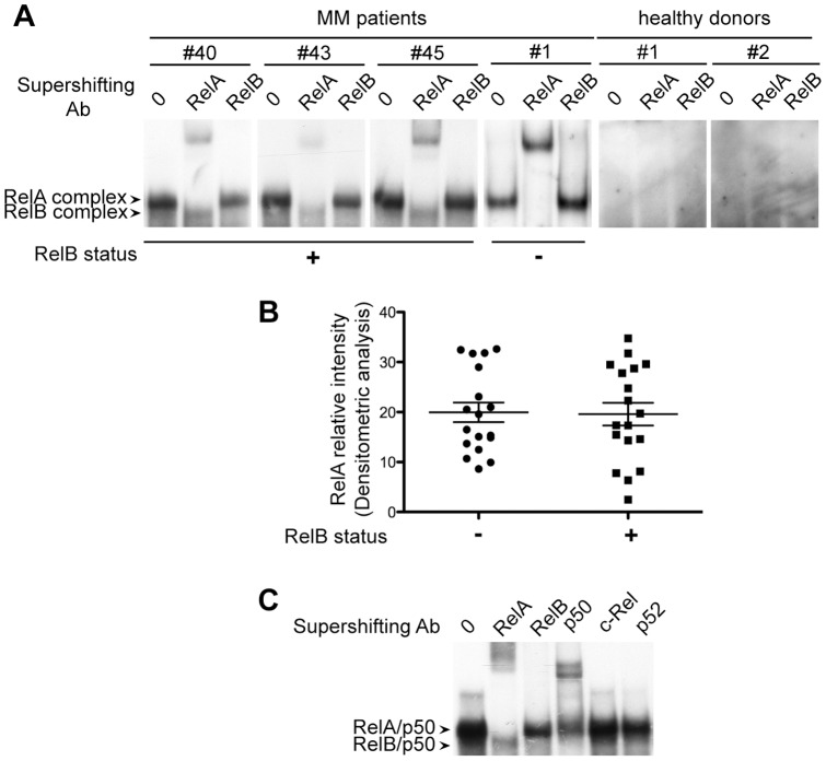
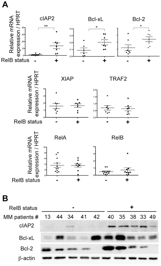
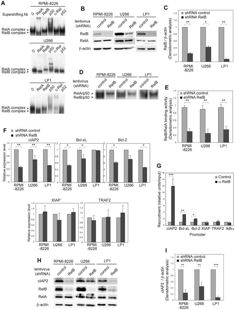
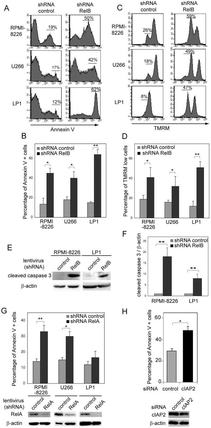
The NF-κB family of transcription factors has emerged as a key player in the pathogenesis of multiple myeloma (MM). NF-κB is activated by at least two major signaling pathways. The classical pathway results in the activation of mainly RelA containing dimers, whereas the alternative pathway leads to the activation of RelB/p52 and RelB/p50 heterodimers. Activating mutations in regulators of the alternative pathway have been identified in 17% of MM patients. However, the status of RelB activation per se and its role in the regulation of cell survival in MM has not been investigated. Here, we reveal that 40% of newly diagnosed MM patients have a constitutive RelB DNA-binding activity in CD138(+) tumor cells, and we show an association with increased expression of a subset of anti-apoptotic NF-κB target genes, such as cIAP2. Furthermore, we demonstrate that RelB exerts a crucial anti-apoptotic activity in MM cells. Our findings indicate that RelB activation is key for promoting MM cell survival through the upregulation of anti-apoptotic proteins. Altogether, our study provides the framework for the development of new molecules targeting RelB in the treatment of MM.


Conflict of interest statement

Competing Interests: The authors have declared that no competing interests exist.

Figures

Figure 1
Figure 1. RelB is constitutively activated in primary CD138+ cells from newly diagnosed MM patients.
(A) Whole cell extracts from primary CD138+ cells isolated from 52 newly diagnosed MM patients and two healthy donors were analyzed for NF-κB activity by EMSA. Four representative MM cases are presented (see Table 1 for tabulated study report). For supershift, whole cell extracts were either left untreated or incubated with either anti-RelA or anti-RelB antibodies for 30 min prior to incubation with the labeled probe. RelA- and RelB-containing complexes are indicated. (B) RelB binding activity is not associated with significant differences in the intensity of RelA DNA-binding activity in MM patient samples. EMSA and supershift analyses were performed from 18 RelB-positive and 18 RelB-negative de novo MM patient samples as in (A). Relative signal intensity of RelA DNA binding activity normalized to a reference patient sample loaded on each gel (means ± SD of two independent experiments for each MM sample) is shown. (C) Subunit composition of RelA- and RelB-containing complexes. Whole cell extracts used in (A) were incubated with the indicated antibodies prior to incubation with the labeled probe. Data of one representative RelB-positive MM patient is shown (#45).
Figure 2
Figure 2. RelB constitutive activation is associated with increased expression of a subset of anti-apoptotic NF-κB target genes in primary CD138+ cells from MM patients.
(A) RelB constitutive activation in CD138+ cell from MM patients is associated with increased anti-apoptotic NF-κB target gene mRNA expression. Quantitative RT-PCR was performed with specific primer pairs for the indicated genes using total RNAs prepared from 14 de novo MM patients either with or without RelB constitutive activation. Results are means ± SD of two independent experiments for each MM sample normalized to the level of HPRT mRNA. (B) RelB constitutive activation in CD138+ cells from MM patients is associated with increased anti-apoptotic NF-κB target gene protein expression. Whole cell extracts from primary CD138+ cells from MM patients tested in (A) were analyzed by immunoblotting for the indicated proteins. *P<0.05, **P<0.01.
Figure 3
Figure 3. RelB promotes anti-apoptotic NF-κB target gene expression in MM cell lines via direct transcriptional activation.
(A) RelB constitutive activation in MM cell lines. Whole cell extracts from RPMI-8226, U266, and LP1 cell lines were analyzed for NF-κB activity by EMSA. For supershift, whole cell extracts were incubated with the indicated antibodies prior to incubation with the labeled probe. RelA- and RelB-containing complexes are indicated. (B, C) RelB protein levels are efficiently knocked down by stable RNA interference. Whole cell extracts from RPMI-8226, U266, and LP1 cell lines transduced with lentiviruses encoding either a shRNA targeting RelB (shRNA RelB) or a scrambled control (shRNA control) were analyzed by immunoblotting for the indicated proteins. Data of a representative experiment are shown (B), and densitometric analysis of RelB protein level normalized to β-actin (means ± SEM of three independent experiments for each MM cell line) is reported (C). (D, E) RelB constitutive DNA-binding activity is efficiently knocked down by stable RNA interference. Whole cell extracts prepared as in (B) were analyzed by EMSA. RelA- and RelB-containing complexes are indicated. Data of one representative experiment are shown (D), and densitometric analysis of RelB DNA-binding activity normalized to RelA DNA-binding activity (means ± SEM of three experiments for each MM cell line) is reported (E). (F) RelB knockdown decreases anti-apoptotic NF-κB target gene expression at the mRNA level in MM cell lines. Quantitative RT-PCR was performed with specific primer pairs for the indicated genes using total RNAs prepared from the three MM cell lines upon RelB knockdown as in (B). Results are means ± SEM of three independent experiments for each MM cell line normalized to the level of HPRT mRNA. (G) RelB is constitutively bound to the cIAP2, Bcl-xL and Bcl-2 promoters in RPMI-8226 MM cells. Recruitment of RelB to the cIAP2, Bcl-xL, Bcl-2, TRAF2, XIAP and IκBα promoters was examined by ChIP experiments followed by quantitative PCR analysis. Results are means ± SEM of four independent experiments normalized to inputs that reflect relative amount of sonicated DNA fragments present before immunoprecipitation, (H, I) RelB knockdown decreases cIAP2 protein expression levels in MM cell lines. Whole cell extracts prepared as in (B) were analyzed by immunoblotting for the indicated proteins. Data of one representative experiment are shown (H), and densitometric analysis of cIAP2 protein level normalized to β-actin (means ± SEM of three independent experiments for each MM cell line) is reported (I). ***P<0.001, **P<0.01, *P<0.05.
Figure 4
Figure 4. RelB is critical for MM cell survival.
(A–D) RelB knockdown induces MM cell apoptosis. RPMI-8226, U266, and LP1 cell lines transduced with lentiviruses encoding either a shRNA targeting RelB or a scrambled control were monitored for apoptosis by annexin V-PE staining followed by FACS analysis. Data of one representative experiment are shown (A), and means ± SEM of three independent experiments for each MM cell line is reported (B). (C, D) RelB knockdown induces loss of mitochondrial transmembrane potential in MM cells. Loss of mitochondrial transmembrane potential was monitored by TMRM staining followed by FACS analysis. Data of one representative experiment are shown (C), and means ± SEM of three independent experiments for each MM cell line is reported (D). (E, F) RelB knockdown induces caspase 3 cleavage in MM cells. Whole cell extracts from RPMI-8226 and LP1 cell lines transduced with lentiviruses encoding either a shRNA targeting RelB or a scrambled control were analyzed by immunoblotting for the indicated proteins. Data of one representative experiment are shown (E), and densitometric analysis of cleaved caspase 3 protein levels normalized to β-actin (means ± SEM of two independent experiments for each MM cell line) is reported (F). (G) RelA knockdown induces apoptosis in RelB-positive MM cell lines. RPMI-8226, U266, and LP1 cell lines transduced with lentiviruses encoding either a shRNA targeting RelA or a scrambled control were monitored for apoptosis by annexin V-PE staining followed by FACS analysis. Means ± SEM of three independent experiments for each MM cell line is reported. Efficient knockdown of RelA protein levels was analyzed by immunoblotting (lower panels) (H) cIAP2 knockdown by RNAi induces MM cell apoptosis. LP1 cells were transfected with either a scrambled control sequence (siRNA control) or a siRNA oligonucleotide targeting cIAP2. Six days after transfection, cell apoptosis was monitored as in (A). Means ± SEM of three independent experiments is reported. Efficient knockdown of cIAP2 protein levels was analyzed by immunoblotting (lower panels) 3 days post-transfection. *P<0.05, **P<0.01.

Similar articles

Cited by

References

    1. Palumbo A, Anderson K (2011) Multiple myeloma. N Engl J Med 364: 1046–1060. - PubMed
    1. Hideshima T, Mitsiades C, Tonon G, Richardson PG, Anderson KC (2007) Understanding multiple myeloma pathogenesis in the bone marrow to identify new therapeutic targets. Nat Rev Cancer 7: 585–598. - PubMed
    1. Laubach J, Richardson P, Anderson K (2011) Multiple myeloma. Annu Rev Med 62: 249–264. - PubMed
    1. Mahindra A, Laubach J, Raje N, Munshi N, Richardson PG, et al. (2012) Latest advances and current challenges in the treatment of multiple myeloma. Nat Rev Clin Oncol 9: 135–143. - PubMed
    1. Hideshima T, Anderson KC (2002) Molecular mechanisms of novel therapeutic approaches for multiple myeloma. Nat Rev Cancer 2: 927–937. - PubMed

Publication types

MeSH terms