. 2013;8(3):e59693.
doi: 10.1371/journal.pone.0059693. Epub 2013 Mar 26.

Human adipose tissue derived mesenchymal stem cells aggravate chronic cyclosporin nephrotoxicity by the induction of oxidative stress

Affiliations

Human adipose tissue derived mesenchymal stem cells aggravate chronic cyclosporin nephrotoxicity by the induction of oxidative stress

Byung Ha Chung et al. PLoS One. 2013.

Abstract

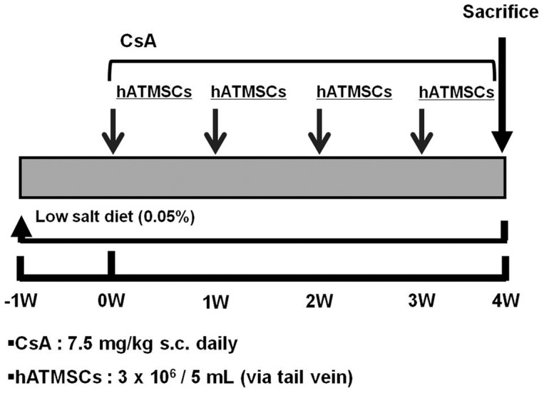
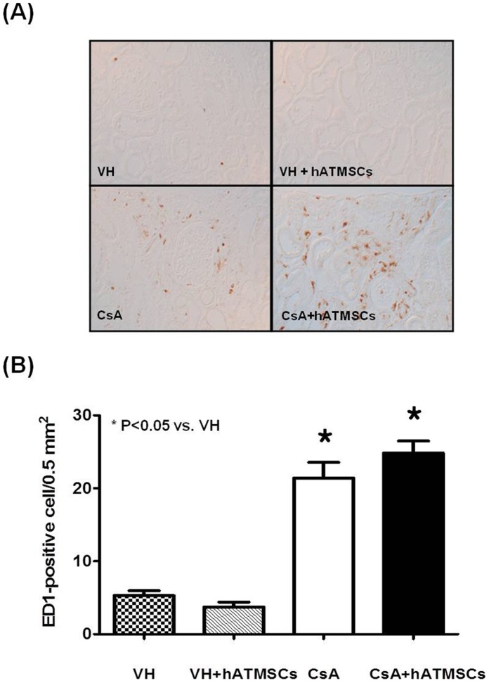
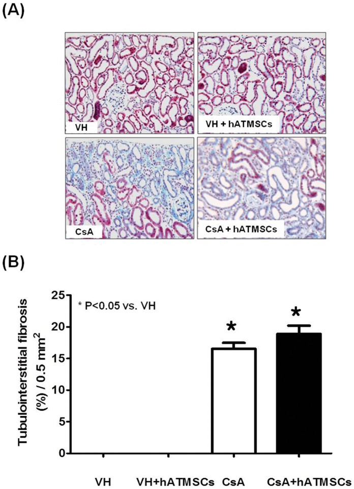
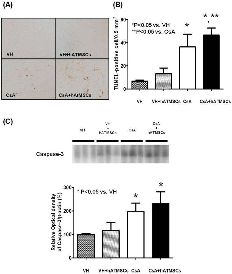
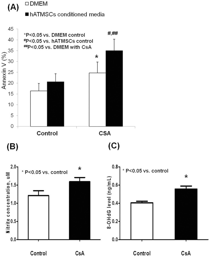
The aim of this study was to investigate whether hATMSCs protect against cyclosporine (CsA)-induced renal injury. CsA (7.5 mg/kg) and hATMSCs (3×10(6)/5 mL) were administered alone and together to rats for 4 weeks. The effect of hATMSCs on CsA-induced renal injury was evaluated by assessing renal function, interstitial fibrosis, infiltration of inflammatory cells, and apoptotic cell death. Four weeks of CsA-treatment produced typical chronic CsA-nephropathy. Combined treatment with CsA and hATMSCs did not prevent these effects and showed a trend toward further renal deterioration. To evaluate why hATMSCs aggravated CsA-induced renal injury, we measured oxidative stress, a major mechanism of CsA-induced renal injury. Both urine and serum 8-hydroxydeoxyguanosine(8-OHdG) levels were higher in the CsA+hATMSCs group than in the CsA group (P<0.05). An in vitro study showed similar results. Although the rate of apoptosis did not differ significantly between HK-2 cells cultured in hATMSCs-conditioned medium and those cultured in DMEM, addition of CsA resulted in greater apoptosis in HK-2 cells cultured in hATMSCs-conditioned medium. Addition of CsA increased oxidative stress in the hATMSCs-conditioned medium. The results of our study suggest that treatment with hATMSCs may aggravate CsA-induced renal injury because hATMSCs cause oxidative stress in the presence of CsA.


Conflict of interest statement

Competing Interests: The authors have declared that no competing interests exist.

Figures

Figure 1
Figure 1. Experimental design in this study.
CsA was administered at a dose of 7.5 mg/kg subcutaneously daily. hATMSCs was infused at 0,1,2 and 3 weeks from the start of CsA with cell number of 3×10⁶/mL via tail vein. hATMSCs; human adipose tissue derived mesenchymal stem cells.
Figure 2
Figure 2. Flowcytometric analysis of surface markers on cultured hATMSCs.
(A) MSC-specific markers CD29, CD73, CD90 and CD105 were expressed on culture hATMSCs. (B) Hematopoietic stem cell markers CD31, CD34 and CD45 were not expressed.
Figure 3
Figure 3. Homing of PKH-26 labeled hATMSCs to the kidney tissue.
(A) The representative of PKH-26 label (B) The staining for nuclei using (DAPI) (C) The overlay of (A) and (B) showed homing of PKH-26 labeled hATMSCs in renal interstitial tissue. PKH-26 staining merged with nuclear staining (in the circle), indicating PKH-26 labeled hATMSCs. (Original magnification x400 in A through C) (D) The quantization of PKH-26 labeled hATMSCs. Labeled cell was more frequently detected in CsA treated group compared to VH group. hATMSCs; human adipose tissue derived mesenchymal stem cells DAPI; 4′,6-Diamidino-2-Phenylindole.
Figure 4
Figure 4. Effect of hATMSCs infusion on inflammatory cell infiltration in rats with chronic CsA nephropathy.
(A) Representative photomicrographs of the immunohistochemical detection of ED1 protein (Original magnification x200). (B) Quantitative analysis of ED1-positive cells in the four groups. CsA administration increased ED1-positive cell number compared to VH and treatment with hATMSCs did not decrease it. hATMSCs; human adipose tissue derived mesenchymal stem cells.
Figure 5
Figure 5. Effect of hATMSCs infusion on interstitial fibrosis in rats with chronic CsA nephropathy.
(A) Representative photomicrographs of trichrome staining. The CsA group showed typical striped interstitial fibrosis, and extracellular matrix deposition in the cortex (Original magnification x200). (B) Quantitative analysis of tubulointerstitial fibrosis. CsA administration increased tubulointerstitial fibrosis compared to VH and it was not reduced by treatment with hATMSCs. hATMSCs; human adipose tissue derived mesenchymal stem cells.
Figure 6
Figure 6. Effect of hATMSCs infusion on apoptosis in rats with chronic CsA nephropathy.
(A) Representative photomicrographs of TUNEL staining in experimental groups (Original magnification x200). (B) Quantitative analysis of TUNEL-positive cells. CsA administration increased TUNEL-positive cell number compared to VH and treatment with hATMSCs further increased it (C) Western blot analysis of caspase-3 protein expression. Caspase-3 was activated after treatment with CsA and treatment with hATMSCs did not reduce it. hATMSCs; human adipose tissue derived mesenchymal stem cells.
Figure 7
Figure 7. Effect of hATMSCs infusion on oxidative damage in rats with chronic CsA nephropathy.
(A) The serum and (B) urinary concentrations of 8-OHdG was increased in the CsA group, and further increased with the treatment of hATMSCs. hATMSCs; human adipose tissue derived mesenchymal stem cells.
Figure 8
Figure 8. The results of in vitro study.
(A) The number of Annexin V positive cell didn’t increase in HK-2 cell cultured in hATMSCs- conditioned medium compared to HK-2 cell cultured in DMEM. Addition of CsA increased Annexin V positive cell both in DMED and hATMSCs-conditioned medium, and it was more frequently detected in HK-2 cell cultured in hATMSCs-conditioned medium compared to HK-2 cells cultured in DMEM. (B) The production of NO was increased in medium from hATMSCs treated with CsA compared with medium from hATMSCs cultured without CsA. (C) The 8-OHdG level was significantly increased in the medium from hATMSCs treated with CsA compared to medium from hATMSCs cultured withCsA-free media as well. hATMSCs; human adipose tissue derived mesenchymal stem cells.

Similar articles

Cited by

References

    1. Bennett WM, DeMattos A, Meyer MM, Andoh T, Barry JM (1996) Chronic cyclosporine nephropathy: the Achilles’ heel of immunosuppressive therapy. Kidney Int 50: 1089–1100. - PubMed
    1. Myers BD, Ross J, Newton L, Luetscher J, Perlroth M (1984) Cyclosporine-associated chronic nephropathy. N Engl J Med 311: 699–705. - PubMed
    1. Wang C, Salahudeen AK (1995) Lipid peroxidation accompanies cyclosporine nephrotoxicity: effects of vitamin E. Kidney Int. 47: 927–934. - PubMed
    1. Yang CW, Faulkner GR, Wahba IM, Christianson TA, Bagby GC, et al. (2002) Expression of apoptosis-related genes in chronic cyclosporine nephrotoxicity in mice. Am J Transplant 2: 391–399. - PubMed
    1. Yoon HE, Yang CW (2009) Established and newly proposed mechanisms of chronic cyclosporine nephropathy. Korean J Intern Med 24: 81–92. - PMC - PubMed

Publication types

MeSH terms

LinkOut - more resources