. 2013;8(3):e59735.
doi: 10.1371/journal.pone.0059735. Epub 2013 Mar 21.

Bapineuzumab alters aβ composition: implications for the amyloid cascade hypothesis and anti-amyloid immunotherapy

Affiliations

Bapineuzumab alters aβ composition: implications for the amyloid cascade hypothesis and anti-amyloid immunotherapy

Alex E Roher et al. PLoS One. 2013.

Abstract

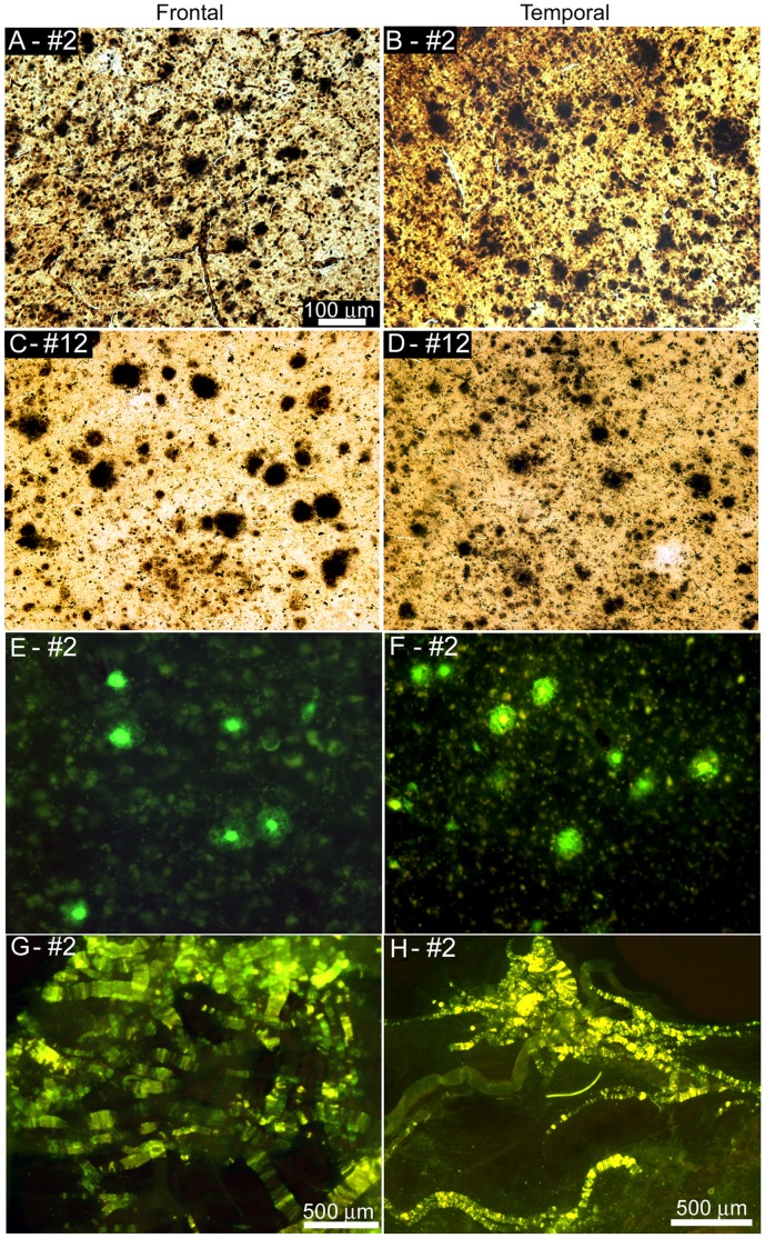
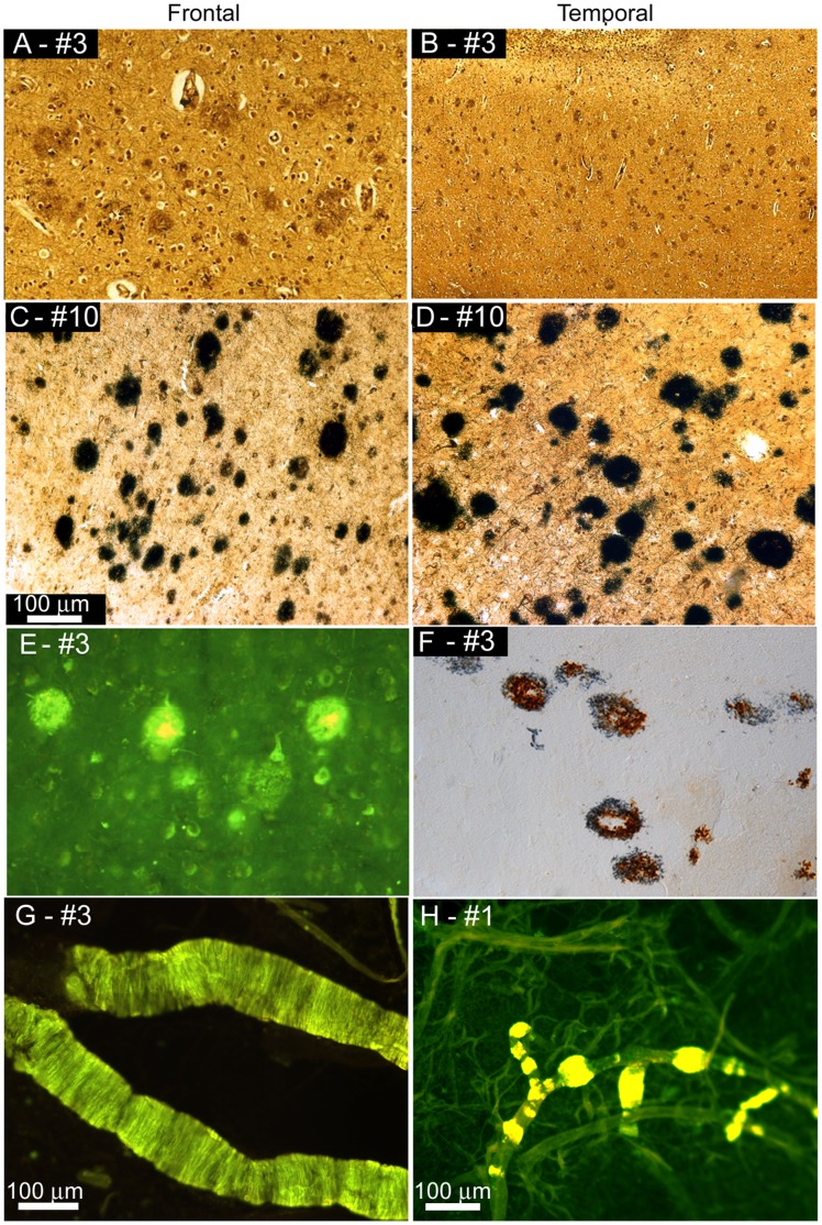
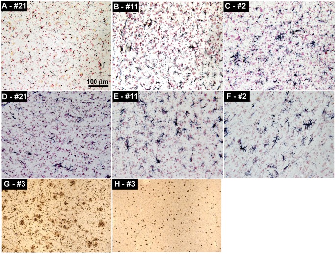
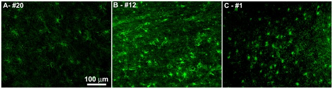
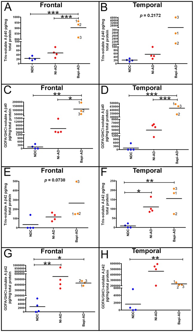
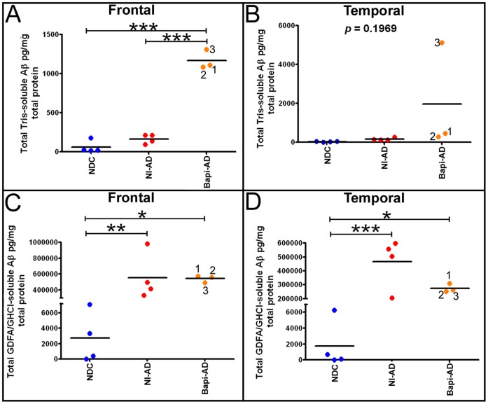
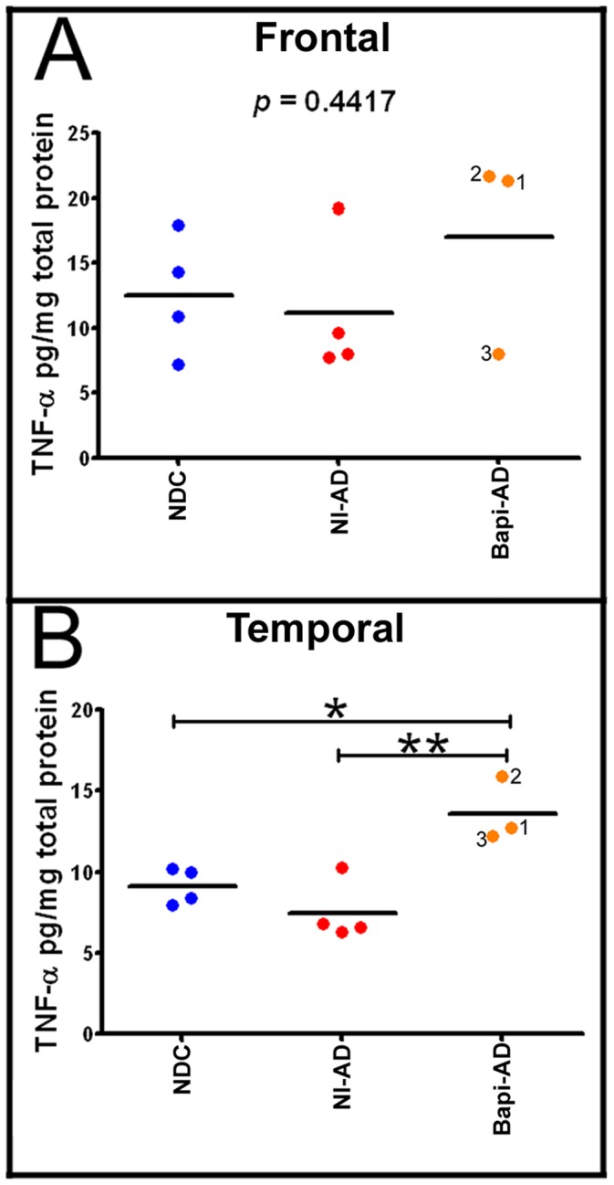
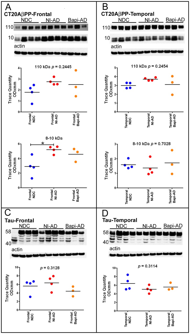
The characteristic neuropathological changes associated with Alzheimer's disease (AD) and other lines of evidence support the amyloid cascade hypothesis. Viewing amyloid deposits as the prime instigator of dementia has now led to clinical trials of multiple strategies to remove or prevent their formation. We performed neuropathological and biochemical assessments of 3 subjects treated with bapineuzumab infusions. Histological analyses were conducted to quantify amyloid plaque densities, Braak stages and the extent of cerebral amyloid angiopathy (CAA). Amyloid-β (Aβ) species in frontal and temporal lobe samples were quantified by ELISA. Western blots of amyloid-β precursor protein (AβPP) and its C-terminal (CT) fragments as well as tau species were performed. Bapineuzumab-treated (Bapi-AD) subjects were compared to non-immunized age-matched subjects with AD (NI-AD) and non-demented control (NDC) cases. Our study revealed that Bapi-AD subjects exhibited overall amyloid plaque densities similar to those of NI-AD cases. In addition, CAA was moderate to severe in NI-AD and Bapi-AD patients. Although histologically-demonstrable leptomeningeal, cerebrovascular and neuroparenchymal-amyloid densities all appeared unaffected by treatment, Aβ peptide profiles were significantly altered in Bapi-AD subjects. There was a trend for reduction in total Aβ42 levels as well as an increase in Aβ40 which led to a corresponding significant decrease in Aβ42:Aβ40 ratio in comparison to NI-AD subjects. There were no differences in the levels of AβPP, CT99 and CT83 or tau species between Bapi-AD and NI-AD subjects. The remarkable alteration in Aβ profiles reveals a dynamic amyloid production in which removal and depositional processes were apparently perturbed by bapineuzumab therapy. Despite the alteration in biochemical composition, all 3 immunized subjects exhibited continued cognitive decline.


Conflict of interest statement

Competing Interests: CB’s salary is supported, in part, by Celgene, Quintiles/Genentech, AVID, Lilly, GE, Wyeth, Bayer, and Pfizer. MNS receives grant support from BMS, Avid, GE, Bayer, Baxter, Wyeth, Janssen, Lilly and Medivation, and is in the consultant/advisory board for Janssen/Pfizer, Amerisciences, Eisai and GSK, and receives royalties from Amerisciences and Wiley. TGB receives funding from AVID-Radiopharmaceuticals, Schering-Bayer Pharmaceuticals and GE Healthcare. None of the above funding was for this specific study. The bapineuzumab used in this study is a Pfizer/Johnson & Johnson/Elan product. There are no further patents, products in development or marketed products to declare. This does not alter the authors’ adherence to all the PLOS ONE policies on sharing data and materials, as detailed online in the guide for authors.

Figures

Figure 1
Figure 1. Amyloid plaques and vascular amyloid in Bapi-AD and NI-AD.
Amyloid plaques in frontal (A, C and E) and temporal (B, D and F) lobes and vascular amyloid (G and H). A) and B) Campell-Switzer staining of Bapi-AD case #2 (89 year old male, APOE ε2/3 genotype). C) and D) Campbell-Switzer staining of NI-AD case #12 (87 year old male, APOE ε2/3). E) and F) Thioflavine-S staining of Bapi-AD case #2. Frequent amyloid plaques are shown in all cases. G) Leptomeninges of Bapi-AD case #2 stained with Thioflavine-S. H) Cortical blood vessels of Bapi-AD case #2 stained with Thioflavine-S. A–F were taken at 100× magnification and the scale bar is shown in caption A. G and H were taken at 25X with the scale bars shown in each caption.
Figure 2
Figure 2. Amyloid plaques and vascular amyloid in Bapi-AD and NI-AD.
Amyloid plaques in frontal (A, C, E) and temporal (B, D, F) lobes and vascular amyloid (G and H). A) and B) Bielschowsky stain of Bapi-AD case #3 (86 year old male, APOE ε3/4 genotype), showing a moderate amyloid plaque accumulation. C) and D) Campbell-Switzer staining of NI-AD case #10 (91 year old female, APOE ε3/4 genotype), exhibiting frequent amyloid plaques. E) Thioflavine-S staining in the frontal cortex of case of Bapi-AD case #3 showing amyloid plaques and NFT in the background. F) Double immunolabeling of amyloid plaques of Bapi-AD case #3 with antibodies against Aβ40 (brown) and Aβ42 (blue) demonstrating that the amyloid plaques contained both peptide species. Cortical blood vessels of Bapi-AD case #3 (G) and Bapi-AD case #1 (H) stained with Thioflavine-S. Magnifications: A, C, D, E, F –100X and B –40x.
Figure 3
Figure 3. Representative images of immunohistochemistry showing microglia and T-lymphocytes.
A) CD68 staining of the microglia in the temporal cortex of NDC case #21. B) CD68 staining of the temporal cortex of NI-AD case #11. C) CD68 staining of the temporal cortex of Bapi-AD case #2. D) HLA-DR staining of microglia in the frontal cortex of NDC case #21. E) HLA-DR staining of the temporal cortex of NI-AD case #11. F) HLA-DR staining of the temporal cortex of Bapi-AD case #2. G) HLA-DR staining of the temporal cortex of Bapi-AD case #3. H) CD3 staining of T-lymphocytes in the temporal cortex of Bapi-AD case #3. Magnifications: A–F –200X and G, H –100X.
Figure 4
Figure 4. Representative images of immunofluoresence staining of GFAP.
A) Frontal cortex of NDC case #20. B) Temporal cortex of NI-AD case #12. C) Temporal cortex of Bapi-AD case #1. For more details, see the Results section. Magnifications: A–C –200X.
Figure 5
Figure 5. ELISA quantification of soluble and insoluble Aβ in frontal and temporal lobes.
A) Frontal cortex Tris-soluble Aβ40. Notice the sharp and significance difference between the Aβ levels of NI-AD and those of the Bapi-AD. B) Temporal cortex Tris-soluble Aβ40. There was a large mean difference between NI-AD and Bapi-AD, however there was not a statistically significant difference due to the spread of values. Frontal (C) and temporal (D) cortices GDFA/GHCl-soluble Aβ40. In both cases there were significant elevations in mean Aβ40 in Bapi-AD relative to NI-AD. Frontal (E) and temporal (F) cortices Tris-soluble Aβ42. In both lobes there were no statistical differences between the NI-AD and Bapi-AD due to the spread in values. Frontal (G) and temporal (H) cortices GDFA/GHCl-soluble Aβ42. There was a mean decrease in Bapi-AD Aβ42 relative to NI-AD more noticeable in the latter than in the former. The numbers in the Bapi-AD columns (1, 2 and 3), correspond to the cases #s given in Table 1 . Statistical analysis was performed using a one-way ANOVA followed by Tukey’s multiple comparison test (*p = 0.05–0.01; **p = 0.01–0.001; ***p<0.0001). Abbreviations: Tris, 20 mM Tris-HCl, 5 mM EDTA, pH 7.8, plus protease inhibitor cocktail; GDFA, glass-distilled formic acid; GHCl, 5 M guanidine hydrochloride, 50 mM Tris-HCl, pH 8.0, plus protease inhibitor cocktail; NDC, non-demented control; NI-AD, non-immunized Alzheimer’s disease; Bapi-AD, bapineuzumab-immunized Alzheimer’s disease.
Figure 6
Figure 6. Total levels (Aβ40+Aβ42) of soluble (Tris) and insoluble (GDFA/GHCl-soluble) Aβ from the frontal and temporal lobes.
A) Frontal cortex total Tris-soluble Aβ. B) Temporal cortex total Tris-soluble Aβ. C) Frontal cortex total GDFA/GHCl-soluble Aβ. D) Temporal cortex total GDFA/GHCl-soluble Aβ. For statistical treatment and abbreviations see legend to Figure 5.
Figure 7
Figure 7. ELISA quantification of tumor necrosis factor-α (TNF-α).
A) Frontal cortex TNF-α levels and B) Temporal cortex TNF-α levels. Notice that there is a significant increase in the amount of this cytokine in the temporal lobe of the Bapi-AD relative to NI-AD. For statistical treatment and abbreviations see legend to Figure 5.
Figure 8
Figure 8. Western blot analyses of AβPP and its C-terminal peptides CT99, CT83 and of the tau isoforms.
Frontal (A) and temporal (B) cortices Western blots of CT20AβPP. The CT20AβPP antibody, raised against the last 20 amino acids of AβPP, was used to detect AβPP and its CT peptides. Frontal (C) and temporal (D) cortices Western blots of tau. The tau isoforms were probed using the HT7 clone made against amino acids 159–163 of the tau molecule. Molecular weights, in kDa, are given on the left of each blot. Actin re-probes are shown below as a total protein loading control. For comparison all Western blots were analyzed against NDC and NI-AD in the same gel. There were no statistical differences between the immunized and non-immunized groups. Statistical analysis and abbreviations are described in the legend to Figure 5.

Similar articles

Cited by

References

    1. Ferri CP, Prince M, Brayne C, Brodaty H, Fratiglioni L, et al. (2005) Global prevalence of dementia: a Delphi consensus study. Lancet 366: 2112–2117. - PMC - PubMed
    1. Pul R, Dodel R, Stangel M (2011) Antibody-based therapy in Alzheimer’s disease. Expert Opin Biol Ther 11: 343–357. - PubMed
    1. Morgan D (2011) Immunotherapy for Alzheimer’s disease. J Intern Med 269: 54–63. - PMC - PubMed
    1. Kerchner GA, Boxer AL (2010) Bapineuzumab. Expert Opin Biol Ther 10: 1121–1130. - PMC - PubMed
    1. Maarouf CL, Daugs ID, Kokjohn TA, Kalback WM, Patton RL, et al. (2010) The biochemical aftermath of anti-amyloid immunotherapy. Mol Neurodegener 5: 39. - PMC - PubMed

Publication types

MeSH terms