. 2013;8(3):e59811.
doi: 10.1371/journal.pone.0059811. Epub 2013 Mar 26.

Development of the mouse dermal adipose layer occurs independently of subcutaneous adipose tissue and is marked by restricted early expression of FABP4

Affiliations

Development of the mouse dermal adipose layer occurs independently of subcutaneous adipose tissue and is marked by restricted early expression of FABP4

Kamila Wojciechowicz et al. PLoS One. 2013.

Abstract

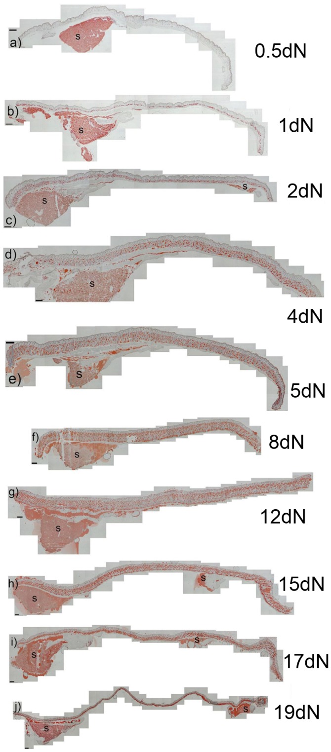
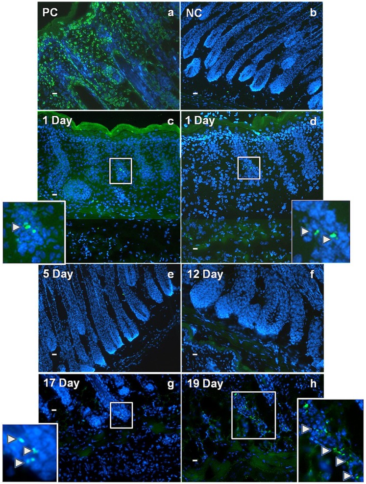
The laboratory mouse is a key animal model for studies of adipose biology, metabolism and disease, yet the developmental changes that occur in tissues and cells that become the adipose layer in mouse skin have received little attention. Moreover, the terminology around this adipose body is often confusing, as frequently no distinction is made between adipose tissue within the skin, and so called subcutaneous fat. Here adipocyte development in mouse dorsal skin was investigated from before birth to the end of the first hair follicle growth cycle. Using Oil Red O staining, immunohistochemistry, quantitative RT-PCR and TUNEL staining we confirmed previous observations of a close spatio-temporal link between hair follicle development and the process of adipogenesis. However, unlike previous studies, we observed that the skin adipose layer was created from cells within the lower dermis. By day 16 of embryonic development (e16) the lower dermis was demarcated from the upper dermal layer, and commitment to adipogenesis in the lower dermis was signalled by expression of FABP4, a marker of adipocyte differentiation. In mature mice the skin adipose layer is separated from underlying subcutaneous adipose tissue by the panniculus carnosus. We observed that the skin adipose tissue did not combine or intermix with subcutaneous adipose tissue at any developmental time point. By transplanting skin isolated from e14.5 mice (prior to the start of adipogenesis), under the kidney capsule of adult mice, we showed that skin adipose tissue develops independently and without influence from subcutaneous depots. This study has reinforced the developmental link between hair follicles and skin adipocyte biology. We argue that because skin adipocytes develop from cells within the dermis and independently from subcutaneous adipose tissue, that it is accurately termed dermal adipose tissue and that, in laboratory mice at least, it represents a separate adipose depot.


Conflict of interest statement

Competing Interests: The authors have declared that no competing interests exist.

Figures

Figure 1
Figure 1. Early accumulation of lipid in cells of the lower dermis follows a spatio-temporal pattern.
Samples of dorsal skin from foetal and early newborn dorsal skin sectioned and stained with Oil Red O to detect lipids (a-h). At e16 no lipid accumulation is detected anywhere in skin (a) note the line of cells representing the precursor of the panniculus carnosus (white arrows). By e18.5–19 the first lipid containing cells are detected, at which point the lipid is often multilocular (b). Sections from the head (c and e) regions of e18.5 dorsal skin show larger numbers of cells with accumulation of lipid in the deep dermis compared with tail regions (d and f) where far fewer lipid producing cells were apparent. In regions between the head and tail, some areas had consistent groups of cells containing lipid (g). Lipid forming cells are occasionally observed in regions where follicle development is apparently less advanced (h). sf = subcutaneous adipose tissue. Scale bars: a, c, d, e, f, g, h = 100 µm, b = 50 µm.
Figure 2
Figure 2. Lipid accumulation in back skin specimens with adipose depots beneath the skin.
Composite image of longitudinally sectioned skin section stained with Oil Red O to show lipid accumulation along the anterior-posterior axis. Samples were prepared from newborn mice at different postnatal time points 0.5 day,1 day, 2 day, 4 day, 5 day, 8 day, 12 day, 15 day, 17 day, 19 day. S = subcutaneous adipose tissue below the anterior and some posterior regions of the skin specimens. (a – j) Scale bar = 300 µm.
Figure 3
Figure 3. Changes in skin thickness and dermal adipose tissue development in lower skin dermis of the newborn mouse.
Skin sections were stained with Oil Red O to detect lipids from birth to 19 days postnatally (a–i). In the 36 hours after the 0.5dN time point (a) there is a rapid increase in both the numbers of stained cells and the size of the lipid droplets (b, c). In association with the downward expansion of follicles the dermal adipose tissue layer thickens until day 12 (f) with the most rapid expansion between about 2 and 5 Days (c, d). As follicles regress, dermal thickness decreases (g, h) until skin thickness is reduced back to that seen in the days shortly after birth, and lipid containing cells accumulate below (i). Scale bar = 30 µm.
Figure 4
Figure 4. No significant apoptosis is observed in the adipocyte layer, during its formation, or at any point in the first hair growth cycle as measured by the TUNEL assay.
TUNEL (terminal deoxynucleotidyl transferase dUTP nick end labelling), a) PC (positive control - nuclease treated 1 day old mouse back skin), b) NC (negative control - 1 day old mouse back skin). In one day specimens labelled cells were occasionally observed in hair follicles (magnified and arrowed in c, d) and in the developing adipose layer (c) but more often they were completely absent from the latter (d). Apart from very occasional follicle-associated labelling virtually no cell death was observed at 5 and 12 days after birth, through the middle stages of the first hair cycle (e, f). Subsequently an increasing amount of cell death was observed associated with regression of the follicles as their growth shut down at 17 and 19 days (magnified and arrowed in g and h) however virtually no cell death labelling was observed in the surrounding adipocyte cells. DNA was counterstained with 4′,6-diamidino-2-phenylindole (DAPI). Scale bar = 30 µm.
Figure 5
Figure 5. Fatty acid binding protein-4 (FABP4) is expressed in the lower dermis of developing mouse back skin from embryonic day 16 (e16). a), e15 (embryonic day 15), b) e16 (embryonic day 16), c) e18 (embryonic day 18), d) P5 (post-natal day 5), e) NC (negative control - no primary antibody).
White dashed line delineates boundary between epidermis and upper dermis. Red dashed line delineates boundary between upper dermis and lower dermis. DNA was counterstained with 4′,6-diamidino-2-phenylindole (DAPI). Scale bar = 50µ m. f) The relative mRNA expression for Fabp4 was analysed from samples at the e17 (17), e18 (18) and e19 (19) time-points from upper (Area “1”, blue) and lower (Area “2”, red) dermis. The baseline (1-fold change) was established for the e19 lower dermis sample (underlined on the figure) and the mRNA levels of the other samples are shown relative to this. Fabp4 mRNA is up-regulated in lower dermis (Area “2”) compared with upper dermis (Area “1”) at all analysed time-points. Data were obtained from triplicate biological replicates. P value refers to the comparison with “19 2” sample (*P≤0.05, **P≤0.01, ***P≤0.001).
Figure 6
Figure 6. Trichorhinophalangeal syndrome I (TRPS1) expression is restricted to upper dermal cells at embryonic day 17 (e17), e18 and e19 in murine dermis.
a), DNA was counterstained with 4′,6-diamidino-2-phenylindole (DAPI). Scale bar = 30 µm. b) The relative mRNA expression for TRPS1 was analysed from samples at the e17 (17), e18 (18) and e19 (19) time-points from upper (Area “1”, blue) and lower (Area “2”, red) dermis. The baseline (1-fold change) was established for the e19 upper dermis sample (underlined on the figure) and the mRNA levels of the other samples are shown relative to this. TRPS1 mRNA is up-regulated in upper dermis (Area “1”) compared with lower dermis (Area “2”) at all analysed time-points. Data were obtained from triplicate biological replicates. P value refers to the comparison with “19 1” sample (*P≤0.05, **P≤0.01, ***P≤0.001).
Figure 7
Figure 7. Relationship between developing skin adipose tissue, the panniculus carnosus and underlying subcutaneous adipose tissue in the dorsal skin of newborn mice.
Mouse dorsal skin sections from the region overlying the anterior (interscapular) subcutaneous adipose depot. Skin sections with underlying subcutaneous adipose tissue were stained with Oil Red O to detect lipids from birth to 19 days postnatally (a–i). The thickness (marked by yellow lines) of the panniculus carnosus layer changes in the specimens over time. An additional thin layer of lipid droplets can be seen under the dermal adipose tissue, for example at day 5 (d - see area close to smaller yellow line). a) 0.5 day old newborn mouse. b) 1 day old newborn mouse. c) 2 day old newborn mouse. d) 5 day old newborn mouse. e) 8 day old newborn mouse. f) 12 day old newborn mouse g) 15 day old newborn mouse. h) 17 day old newborn mouse. i) 19 day old newborn mouse. (a - i) Scale bar = 65 µm.
Figure 8
Figure 8. The lower dermis becomes the skin adipose layer independently of subcutaneous influence.
External view of green fluorescent protein (GFP)-labelled E14 mouse back skin, microdissected and cultured for 24 hours on agar (a and a′). Fluorescent microscopy of mouse back skin grafts showing presence of GFP 10 days after grafting to the kidney capsule of a non-GFP nude mouse. Both hair follicles and the surrounding adipose tissue express GFP (b and b′) Haematoxylin and eosin (H+E) staining of E14.5 mouse back skin showing absence of skin adipose tissue(c and c′) and 10 days post-grafting to the kidney capsule showing the presence of a skin adipose layer in the lower dermis (d and d′). Confirmation of lipid production only in the lower dermis of skin grafts by Oil Red O staining (e) and lipidTOX staining (f-h). Fatty acid binding protein-4 (FABP4) is highly expressed in the lower but not the upper dermis of grafted skin after 10 days (i) Antibody staining for GFP in the dermal adipose layer of the grafted mouse back skin confirmed donor origin (j). Negative controls, (no primary antibody) for FABP4 and GFP antibody staining, respectively k and l. White dashed line delineates boundary between epidermis and upper dermis. Red dashed line delineates boundary between upper dermis and lower dermis. Black arrowheads in figure e (sebaceous glands). In figures f, g, h, i, j, k and l DNA was counterstained with 4′,6-diamidino-2-phenylindole (DAPI). Scale bar = 30 µm.

Similar articles

Cited by

References

    1. Cinti S (2007) Nutrition and Health. In: Fantuzzi G, Mazzone T, editors. Adipose Tissue and Adipokines in Health and Disease. Humana Press Incorporated, Totowa, New Jersey. 3–19.
    1. Gesta S, Tseng YH, Kahn CR (2007) Developmental origin of fat: tracking obesity to its source. Cell 131: 242–256. - PubMed
    1. Billon N, Iannarelli P, Monteiro MC, Glavieux-Pardanaud C, Richardson WD, et al. (2007) The generation of adipocytes by the neural crest. Development 134: 2283–2292. - PMC - PubMed
    1. Billon N, Monteiro MC, Dani C (2008) Developmental origin of adipocytes: new insights into a pending question. Biology of the cell 100: 563–575. - PubMed
    1. Billon N, Dani C (2012) Developmental origins of the adipocyte lineage: new insights from genetics and genomics studies. Stem cell reviews 8: 55–66. - PubMed

Publication types

MeSH terms

LinkOut - more resources