. 2013;8(3):e59812.
doi: 10.1371/journal.pone.0059812. Epub 2013 Mar 28.

Specific in vivo labeling of tyrosinated α-tubulin and measurement of microtubule dynamics using a GFP tagged, cytoplasmically expressed recombinant antibody

Affiliations

Specific in vivo labeling of tyrosinated α-tubulin and measurement of microtubule dynamics using a GFP tagged, cytoplasmically expressed recombinant antibody

Lynne Cassimeris et al. PLoS One. 2013.

Abstract

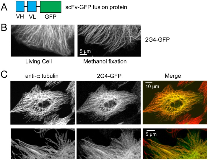
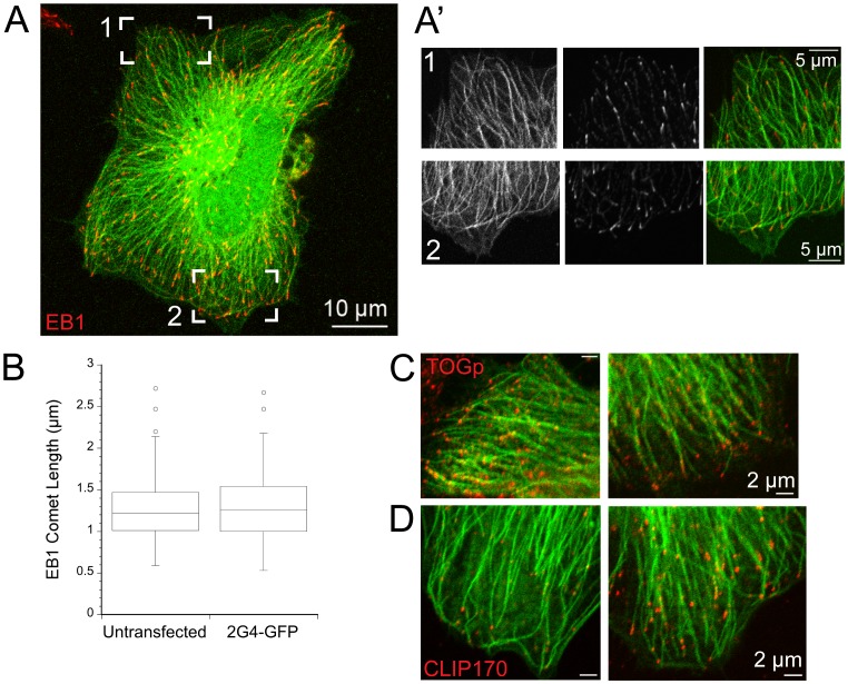
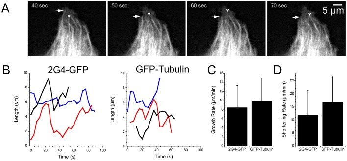
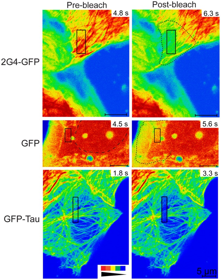
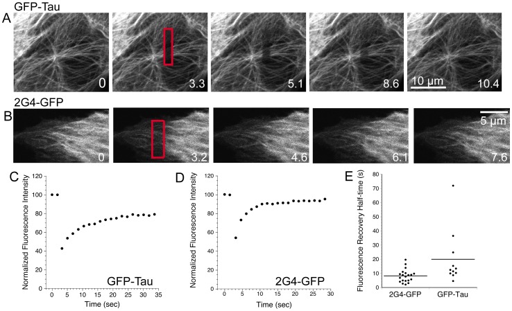
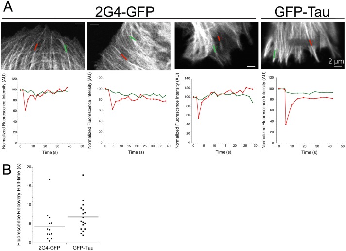
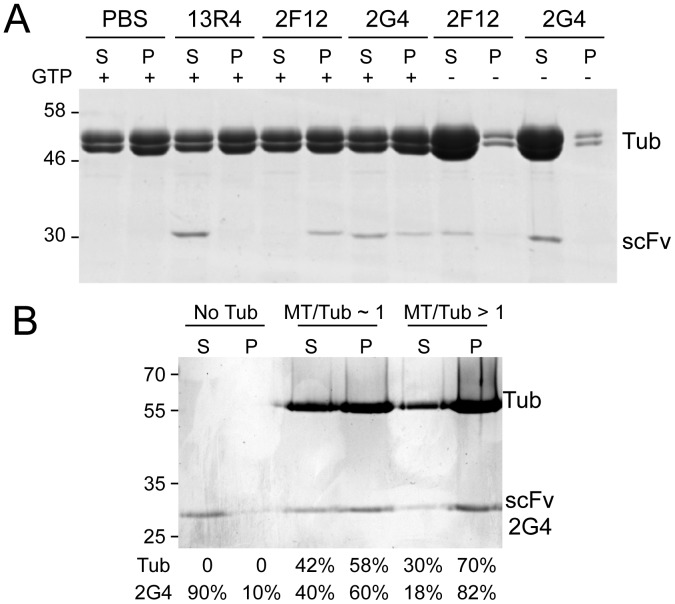
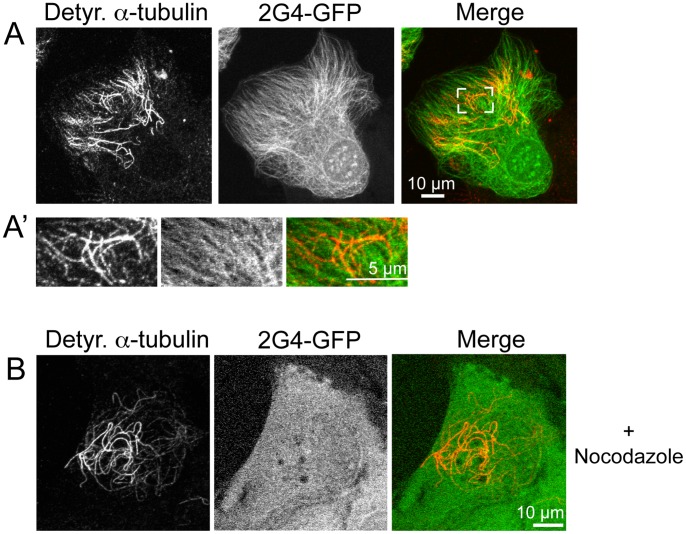
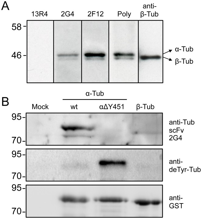
GFP-tagged proteins are used extensively as biosensors for protein localization and function, but the GFP moiety can interfere with protein properties. An alternative is to indirectly label proteins using intracellular recombinant antibodies (scFvs), but most antibody fragments are insoluble in the reducing environment of the cytosol. From a synthetic hyperstable human scFv library we isolated an anti-tubulin scFv, 2G4, which is soluble in mammalian cells when expressed as a GFP-fusion protein. Here we report the use of this GFP-tagged scFv to label microtubules in fixed and living cells. We found that 2G4-GFP localized uniformly along microtubules and did not disrupt binding of EB1, a protein that binds microtubule ends and serves as a platform for binding by a complex of proteins regulating MT polymerization. TOGp and CLIP-170 also bound microtubule ends in cells expressing 2G4-GFP. Microtubule dynamic instability, measured by tracking 2G4-GFP labeled microtubules, was nearly identical to that measured in cells expressing GFP-α-tubulin. Fluorescence recovery after photobleaching demonstrated that 2G4-GFP turns over rapidly on microtubules, similar to the turnover rates of fluorescently tagged microtubule-associated proteins. These data indicate that 2G4-GFP binds relatively weakly to microtubules, and this conclusion was confirmed in vitro. Purified 2G4 partially co-pelleted with microtubules, but a significant fraction remained in the soluble fraction, while a second anti-tubulin scFv, 2F12, was almost completely co-pelleted with microtubules. In cells, 2G4-GFP localized to most microtubules, but did not co-localize with those composed of detyrosinated α-tubulin, a post-translational modification associated with non-dynamic, more stable microtubules. Immunoblots probing bacterially expressed tubulins confirmed that 2G4 recognized α-tubulin and required tubulin's C-terminal tyrosine residue for binding. Thus, a recombinant antibody with weak affinity for its substrate can be used as a specific intracellular biosensor that can differentiate between unmodified and post-translationally modified forms of a protein.


Conflict of interest statement

Competing Interests: The authors have declared that no competing interests exist.

Figures

Figure 1
Figure 1. The scFv 2G4-GFP colocalizes with the majority of microtubules.
(A) Diagram outlining the recombinant antibody. The combined mw of the VH and VL regions approximately equals that of EGFP. (B) 2G4-GFP localizes to linear filaments in either living cells or in cells fixed in −20°C methanol. Images shown are from the edges of two different LLCPK cells (scale bar = 5 µm). (C) 2G4-GFP localizes to the majority of microtubules. LLCPKs were transfected with plasmid encoding 2G4-GFP and fixed 24 h later. Microtubules were stained with an antibody to a-tubulin. Images in the top row show a maximum intensity projection from a Z series (scale bar = 10 µm). The bottom row shows single optical section from the edge of a second LLCPK cell (scale bar = 5 µm).
Figure 2
Figure 2. Expression of scFv 2G4-GFP does not disrupt protein binding to microtubule plus ends.
(A) 2G4-GFP binding extends to the distal ends of microtubules, as marked by EB1. A maximum intensity projection from optical sections through a Hela cell is shown (scale bar = 10 µm). (A′) Single optical sections from bracketed regions of the cell shown in (A) (scale bar = 5 µm). 2G4-GFP binds uniformly along microtubules and extends to the ends of these microtubules. (B) Box plot of EB1 comet lengths at microtubule plus ends. Expression of 2G4-GFP did not change the length of EB1 comets. (C) TOGp, another microtubule plus end binding protein, is localized to microtubules labeled by 2G4-GFP. (D) CLIP-170 was also localized to microtubule plus ends in cells expressing 2G4-GFP. Note that CLIP-170 was localized to only a subset of microtubule ends. We observed a similar pattern in Hela cells expressing GFP- a-tubulin (data not shown). Scale bars in C, D = 2 µm.
Figure 3
Figure 3. Microtubule length changes detected by 2G4-GFP or GFP- α-tubulin.
(A) Sequential images from the periphery of an LLCPK cell expressing 2G4-GFP. Arrows note length changes of several microtubules. Scale bar = 5 µm. The video sequence is presented in Movie S1. (B) Plots of microtubule length changes over time for three microtubules labeled by 2G4-GFP or by GFP-α-tubulin. Length changes over time were determined from image series as described in Methods (C,D). Microtubule growth (C) and shortening velocities (D) measured by 2G4-GFP or GFP-α-tubulin. Data shown are means ± sd. Additional parameters of dynamic instability are summarized in Table 1.
Figure 4
Figure 4. Photobleached 2G4-GFP spreads beyond the region targeted by the bleaching laser.
Pre-bleach and post-bleach images are shown for LLCPK cells expressing 2G4-GFP, GFP or GFP-tau as noted. Pre-bleach images were collected immediately prior to photobleaching an area marked by the black rectangular boxes. Post-bleach images were collected immediately after photobleaching. Time in each frame is given in seconds from the start of an imaging experiment. Fluorescence intensity is represented by a rainbow palette from red to blue (see inset at bottom left). For both 2G4-GFP and GFP, the region of dimmed fluorescence has spread beyond that targeted by the bleaching laser. This spread is highlighted by dotted lines in the post-bleach images. The spread of photobleached proteins is likely due to rapid diffusion in and out of the photobleached area. For the GFP pre-bleach image, the nucleus is outlined by a dashed line. Note that fluorescence within the nucleus does not appear to exchange significantly with that in the cytoplasm over the ∼2 s interval between images. In contrast to 2G4-GFP and GFP, GFP-tau photobleaching yields an area of dimmed fluorescence that closely matches the region targeted by the laser. Scale bars for all images are 5 µm.
Figure 5
Figure 5. Rapid recovery of GFP-tau or 2G4-GFP after fluorescence photobleaching.
(A,B) Images of GFP-tau or 2G4-GFP expressing LLCKP cells before (time 0) and after photobleaching rectangular boxes (bleached regions indicated in red) are shown. Time is given in s. (C,D) Typical fluorescence recovery curves for photobleached rectangles in cells expressing GFP-tau or 2G4-GFP. Fluorescence was normalized as described in Methods. (E) Dot plot showing the half times of fluorescence recovery within boxed regions for 2G4-GFP or GFP-tau. Video sequences are available as Movie S2, S3, S4.
Figure 6
Figure 6. 2G4-GFP turns over rapidly on microtubules.
(A) Images shown were recorded immediately after photobleaching. Line scans along individual photobleached microtubules within LLCPK cells expressing either 2G4-GFP or GFP-tau are positioned as indicated. Line scan 1 (red) follows a microtubule region within the photobleached area and line scan 2 (green) follows a microtubule outside the photobleached rectangle. Normalized fluorescence recovery, integrated over each line, is shown below each example. (B) Dot plot showing fluorescence recovery half times.
Figure 7
Figure 7. Purified 2G4 co-pellets with microtubules.
Purified porcine brain tubulin was polymerized by addition of GTP and warming to 37°C. After incubation with purified scFv’s, the microtubule fraction was isolated by pelleting through a 40% sucrose cushion and the supernatants and pellets resolved on SDS-PAGE gels. The two anti-tubulin scFv’s, 2F12 and 2G4, co-pelleted with microtubules, but a greater fraction of 2F12 was pelleted compared to 2G4, consistent with comparatively weaker binding of 2G4 to microtubules. 13R4, an anti-ß-galactosidase scFv, did not co-pellet with microtubules, indicating that scFv are not trapped in the microtubule pellet. To confirm that co-pelleting represents binding to microtubules, GTP was omitted from the assembly mixture to significantly reduced tubulin polymerization. Under these conditions 2F12 and 2G4 are found in the supernatant fraction. B) To confirm that the fraction of scFv 2G4 present in the supernatant was active, we stabilized polymerized microtubules with Taxol in order to obtain a higher ratio of microtubules to soluble tubulin. The same experiment described in (A) was repeated in the absence (labeled “No Tub”), and in the presence of about equal concentrations of microtubules and tubulin (labeled “MT/Tub∼1”) and most tubulin polymerized into microtubules (labeled “MT/Tub>1”). The bands were quantified and the percentage of soluble tubulin, microtubules, and soluble and pelleted scFv are indicated below the lanes. Because of saturation of the Coomassie signal, tubulin quantitations are approximate and the percentage of tubulin in the pellet fraction is underestimated in the last lane.
Figure 8
Figure 8. Recombinant scFv 2G4-GFP does not co-localize with de-tyrosinated α-tubulin.
(A) LLCPK cells were fixed 24 h after transfection and stained with antibodies specific for de-tyrosinated α-tubulin. The area bracketed in white is enlarged in (A′). 2G4-GFP does not show detectable binding to microtubules recognized by an antibody specific for detyrosinated α-tubulin. (B) LLCPK cells expressing 2G4-GFP were incubated in 33 µM nocodazole for 15 m prior to fixation and localization of detyrosinated α-tubulin. Depolymerization of the majority of microtubules shifted 2G4-GFP to a soluble protein present uniformly throughout the cell and it did not colocalize with microtubules composed of detyrosinated α-tubulin. Scale bars = 10 µm (whole cell images) and 5 µm (enlarged region). Images shown are maximum intensity projections from optical sections.
Figure 9
Figure 9. Recombinant antibody 2G4 recognizes tyrosinated, but not detyrosinated α-tubulin.
(A) Immunoblot of pig brain extracts probed with scFv’s or anti-tubulin antibodies. Results are shown for the purified recombinant antibodies 13R4 (irrelevant anti-ß-galactosidase scFv), 2G4 or 2F12 as described in Methods. Signals from commercial anti-tubulin polyclonal and monoclonal anti-ß-tubulin antibodies are shown for comparison. (B) Total E. coli extracts from cells expressing GST-tubulin fusion proteins were probed with the scFv 2G4 (10 µg/ml), anti-detyrosinated tubulin, or with an anti-GST antibody (loading control). The terminal tyrosine of α-tubulin is deleted from the delta Y451 fusion protein. See methods section for sequence accession numbers.

References

    1. Hunter AW, Barker RJ, Zhu C, Gourdie RG (2005) Zonula Occludens-1 alters connexin43 gap junction size and organization by influencing channel accretion. Mol Biol Cell 16: 5686–5698. - PMC - PubMed
    1. Nizak C, Monier S, del Nery E, Moutel S, Goud B, et al. (2003) Recombinant antibodies to the small GTPase Rab6 as conformation sensors. Science 300: 984–987. - PubMed
    1. Philibert P, Stoessel A, Wang W, Sibler A-P, Bec N, et al.. (2007) A focused antibody library for selecting scFvs expressed at high levels in the cytoplasm. BMC Biotechnology 7:81. Available: http://www.biomedcentral.com/1472-6750/7/81. Accessed 1 August 1 2012. - PMC - PubMed
    1. Dimitrov A, Quesnoit M, Moutel S, Cantaloube I, Pous C, et al. (2008) Detection of GTP-tubulin conformation in vivo reveals a role for GTP remnants in microtubule rescues. Science 322: 1353–1356. - PubMed
    1. Glockshuber R, Schmidt T, Pluckthun A (1992) The disulfide bonds in antibody variable domains: effects on stability, folding in vitro, and functional expression in Escherichia coli. Biochem 11: 1270–1279. - PubMed

Publication types

LinkOut - more resources