. 2013;8(3):e59842.
doi: 10.1371/journal.pone.0059842. Epub 2013 Mar 26.

BANK1 and BLK act through phospholipase C gamma 2 in B-cell signaling

Affiliations

BANK1 and BLK act through phospholipase C gamma 2 in B-cell signaling

Manuel Bernal-Quirós et al. PLoS One. 2013.

Abstract

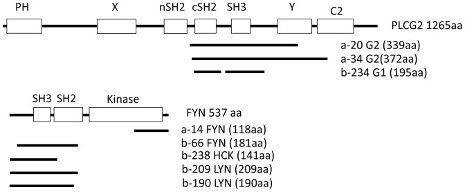
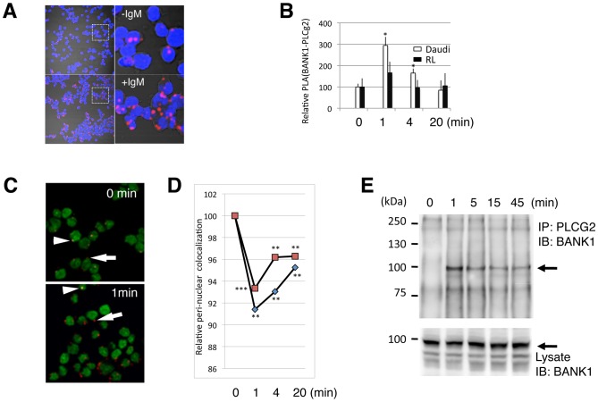
The B cell adaptor protein with ankyrin repeats (BANK1) and the B lymphoid tyrosine kinase (BLK) have been genetically associated with autoimmunity. The proteins of these genes interact physically and work in concert during B-cell signaling. Little is know about their interactions with other B-cell signaling molecules or their role in the process. Using yeast two hybrid (Y2H) we sought for factors that interact with BANK1. We found that the molecular switch PLCg2 interacts with BANK1 and that the interaction is promoted by B-cell receptor (BCR) stimulation. We found further that the kinase activity of BLK enhanced BANK1- PLCg2 binding and that the interaction was suppressed upon BLK depletion. Immunoprecipitation and mutational analysis demonstrated that the interaction between BANK1 and PLCg2 was dependent on specific tyrosine and proline residues on the adaptor protein. Our results provide new information important to understand the role of these two genes in basic B-cell physiology and immune-related diseases.


Conflict of interest statement

Competing Interests: MBQ, MEAR and CCL are affiliated to Pfizer - Universidad de Granada - Junta de Andalucía Centre for Genomics and Oncological Research. Patent application: European patent n° EP13382041.5 " METHOD FOR THE DIAGNOSIS AND MONITORING OF AUTOINMUNE DISEASES ", by FUNDACIÓN PÚBLICA ANDALUZA PROGRESO Y SALUD. The inventors are MBQ, MEAR and CCL. There are no further patents, products in development or marketed products to declare. This does not alter the authors' adherence to all the PLOS ONE policies on sharing data and materials, as detailed online in the guide for authors.

Figures

Figure 1
Figure 1. Clones belonging to the phosphoinositide-specific phospholipase C and the family of Src kinases isolated in two yeast two-hybrid screens.
(A) Representation of PLCg2 modular structure and the coding region of the clones identified in the Y2H. Clones named a- correspond to the first screen using as bait the full-length BANK1, clones starting with b- are the ones identified in the screen with the non autoactivating truncated protein BANK1 (331–785). The clone b-234 has a deletion of 25 aa between the cSH2 and SH3 domains. (B) Structure of the Src kinase FYN and the isolated clones belonging to this family of non- receptor kinase. PH: Pleckstrin homology domain, involved in the recruitment to membranes by binding to phosphatidylinositol containing lipids. X and Y are the two halves of the catalytic isomerase. SH2 (Src homology 2) conserved domain that typically binds to phosphorylated tyrosine residues. SH3 (Src homology 3) usually binds to proline-rich motifs. The C2 motif is present in many proteins that interact with membranes and are frequently involved in calcium dependent phospholipid binding and membrane targeting processes.
Figure 2
Figure 2. BANK1 and PLCG2 co-localize in cytoplasmic compartments.
Confocal images of HEK293 cells co-expressing fluorescently tagged proteins. BANK1 is a cytoplasmic adaptor protein that shows a punctate and homogeneous pattern of distribution. PLCg2 shares the same sub-cellular compartments when co-expressed with BANK1. Upper row, co-expression of PLCg2 and BANK1 showing sub-cellular co-localization in punctate structures. Although a perfect co-localization between the two proteins is observed, there are dots (indicated by arrows, both in the merge image and in the amplification at the right upper corner) in which the ratio of the two proteins is reversed. Cross-talks between light channels were further ruled out by assessing the emission on cells expressing only one fluoresce protein and excitated sequencially with both lasers. Middle row, images showing co-localization between PLCg2 and BANK1 in a homogeneous cytoplasmic distribution. Lower row, images showing only partial co-localization between the cytoplasmic scavenger receptor, CD163 and BANK1. Scale bar: 10 µm.(-yfp; yellow fluorescence protein. –cfp; cyan fluorescence protein. –chr; cherry fluorescent protein. –gfp; green fluorescent protein).
Figure 3
Figure 3. BANK1-PLCG2 complex formation is transient and induced by IgM stimulation.
(A) Confocal images of Daudi B cells showing increase molecular proximity between endogenous BANK1 and PLCg2 proteins upon anti-IgM stimulation. The staining was done using in situ a PLA protocol with rabbit anti-BANK1 (ET-52) and mouse anti PLCg2 (Abcam). Nuclei were stained with DAPI in blue. The confocal images (PLA signals) were taken with a pinhole of 2.5 (Zeiss Plan-Apochromat 63× oil objective). Upper panel, non-stimulated cells. Low panel, cells stimulated for 1 minute with anti-human IgM-F(ab')2. To the right are shown digital magnifications. (B) Time variation of PLA BANK1-PLCg2 interaction upon stimulation with anti-IgM in two human B-cell lines, the Burkitt´s derived Daudi and the non-Hodgkin´s lymphoma derived RL. Results are shown as the mean from three independent experiments. Error bars represent the SD from the mean. *P<0,05 based on Student´s test comparing stimulated cells versus time 0 (non stimulated cells).(C) Merge confocal images (pinhole = 1) of Daudi cells showing the nuclei in green and the BANK1-PLCg2 PLA interaction in red to permit co-location analysis. PLA signals close to the nucleus appeared as yellow dots (arrowheads) and when localize in the periphery of the cell as red dots (arrows). (D) Time course upon anti-IgM stimulation of co-localization between the PLA signal and the nucleus. Quantification was done using the overlap coefficient after Manders because this coefficient is insensitive to differences in signal intensities between channels. After one minute of stimulation the co-localization decreases suggesting a translocation of the BANK1-PLCg2 complex. The graft shows two independent experiments, each point represents the analysis of at least 300 cells.**,P<0,01;***P<0,001 based on Student´s test comparing stimulated cells versus time 0 (non stimulated). (E) Immunoprecipitation of the BANK1-PLCg2 complex in Daudi cells. Anti-PLCg2 immunoprecipitates (above) and total cell lysate (below) were analyzed by immunoblotting with anti-BANK1 antibody (BANK1-ET-52). The position of the BANK1 protein is indicated by arrows.
Figure 4
Figure 4. The constitutive-active form of BLK enhances the binding between BANK1 and PLCG2.
(A) Schematic representation of the constructs used to study the association between BANK1 and PLCg2 in transfected HEK293 cells. The constructs coding for wild-type forms of BLK, LYN, GFP and PLCg2 or the indicated mutated forms were fused to the epitope V5 at the C-termini. BANK1 was targeted with the Flag epitope at the N- terminus. The catalytic domains of BLK and PLCg2 are shown in red. The kinase dead form (BLK-KL-v5) has a substitution K (lysine) to L (leucine) at position 269 and the constitutively active form (BLK-YF-v5) has a Y501F substitution that prevents the phosphorylation of the inhibitory tyrosine. The lipidation in the amino terminal of BLK is indicated as back line. The myristoylation site was deleted by G2V substitution (glycine to valine) and the addition of an extra by palmitoylated site by L3C substitution (leucine to cysteine). The Src homology 3 domains (SH3) that bind to proline-rich motifs are drawn in orange and the SH2 domains in yellow. The Pleckstrin homology domain (PH) that binds to phosphatidylinositol lipids is shown in blue. In BANK1 are shown the Dof/BCAP/BANK (DBB) motif (amino acids 199–327), the double ankyrin repeat-like (ANK) motifs (amino acids 339-402) and the putative coiled coil (CC) region (amino acids 677–705). (B) HEK293 cells were transiently co-transfected with plasmids coding for the wild-type form of BLK, its functionally mutated forms (KL and YF), LYN or GFP in addition to plasmids expressing BANK1 and PLCg2. The lysates were immunoprecipitated using anti-PLCg2 antibody (above) and immunoblotted sequencially with anti-BANK1 antibody, anti-V5 to detect PLCg2, Srcs kinases and GFP and anti-phosphotyrosine antibody. (C). Mutation of lipidation sites of the kinases influence the formation of the BANK1-PLCg2 complex and the overall tyrosine phosphorylation on PLCg2. The blots were interrogated as in B.
Figure 5
Figure 5. Silencing of the BLK kinase leads to alteration of the association between BANK1 and PLCg2.
(A) Immunoblot of extracts derived from human Daudi B-cells showing efficient silencing of endogenous BLK protein. The cell line shBLK was transduced with lentivirus coding for small hairpin RNAs targeting the BLK kinase while shControl lentivirus codes for unrelated sequences. Top, western blot analysis using antibody to BLK; bottom, western blot analysis using antibody to GAPDH as loading control. (B) The relative BLK mRNA is reduced to half in the silencing line (shBLK)(***P<0,001 based on Student´s test comparing mRNA expression between Daudi cell versus cBLK silencing cells and control silenced cells versus BLK silencing cells). (C) Immunoprecipitates (IPs) of stimulated silenced shBLK cells with anti-IgM, using anti-PLCg2 antibody and interrogated with anti-BANK1 to assay BANK1-PLCg2 association. Quantification of the immunoprecipitates normalized with BANK1 is displayed below the bands. Silencing of BLK leads to less association between BANK1-PLCg2. (D) Kinetics of the association between BANK1-PLCg2 assayed with in situ Proximity Ligation (PLA) in control and BLK-silenced cell lines. The association between BANK1-PLCg2 is reduced in silenced cells during resting conditions and after 15 minutes of IgM stimulation (***P≤0,001 based on Student´s test comparing control cells versus BLK silencing cells). The difference of BANK1-PLCg2 association between control and the silenced lines was not significant at 1 minute after stimulation.
Figure 6
Figure 6. Mutations of specific tyrosine residues and proline rich domains of BANK1 abrogated the association with PLCg2.
(A) Schematic representation of the motifs and the structure of the human BANK1 splicing variants and analysis of the motifs affecting the BANK1-PLCg2 binding. The Dof/BCAP/BANK (DBB) motif (amino acids 199–327), the double ankyrin repeat-like (ANK) motifs (amino acids 339–402) and the presumptive coiled coils (CC) region (amino acids 677–705) are indicated. Tyrosine residues susceptible to be phosphorylated are shown by Y. Residues Y125, Y146, Y161, Y416, Y484 and Y488 that predict the putative SH2 binding sites are indicated as bold Y (http://scansite.mit.edu/motifscan_seq.phtml). The positions of the mutated amino acids are indicated above the BANK1 drawing. (B) Alignment of the BANK1 amino acid motifs in different species indicating the mutated residues corresponding to putative SH2 and SH3 binding domains, alignment was done using data downloaded from www.ensembl.org. (C) Phosphorylation and immunoprecipitation analysis of the wild type and mutated forms of BANK1 co-expressed with the constitutively active form of BLK (BLK-YF) and PLCg2. Relative expression of the constructs was monitored by western blot (IB) of an aliquot (1/10) of the transfected HEK293T cell extract. The rest of the lysates were used for immunoprecipitation (IP) using anti-PLCg2 (Abcam) and it is shown in the lower row. Lane 0 represents immunoprecipitation without the IP antibody. (D) Quantification of BANK1 phosphorylation and immunoprecipitation using anti-PLCg2 antibody. Bands of the western blots were quantified using ImageJ program. Values of expression of BANK1 in the lysates (IB:Anti-BANK1) were taken to normalize the results of phosphorylation and immunoprecipitation of BANK1. The relative expression of BANK1 was: 1, 1.03, 0.93, 0.85,0.69, 0,81, and 0.73 from lane 1 to 7 in the lysates interrogated with anti-BANK1. The Y484-8F mutation significantly reduced both the tyrosine phosphorylation of BANK1 and the binding to PLCg2. The PP513LL mutation does not affect the tyrosine phosphorylation of BANK1, however it leads to a decrease in binding to PLCg2. (E) Co-expression in HEK293T cells of the two isoforms of BANK1 renders equivalent recovery of both isoforms in the anti-PLCg2 immunoprecipitate, indicating that exon 2 did not participate in the binding between BANK1 and PLCg2.

References

    1. Takata M, Sabe H, Hata A, Inazu T, Homma Y, et al. (1994) Tyrosine kinases Lyn and Syk regulate B cell receptor-coupled Ca2+ mobilization through distinct pathways. EMBO J 13: 1341–1349. - PMC - PubMed
    1. Nishizumi H, Horikawa K, Mlinaric-Rascan I, Yamamoto T (1998) A double-edged kinase Lyn: a positive and negative regulator for antigen receptor-mediated signals. J Exp Med 187: 1343–1348. - PMC - PubMed
    1. Chan VW, Lowell CA, DeFranco AL (1998) Defective negative regulation of antigen receptor signaling in Lyn-deficient B lymphocytes. Curr Biol 8: 545–553. - PubMed
    1. Sato S, Hasegawa M, Fujimoto M, Tedder TF, Takehara K (2000) Quantitative genetic variation in CD19 expression correlates with autoimmunity. J Immunol 165: 6635–6643. - PubMed
    1. Majeti R, Xu Z, Parslow TG, Olson JL, Daikh DI, et al. (2000) An inactivating point mutation in the inhibitory wedge of CD45 causes lymphoproliferation and autoimmunity. Cell 103: 1059–1070. - PubMed

Publication types