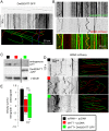
Huntingtin's function in axonal transport is conserved in Drosophila melanogaster
- PMID: 23555909
- PMCID: PMC3610688
- DOI: 10.1371/journal.pone.0060162
Huntingtin's function in axonal transport is conserved in Drosophila melanogaster
Abstract
Huntington's disease (HD) is a devastating dominantly inherited neurodegenerative disorder caused by an abnormal polyglutamine expansion in the N-terminal part of the huntingtin (HTT) protein. HTT is a large scaffold protein that interacts with more than a hundred proteins and is probably involved in several cellular functions. The mutation is dominant, and is thought to confer new and toxic functions to the protein. However, there is emerging evidence that the mutation also alters HTT's normal functions. Therefore, HD models need to recapitulate this duality if they are to be relevant. Drosophila melanogaster is a useful in vivo model, widely used to study HD through the overexpression of full-length or N-terminal fragments of mutant human HTT. However, it is unclear whether Drosophila huntingtin (DmHTT) shares functions similar to the mammalian HTT. Here, we used various complementary approaches to analyze the function of DmHTT in fast axonal transport. We show that DmHTT interacts with the molecular motor dynein, associates with vesicles and co-sediments with microtubules. DmHTT co-localizes with Brain-derived neurotrophic factor (BDNF)-containing vesicles in rat cortical neurons and partially replaces mammalian HTT in a fast axonal transport assay. DmHTT-KO flies show a reduced fast axonal transport of synaptotagmin vesicles in motoneurons in vivo. These results suggest that the function of HTT in axonal transport is conserved between flies and mammals. Our study therefore validates Drosophila melanogaster as a model to study HTT function, and its dysfunction associated with HD.
Conflict of interest statement
Figures


Similar articles
-
Huntingtin-mediated axonal transport requires arginine methylation by PRMT6.Cell Rep. 2021 Apr 13;35(2):108980. doi: 10.1016/j.celrep.2021.108980. Cell Rep. 2021. PMID: 33852844 Free PMC article.
-
pARIS-htt: an optimised expression platform to study huntingtin reveals functional domains required for vesicular trafficking.Mol Brain. 2010 Jun 1;3:17. doi: 10.1186/1756-6606-3-17. Mol Brain. 2010. PMID: 20515468 Free PMC article.
-
Excess Rab4 rescues synaptic and behavioral dysfunction caused by defective HTT-Rab4 axonal transport in Huntington's disease.Acta Neuropathol Commun. 2020 Jul 1;8(1):97. doi: 10.1186/s40478-020-00964-z. Acta Neuropathol Commun. 2020. PMID: 32611447 Free PMC article.
-
[Huntington's disease: intracellular signaling pathways and neuronal death].J Soc Biol. 2005;199(3):247-51. doi: 10.1051/jbio:2005026. J Soc Biol. 2005. PMID: 16471265 Review. French.
-
Choosing and using Drosophila models to characterize modifiers of Huntington's disease.Biochem Soc Trans. 2012 Aug;40(4):739-45. doi: 10.1042/BST20120072. Biochem Soc Trans. 2012. PMID: 22817726 Review.
Cited by
-
Huntingtin differentially regulates the axonal transport of a sub-set of Rab-containing vesicles in vivo.Hum Mol Genet. 2015 Dec 20;24(25):7182-95. doi: 10.1093/hmg/ddv415. Epub 2015 Oct 8. Hum Mol Genet. 2015. PMID: 26450517 Free PMC article.
-
Do Changes in Synaptic Autophagy Underlie the Cognitive Impairments in Huntington's Disease?J Huntingtons Dis. 2021;10(2):227-238. doi: 10.3233/JHD-200466. J Huntingtons Dis. 2021. PMID: 33780373 Free PMC article. Review.
-
Characterization of axonal transport defects in Drosophila Huntingtin mutants.J Neurogenet. 2016 Sep-Dec;30(3-4):212-221. doi: 10.1080/01677063.2016.1202950. Epub 2016 Jul 22. J Neurogenet. 2016. PMID: 27309588 Free PMC article.
-
Role of context in RNA structure: flanking sequences reconfigure CAG motif folding in huntingtin exon 1 transcripts.Biochemistry. 2013 Nov 19;52(46):8219-25. doi: 10.1021/bi401129r. Epub 2013 Nov 7. Biochemistry. 2013. PMID: 24199621 Free PMC article.
-
Huntingtin and the Synapse.Front Cell Neurosci. 2021 Jun 15;15:689332. doi: 10.3389/fncel.2021.689332. eCollection 2021. Front Cell Neurosci. 2021. PMID: 34211373 Free PMC article. Review.
References
-
- Duffy JB (2000) GAL4 system in Drosophila: a fly geneticist’s Swiss army knife. Genesis 34: 1–15. - PubMed
-
- The Huntington’s Disease Collaborative Research Group (1993) A novel gene containing a trinucleotide repeat that is expanded and unstable on Huntington’s disease chromosomes. The Huntington's Disease Collaborative Research Group. Cell 72: 971–983. - PubMed
-
- Steffan JS, Bodai L, Pallos J, Poelman M, McCampbell A, et al. (2001) Histone deacetylase inhibitors arrest polyglutamine-dependent neurodegeneration in Drosophila. Nature 413: 739–743. - PubMed
-
- Jackson GR, Salecker I, Dong X, Yao X, Arnheim N, et al. (1998) Polyglutamine-Expanded Human Huntingtin Transgenes Induce Degeneration of Drosophila Photoreceptor Neurons. Cell 21: 633–642. - PubMed
Publication types
MeSH terms
Substances
LinkOut - more resources
Full Text Sources
Other Literature Sources
Molecular Biology Databases
Research Materials

