

Interhemispheric functional connectivity and its relationships with clinical characteristics in major depressive disorder: a resting state fMRI study
- PMID: 23555920
- PMCID: PMC3612036
- DOI: 10.1371/journal.pone.0060191
Interhemispheric functional connectivity and its relationships with clinical characteristics in major depressive disorder: a resting state fMRI study
Abstract
Background: Abnormalities in large-scale, structural and functional brain connectivity have been increasingly reported in patients with major depressive disorder (MDD). However, MDD-related alterations in functional interaction between the cerebral hemispheres are still not well understood. Resting state fMRI, which reveals spontaneous neural fluctuations in blood oxygen level dependent signals, provides a means to detect interhemispheric functional coherence. We examined the resting state functional connectivity (RSFC) between the two hemispheres and its relationships with clinical characteristics in MDD patients using a recently proposed measurement named "voxel-mirrored homotopic connectivity (VMHC)".
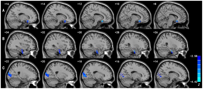
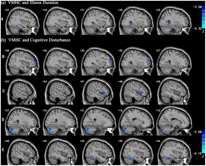
Methodology/principal findings: We compared the interhemispheric RSFC, computed using the VMHC approach, of seventeen first-episode drug-naive patients with MDD and seventeen healthy controls. Compared to the controls, MDD patients showed significant VMHC decreases in the medial orbitofrontal gyrus, parahippocampal gyrus, fusiform gyrus, and occipital regions including the middle occipital gyrus and cuneus. In MDD patients, a negative correlation was found between VMHC of the fusiform gyrus and illness duration. Moreover, there were several regions whose VMHC showed significant negative correlations with the severity of cognitive disturbance, including the prefrontal regions, such as middle and inferior frontal gyri, and two regions in the cereballar crus.
Conclusions/significance: These findings suggest that the functional coordination between homotopic brain regions is impaired in MDD patients, thereby providing new evidence supporting the interhemispheric connectivity deficits of MDD. The significant correlations between the VMHC and clinical characteristics in MDD patients suggest potential clinical implication of VMHC measures for MDD. Interhemispheric RSFC may serve as a useful screening method for evaluating MDD where neural connectivity is implicated in the pathophysiology.
Conflict of interest statement
Figures
References
-
- Garrett A, Kelly R, Gomez R, Keller J, Schatzberg AF, et al. (2011) Aberrant brain activation during a working memory task in psychotic major depression. Am J Psychiatry 168: 173–182. - PubMed
-
- Schlosser RG, Wagner G, Koch K, Dahnke R, Reichenbach JR, et al. (2008) Fronto-cingulate effective connectivity in major depression: a study with fMRI and dynamic causal modeling. Neuroimage 43: 645–655. - PubMed
-
- van Wingen GA, van Eijndhoven P, Tendolkar I, Buitelaar J, Verkes RJ, et al. (2011) Neural basis of emotion recognition deficits in first-episode major depression. Psychol Med 41: 1397–1405. - PubMed
-
- Chantiluke K, Halari R, Simic M, Pariante CM, Papadopoulos A, et al. (2012) Fronto-striato-cerebellar dysregulation in adolescents with depression during motivated attention. Biol Psychiatry 71: 59–67. - PubMed
-
- Zhu X, Wang X, Xiao J, Liao J, Zhong M, et al. (2012) Evidence of a dissociation pattern in resting-state default mode network connectivity in first-episode, treatment-naive major depression patients. Biol Psychiatry 71: 611–617. - PubMed
Publication types
MeSH terms
LinkOut - more resources
Full Text Sources
Other Literature Sources
Medical
