. 2013;8(3):e60347.
doi: 10.1371/journal.pone.0060347. Epub 2013 Mar 28.

Using multivalent adenoviral vectors for HIV vaccination

Affiliations

Using multivalent adenoviral vectors for HIV vaccination

Linlin Gu et al. PLoS One. 2013.

Abstract

Adenoviral vectors have been used for a variety of vaccine applications including cancer and infectious diseases. Traditionally, Ad-based vaccines are designed to express antigens through transgene expression of a given antigen. For effective vaccine development it is often necessary to express or present multiple antigens to the immune system to elicit an optimal vaccine as observed preclinically with mosaic/polyvalent HIV vaccines or malaria vaccines. Due to the wide flexibility of Ad vectors they are an ideal platform for expressing large amounts of antigen and/or polyvalent mosaic antigens. Ad vectors that display antigens on their capsid surface can elicit a robust humoral immune response, the "antigen capsid-incorporation" strategy. The adenoviral hexon protein has been utilized to display peptides in the majority of vaccine strategies involving capsid incorporation. Based on our abilities to manipulate hexon HVR2 and HVR5, we sought to manipulate HVR1 in the context of HIV antigen display for the first time ever. More importantly, peptide incorporation within HVR1 was utilized in combination with other HVRs, thus creating multivalent vectors. To date this is the first report where dual antigens are displayed within one Ad hexon particle. These vectors utilize HVR1 as an incorporation site for a seven amino acid region of the HIV glycoprotein 41, in combination with six Histidine incorporation within HVR2 or HVR5. Our study illustrates that these multivalent antigen vectors are viable and can present HIV antigen as well as His6 within one Ad virion particle. Furthermore, mouse immunizations with these vectors demonstrate that these vectors can elicit a HIV and His6 epitope-specific humoral immune response.


Conflict of interest statement

Competing Interests: The authors have declared that no competing interests exist.

Figures

Figure 1
Figure 1. KWAS and His6 genetically incorporated into the HVR1 location as well as His6 within HVR2 or HVR5.
Rescued vectors were amplified and viral DNA analyzed to confirm stable modification of relevant genes. A) Hexon-specific PCR primers confirmed the presence of the hexon gene in all of the modified vectors. Lane 1, Ad5; lane 2, Ad5/HVR2-MPER-L15ΔE1; lane 3, Ad/H5-HVR1-His6; lane 4, Ad5/H5-HVR1-KWAS-HVR2-His6; and lane 5, Ad5/H5-HVR1-KWAS-HVR5-His6. B) KWAS-specific primers confirmed the incorporation of coding regions for KWAS inserts in multivalent vectors. Lane 1, Ad5; lane 2, Ad5/HVR2-MPER-L15ΔE1; lane 3, Ad5/H5-HVR1-KWAS-HVR2-His6; and lane 4, Ad5/H5-HVR1-KWAS-HVR5-His6. C) His6-specific primers confirmed the incorporation of coding regions for His6 inserts in multivalent vectors. Lane 1, Ad5; lane 2, Ad/H5-HVR1-His6; lane 3, Ad5/H5-HVR1-KWAS-HVR2-His6; and lane 4, Ad5/H5-HVR1-KWAS-HVR5-His6. These vectors have been described in Table # 1.
Figure 2
Figure 2. Western blotting confirmed the presence of His6, KWAS, and fiber on multivalent vaccine vectors.
A) Western blotting confirmed the presence of His6 incorporation within the dual modified vectors. In this assay, 5×10⁹ VP of Ad5 (lane 1), Ad/H5-HVR1-His6 (lane 2), Ad5/H5-HVR1-KWAS-HVR2-His6 (lane 3), and Ad5/H5-HVR1-KWAS-HVR5-His6 (lane 4) were separated on 4 to 15% polyacrylamide gradient SDS-PAGE gel. The proteins were transferred to polyvinylidene fluoride (PVDF) membrane then incubated with anti-His antibody. The arrow indicates the His tag is genetically incorporated into the hexon protein. B) Western blotting confirmed the presence of KWAS incorporation within the dual modified vectors. In this assay, 5×10⁹ VP of Ad5 (lane 1), Ad5/HVR2-MPER-L15ΔE1 (lane 2), Ad5/H5-HVR1-KWAS-HVR2-His6 (lane 3), and Ad5/H5-HVR1-KWAS-HVR5-His6 (lane 4) were separated on 4 to 15% polyacrylamide gradient SDS-PAGE gel. The proteins were transferred to PVDF membrane then incubated with anti-KWAS antibody. The arrow indicates the KWAS epitope is genetically incorporated into the hexon protein. C) Western blotting confirmed the presence of fiber incorporation within the dual modified vectors. In this assay, 5×10⁹ VP of Ad5 (lane 1), Ad5/HVR2-MPER-L15ΔE1 (lane 2), Ad/H5-HVR1-His6 (lane 3), Ad5/H5-HVR1-KWAS-HVR2-His6 (lane 4), and Ad5/H5-HVR1-KWAS-HVR5-His6 (lane 5) were separated on 4 to 15% polyacrylamide gradient SDS-PAGE gel. The proteins were transferred to PVDF membrane then incubated with anti-Fiber antibody. The arrow indicates detection of the fiber protein.
Figure 3
Figure 3. HIV and His6 epitopes in the HVRs are exposed on the virion surface.
A) In the assay, varying amounts of Ad5, Ad/H5-HVR1-His6, Ad5/H5-HVR1-KWAS-HVR2-His6, or Ad5/H5-HVR1-KWAS-HVR5-His6 were immobilized in the wells of ELISA plates and incubated with anti-His6 antibody. The binding was detected with an HRP-conjugated secondary antibody. B) In the assay, varying amounts of Ad5, Ad5/HVR2-MPER-L15ΔE1, Ad5/H5-HVR1-KWAS-HVR2-His6, or Ad5/H5-HVR1-KWAS-HVR5-His6 were immobilized in the wells of ELISA plates and incubated with anti-HIV antibody. The binding was detected with an HRP-conjugated secondary antibody. C) In the assay 6×10⁸ VP of Ad5, Ad/H5-HVR1-His6, Ad5/H5-HVR1-KWAS-HVR2-His6, or Ad5/H5-HVR1-KWAS-HVR5-His6 were immobilized on an ELISA plate followed by varying dilutions of His6 antibody. The binding was detected with an HRP-conjugated secondary antibody. D) In the assay 6 x 10⁸ VP of Ad5, Ad5/HVR2-MPER-L15ΔE1, Ad5/H5-HVR1-KWAS-HVR2-His6, or Ad5/H5-HVR1-KWAS-HVR5-His6 were immobilized on an ELISA plate followed by varying dilutions of HIV antibody. The binding was detected with an HRP-conjugated secondary antibody.
Figure 4
Figure 4. Multivalent vectors elicit an in vivo anti-His6 immune response.
BALB/c mice (n = 8) were primed, boosted, and reboosted with 1×10¹⁰ VP of Ad vectors. A) Immunization timeline showing when immunizations were performed (solid arrows); serum was collected (dashed arrows). B–C) Post-prime and post-reboost serum was collected for ELISA binding assays. 1 µM of purified His6 (LGSHHHHHHLGS) antigenic peptide was bound to ELISA plates. Residual unbound peptide was washed from the plates. The plates were then incubated with varying concentrations of immunized mice serum and the binding was detected with HRP conjugated secondary antibody. OD absorbance at 450 nm represents His6 antibody levels in serum. The values are expressed as the mean ± standard deviation. The asterisks (***) indicate a P value ≤ 0.001, ** indicate a P value ≤ 0.01, and * indicate a P value ≤ 0.05.
Figure 5
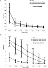
Figure 5. Multivalent vectors elicit an in vivo anti-KWAS immune response.
BALB/c mice (n = 8) were primed, boosted, and reboosted with 1×10¹⁰ VP of Ad vectors (Figure 4A). A–B) Post-prime and post-reboost serum was collected at various time points for ELISA binding assays. 1 µM of purified KWAS (PCEWDEAELDKWASNLEEEDDDNE) antigenic peptide was bound to ELISA plates. Residual unbound peptide was washed from the plates. The plates were then incubated with immunized mice serum and the binding was detected with HRP conjugated secondary antibody. OD absorbance at 450 nm represents KWAS antibody levels in sera. The values are expressed as the mean ± standard deviation. The *** indicate a P value ≤ 0.001, and ** indicate a P value ≤ 0.01.
Figure 6
Figure 6. Multivalent vectors elicit in vivo His6 and KWAS isotype-specific responses.
BALB/c mice (n = 8) were primed, boosted, and reboosted with 1×10¹⁰ VP of Ad vectors (Figure 4A). 10 days post-reboost serum was used for the isotype-specific assays. A–B) 1 µM of purified His6 (LGSHHHHHHLGS) antigenic peptide or C–D) 1 µM of purified KWAS (PCEWDEAELDKWASNLEEEDDDNE) antigenic peptide was bound to ELISA plates. Residual unbound peptide was washed from the plates. The plates were then incubated with immunized mice serum followed by isotype-specific antibodies. The binding was detected with HRP conjugated secondary. OD at 450 nm represents isotype-specific His6 or KWAS antibody levels in sera. The values are expressed as the mean ± standard deviation. The *** indicate a P value ≤ 0.001, ** indicate a P value ≤ 0.01, and * indicate a P value ≤ 0.05.

References

    1. Crompton J, Toogood CI, Wallis N, Hay RT (1994) Expression of a foreign epitope on the surface of the adenovirus hexon. JGenVirol 75 (Pt 1): 133–139. - PubMed
    1. McConnell MJ, Danthinne X, Imperiale MJ (2006) Characterization of a permissive epitope insertion site in adenovirus hexon. JVirol 80: 5361–5370. - PMC - PubMed
    1. Worgall S, Krause A, Rivara M, Hee KK, Vintayen EV, et al. (2005) Protection against P. aeruginosa with an adenovirus vector containing an OprF epitope in the capsid. JClinInvest 115: 1281–1289. - PMC - PubMed
    1. Matthews QL, Yang P, Wu Q, Belousova N, Rivera AA, et al. (2008) Optimization of capsid-incorporated antigens for a novel adenovirus vaccine approach. Virol J 5: 98. - PMC - PubMed
    1. Matthews QL, Fatima A, Tang Y, Perry BA, Tsuruta Y, et al. (2010) HIV antigen incorporation within adenovirus hexon hypervariable 2 for a novel HIV vaccine approach. PLoS One 5: e11815. - PMC - PubMed

Publication types

MeSH terms