

Prevention of LPS-induced microglia activation, cytokine production and sickness behavior with TLR4 receptor interfering peptides
- PMID: 23555964
- PMCID: PMC3610686
- DOI: 10.1371/journal.pone.0060388
Prevention of LPS-induced microglia activation, cytokine production and sickness behavior with TLR4 receptor interfering peptides
Abstract
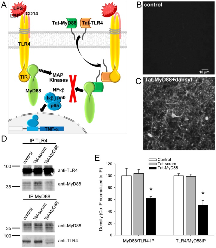
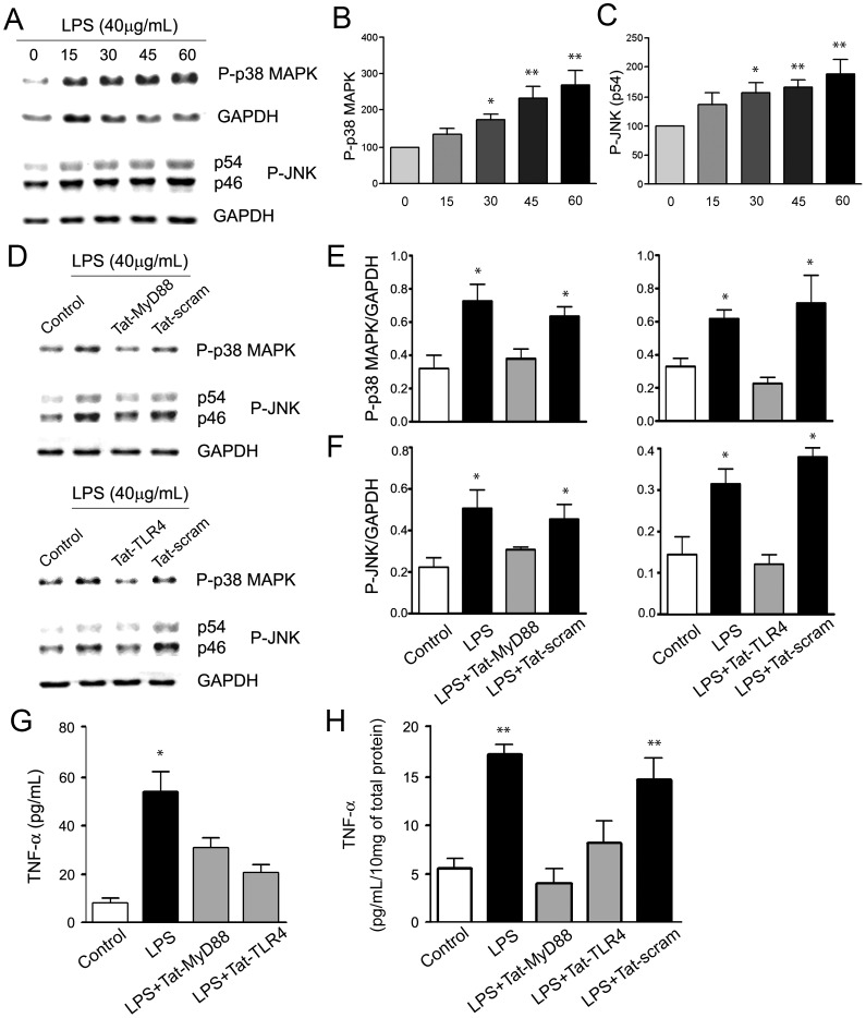
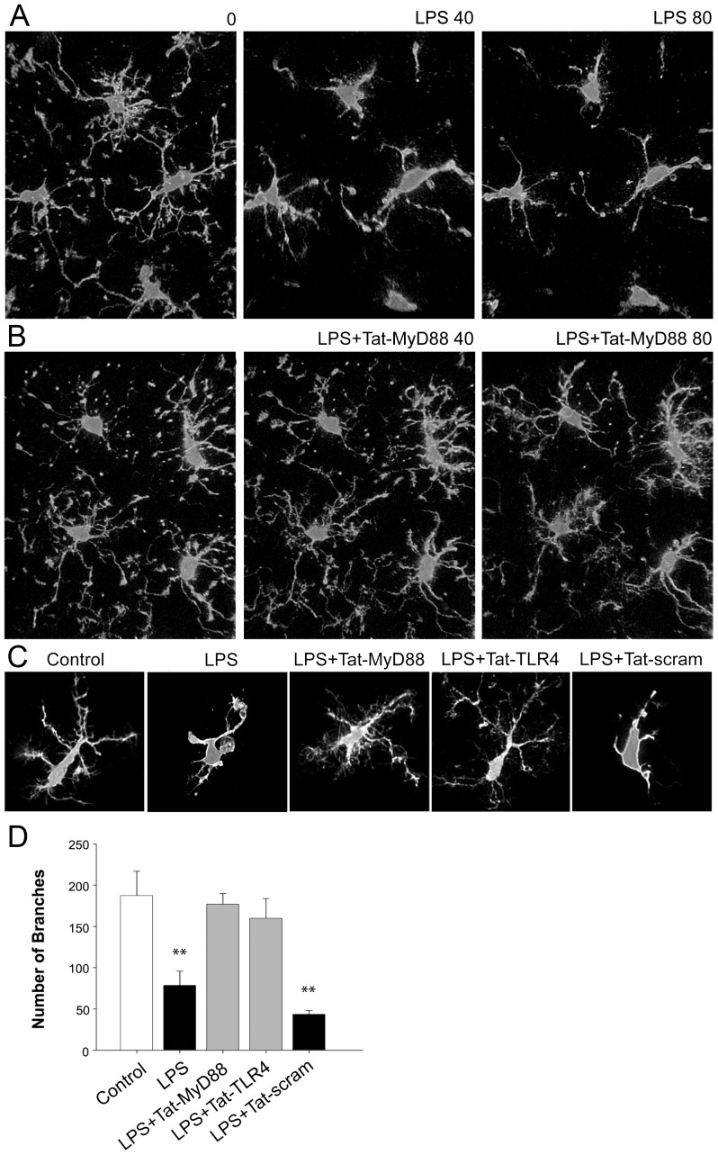
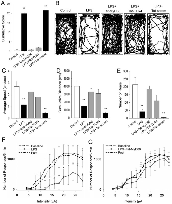
The innate immune receptor Toll-like 4 (TLR4) is the receptor activated by lipopolysaccharide (LPS), and TLR4-LPS interaction is well known to induce an innate immune response, triggering sickness behavior. Within the brain, TLR4 is highly expressed in brain microglia, and excessive inflammation resulting from activation of this pathway in the brain has been implicated in depressive disorders and neurodegenerative pathologies. We hypothesized that blocking LPS-induced activation of TLR4 would prevent downstream immune signaling in the brain and suppress the induction of sickness behavior. We used interfering peptides to block TLR4 activation and confirmed their efficacy in preventing second messenger activation and cytokine production normally induced by LPS treatment. Further, these peptides blocked morphological changes in microglia that are typically induced by LPS. We also demonstrated that intraperitoneal (i.p.) injection of Tat-TLR4 interfering peptides prevented LPS-induced sickness behavior, as assessed in home cage behavior and with the intracranial self-stimulation paradigm. These newly synthesised peptides inhibit TLR4 signaling thereby preventing changes in behavior and motivation caused by inflammatory stimuli. These peptides highlight the roll of TLR4 and microglia morphology changes in sickness behavior, and thus may be of therapeutic value in limiting the deleterious impact of excessive inflammation in specific CNS pathologies.
Conflict of interest statement
Figures
Similar articles
-
Methamphetamine alters the TLR4 signaling pathway, NF-κB activation, and pro-inflammatory cytokine production in LPS-challenged NR-9460 microglia-like cells.Mol Immunol. 2020 May;121:159-166. doi: 10.1016/j.molimm.2020.03.013. Epub 2020 Mar 26. Mol Immunol. 2020. PMID: 32222586 Free PMC article.
-
Synergistic effects of NOD1 or NOD2 and TLR4 activation on mouse sickness behavior in relation to immune and brain activity markers.Brain Behav Immun. 2015 Feb;44:106-20. doi: 10.1016/j.bbi.2014.08.011. Epub 2014 Sep 11. Brain Behav Immun. 2015. PMID: 25218901 Free PMC article.
-
Persistent Toll-like receptor 7 stimulation induces behavioral and molecular innate immune tolerance.Brain Behav Immun. 2019 Nov;82:338-353. doi: 10.1016/j.bbi.2019.09.004. Epub 2019 Sep 6. Brain Behav Immun. 2019. PMID: 31499172 Free PMC article.
-
Ciprofloxacin and levofloxacin attenuate microglia inflammatory response via TLR4/NF-kB pathway.J Neuroinflammation. 2019 Jul 18;16(1):148. doi: 10.1186/s12974-019-1538-9. J Neuroinflammation. 2019. PMID: 31319868 Free PMC article. Review.
-
Therapeutic targeting of innate immunity with Toll-like receptor 4 (TLR4) antagonists.Biotechnol Adv. 2012 Jan-Feb;30(1):251-60. doi: 10.1016/j.biotechadv.2011.05.014. Epub 2011 Jun 6. Biotechnol Adv. 2012. PMID: 21664961 Review.
Cited by
-
CRF-amplified neuronal TLR4/MCP-1 signaling regulates alcohol self-administration.Neuropsychopharmacology. 2015 May;40(6):1549-59. doi: 10.1038/npp.2015.4. Epub 2015 Jan 8. Neuropsychopharmacology. 2015. PMID: 25567426 Free PMC article.
-
Physcion Mitigates LPS-Induced Neuroinflammation, Oxidative Stress, and Memory Impairments via TLR-4/NF-кB Signaling in Adult Mice.Pharmaceuticals (Basel). 2024 Sep 11;17(9):1199. doi: 10.3390/ph17091199. Pharmaceuticals (Basel). 2024. PMID: 39338361 Free PMC article.
-
Myalgic encephalomyelitis or chronic fatigue syndrome: how could the illness develop?Metab Brain Dis. 2019 Apr;34(2):385-415. doi: 10.1007/s11011-019-0388-6. Epub 2019 Feb 13. Metab Brain Dis. 2019. PMID: 30758706 Free PMC article. Review.
-
In Vitro Analysis of LPS-Induced miRNA Differences in Bovine Endometrial Cells and Study of Related Pathways.Animals (Basel). 2024 Nov 22;14(23):3367. doi: 10.3390/ani14233367. Animals (Basel). 2024. PMID: 39682333 Free PMC article.
-
Identification of a specific α-synuclein peptide (α-Syn 29-40) capable of eliciting microglial superoxide production to damage dopaminergic neurons.J Neuroinflammation. 2016 Jun 21;13(1):158. doi: 10.1186/s12974-016-0606-7. J Neuroinflammation. 2016. PMID: 27329107 Free PMC article.
References
-
- Dantzer R (2001) Cytokine-induced sickness behavior: mechanisms and implications. Ann N Y Acad Sci 933: 222–234. - PubMed
-
- Hart BL (1988) Biological basis of the behavior of sick animals. Neurosci Biobehav Rev 12: 123–137. - PubMed
-
- Perry VH (2004) The influence of systemic inflammation on inflammation in the brain: implications for chronic neurodegenerative disease. Brain Behav Immun 18: 407–413. - PubMed
-
- Kettenmann H (2007) Neuroscience: the brain's garbage men. Nature 446: 987–989. - PubMed
Publication types
MeSH terms
Substances
Grants and funding
LinkOut - more resources
Full Text Sources
Other Literature Sources
Molecular Biology Databases
