

Population of anatomically variable 4D XCAT adult phantoms for imaging research and optimization
- PMID: 23556927
- PMCID: PMC3612121
- DOI: 10.1118/1.4794178
Population of anatomically variable 4D XCAT adult phantoms for imaging research and optimization
Abstract
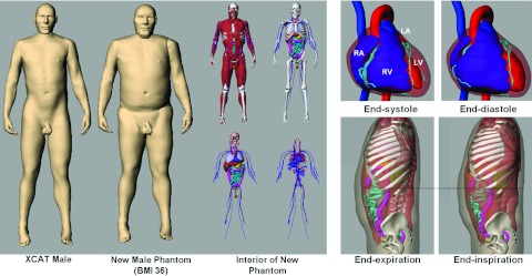
Purpose: The authors previously developed the 4D extended cardiac-torso (XCAT) phantom for multimodality imaging research. The XCAT consisted of highly detailed whole-body models for the standard male and female adult, including the cardiac and respiratory motions. In this work, the authors extend the XCAT beyond these reference anatomies by developing a series of anatomically variable 4D XCAT adult phantoms for imaging research, the first library of 4D computational phantoms.
Methods: The initial anatomy of each phantom was based on chest-abdomen-pelvis computed tomography data from normal patients obtained from the Duke University database. The major organs and structures for each phantom were segmented from the corresponding data and defined using nonuniform rational B-spline surfaces. To complete the body, the authors manually added on the head, arms, and legs using the original XCAT adult male and female anatomies. The structures were scaled to best match the age and anatomy of the patient. A multichannel large deformation diffeomorphic metric mapping algorithm was then used to calculate the transform from the template XCAT phantom (male or female) to the target patient model. The transform was applied to the template XCAT to fill in any unsegmented structures within the target phantom and to implement the 4D cardiac and respiratory models in the new anatomy. Each new phantom was refined by checking for anatomical accuracy via inspection of the models.
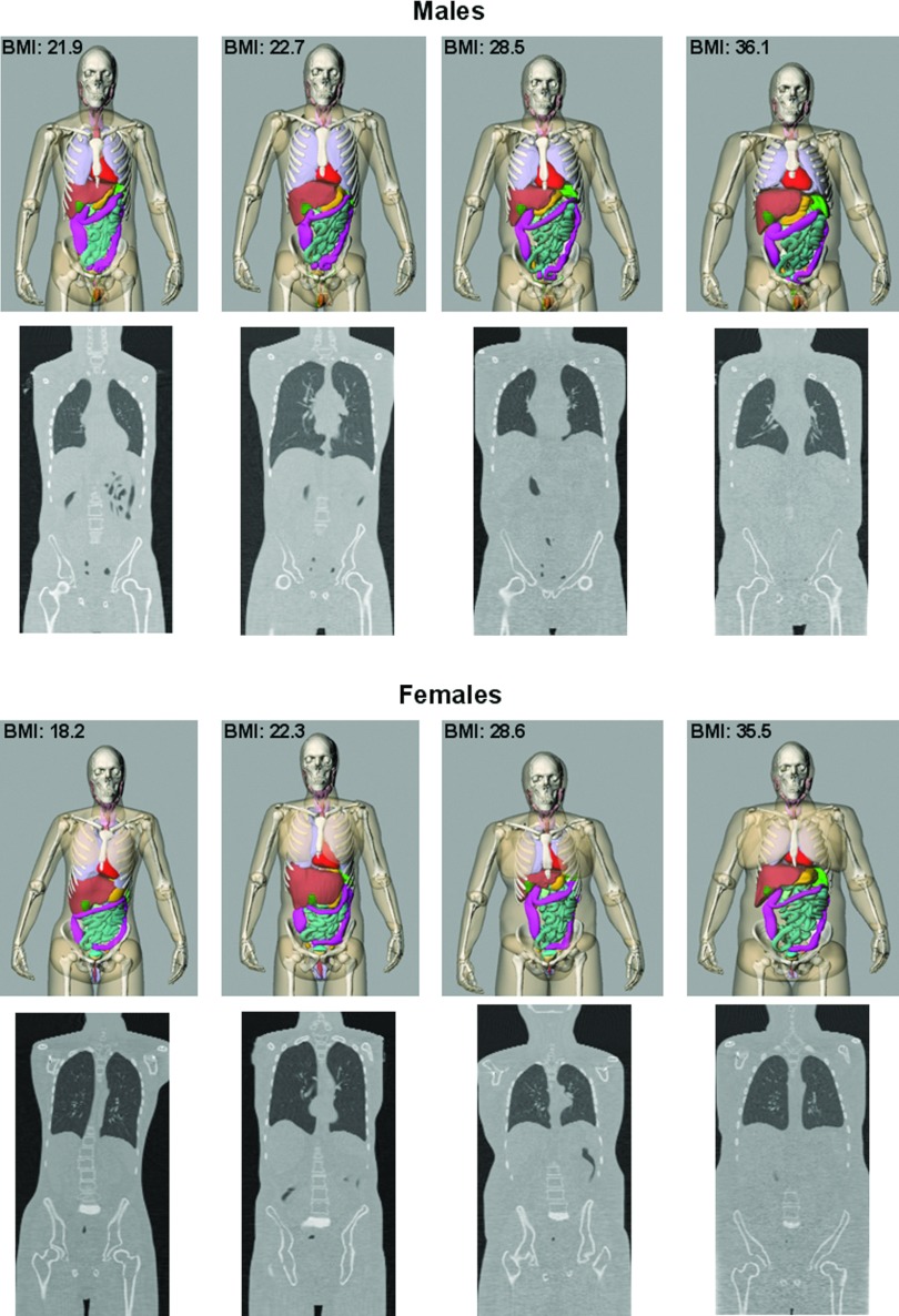
Results: Using these methods, the authors created a series of computerized phantoms with thousands of anatomical structures and modeling cardiac and respiratory motions. The database consists of 58 (35 male and 23 female) anatomically variable phantoms in total. Like the original XCAT, these phantoms can be combined with existing simulation packages to simulate realistic imaging data. Each new phantom contains parameterized models for the anatomy and the cardiac and respiratory motions and can, therefore, serve as a jumping point from which to create an unlimited number of 3D and 4D variations for imaging research.
Conclusions: A population of phantoms that includes a range of anatomical variations representative of the public at large is needed to more closely mimic a clinical study or trial. The series of anatomically variable phantoms developed in this work provide a valuable resource for investigating 3D and 4D imaging devices and the effects of anatomy and motion in imaging. Combined with Monte Carlo simulation programs, the phantoms also provide a valuable tool to investigate patient-specific dose and image quality, and optimization for adults undergoing imaging procedures.
Figures


References
-
- Zankl M., Panzer W., Petoussihenss N., and Drexler G., “Organ doses for children from computed tomographic examinations,” Radiat. Prot. Dosim. 57(1–4), 393–396 (1995).
-
- Caon M., Bibbo G., and Pattison J., “Monte Carlo calculated effective dose to teenage girls from computed tomography examinations,” Radiat. Prot. Dosim. 90(4), 445–448 (2000). 10.1093/oxfordjournals.rpd.a033172 - DOI
-
- Karabulut N., Toru M., Gelebek V., Gulsun M., and Ariyurek O. M., “Comparison of low-dose and standard-dose helical CT in the evaluation of pulmonary nodules,” Eur. Radiol. 12(11), 2764–2769 (2002). - PubMed
Publication types
MeSH terms
Grants and funding
LinkOut - more resources
Full Text Sources
Other Literature Sources
