. 2013 Apr 23;52(16):2821-7.
doi: 10.1021/bi400212m. Epub 2013 Apr 15.

The insoluble TGFBIp fraction of the cornea is covalently linked via a disulfide bond to type XII collagen

Affiliations

The insoluble TGFBIp fraction of the cornea is covalently linked via a disulfide bond to type XII collagen

Kasper Runager et al. Biochemistry. .

Abstract

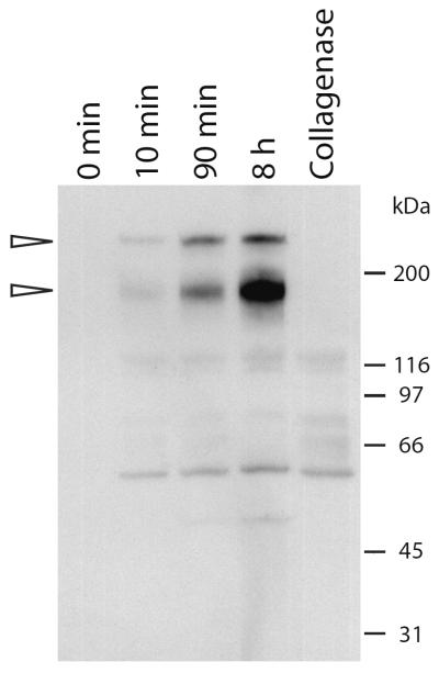
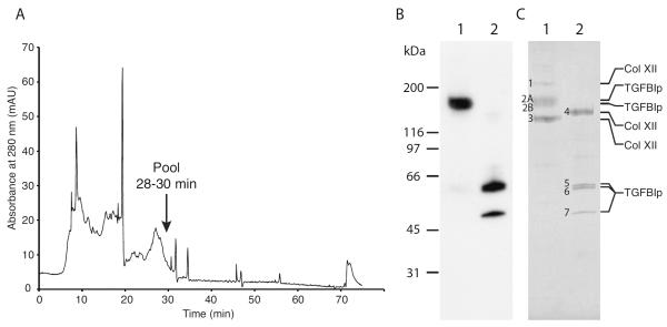
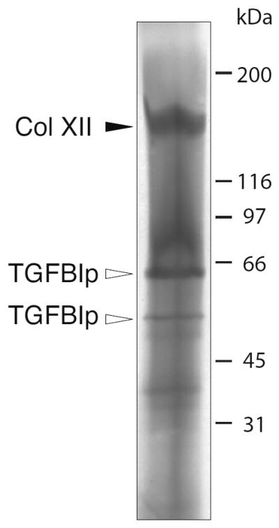
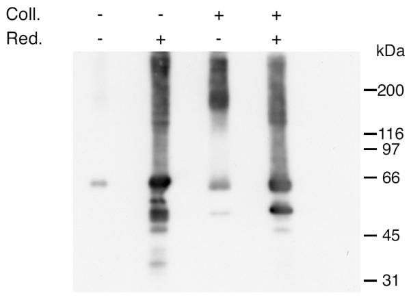
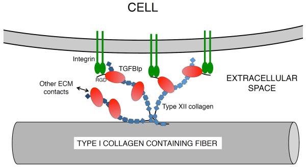
TGFBIp, also known as keratoepithelin and βig-h3, is among the most abundant proteins in the human cornea, and approximately 60% is associated with the insoluble fraction following extraction in sodium dodecyl sulfate (SDS) sample buffer. TGFBIp is of particular interest because a wide range of mutations causes amyloid or fuchsinophilic crystalloid deposits in the cornea leading to visual impairment. We show that the SDS-insoluble fraction of TGFBIp from porcine and human corneas is covalently linked via a reducible bond to the NC3 domain of type XII collagen in a TGFBIp:type XII collagen stoichiometric ratio of 2:1. Because type XII collagen is anchored to striated collagen fibers of the extracellular matrix, its interaction with TGFBIp is likely to provide anchoring for cells to the extracellular matrix through the integrin binding capability of TGFBIp. Furthermore, the TGFBIp-type XII collagen molecule will affect our understanding of the molecular pathogenesis of the TGFBI-linked corneal dystrophies.


Figures

Figure 1
Figure 1. Collagenase I-digestion releases a 170-kDa TGFBIp-containing complex from the SDS-insoluble fraction of porcine cornea
Porcine cornea powder was washed extensively and digested with collagenase I for different periods of time at 37°C. The supernatant from the first wash (0 min), and supernatants after digestion for 10 min, 90 min, and 8 hours were analyzed by anti-TGFBIp immunoblotting following non-reducing SDS-PAGE. Collagenase auto digest (no cornea powder) was loaded in the final lane (Collagenase). Molecular weights are indicated on the right. The immunoblot shows that there is a time-dependent release of a major 170-kDa and a minor 210-kDa TGFBIp-containing molecule when corneal tissue is incubated with collagenase I.
Figure 2
Figure 2. Purification of the high-molecular-weight TGFBIp-containing complex from porcine cornea
(A) Anion exchange chromatogram from the final step of purification of the high-molecular-weight TGFBIp-containing complex. Collagenase I-digested cornea was initially applied to a heparin affinity column from which all bound protein was dialyzed and applied to an anion exchange column. Fractions 28–30 were pooled and further analyzed. (B) Immunoblot of non-reducing (lane 1) and reducing (lane 2) SDS-PAGE of the pooled fractions containing the 170-kDa TGFBIp-containing complex. (C) Coomassie-stained SDS-PAGE gel of non-reduced (lane 1) and reduced (lane 2) samples of the purified 170-kDa TGFBIp-containing complex. Protein bands that were analyzed by MALDI-MS and N-terminal sequencing are indicated with numbers 1 to 8 and the identity based on MS-analysis is shown on the right. The characterization of the TGFBIp-containing complex shows that non-covalently bound type XII collagen molecules migrating at 210 kDa and 138 kDa co-purifies with the covalent and reducible 170-kDa TGFBIp:collagen XII complex.
Figure 3
Figure 3. Electro-elution of the 170-kDa TGFBIp-containing band
The 170-kDa TGFBIp-containing band observed by SDS-PAGE under non-reducing conditions was electro-eluted and analyzed by reducing SDS-PAGE. MS analysis of the resulting marked bands show that the 170-kDa TGFBIp-containing complex contains one isoform of type XII collagen of 151 kDa (solid arrowhead marked Col XII) and TGFBIp isoforms migrating at 62 kDa and 50 kDa (open arrowheads marked TGFBIp).
Figure 4
Figure 4. Collagenase I digestion releases a 170-kDa TGFBIp-containing complex in human corneas
Dried human cornea was analyzed for the presence of high-molecular-weight TGFBIp-containing complexes before and after digestion with collagenase I. Collagenase digested (Coll.; +) and non-digested (Coll.; −) samples were analyzed by reducing (Red.; +) and non-reducing (Red.; −) conditions followed by immunoblotting to detect TGFBIp-containing bands. The immunoblot shows that in the non-digested cornea only little TGFBIp is visible on the gel without reducing agent present compared to the massive band observed in the reducing lane. When the collagenase I-digested sample is analyzed a TGFBIp-containing band of 170 kDa is observed in the non-reducing lane as seen for the porcine tissue (Figures 1 and 2). Upon reduction with DTT the most prominent signal migrates as a TGFBIp monomer at 66 kDa. Collectively, these data suggest that TGFBIp in the human cornea is also covalently linked to type XII collagen through a disulfide bridge.
Figure 5
Figure 5. Schematic illustration of type XII collagen, TGFBIp, and the species purified from porcine cornea
Human type XII collagen exists as a long and a short splice-variant. Porcine type XII collagen (accession no. XP_001927071) has not been characterized at the protein level at this point, but the sequence of the predicted protein is more than 90% identical to that of human type XII collagen. The fragment of porcine type XII collagen purified in the present study (protein band 4 (151 kDa) in Figure 2) is shown. The C-terminal part of this fragment (indicated with dashed lines) was not identified by MALDI-MS but is likely to be present due to the size of the band in SDS-PAGE (151 kDa).
Figure 6
Figure 6. Model for the proposed biological function of the covalent TGFBIp-type XII collagen interaction in the cornea
Type XII collagen has been shown to interact noncovalently with type I collagen fibers of the corneal ECM. The non-collagenous domains of type XII collagen trimer extents into the extracellular space and binds TGFBIp. TGFBIp is consequently able to interact with other macromolecules of the ECM. The immobilized TGFBIp may function as an ECM attachment anchor for the cells of the cornea.

References

    1. Kitahama S, Gibson MA, Hatzinikolas G, Hay S, Kuliwaba JL, Evdokiou A, Atkins GJ, Findlay DM. Expression of fibrillins and other microfibril-associated proteins in human bone and osteoblast-like cells. Bone. 2000;27:61–67. - PubMed
    1. Ferguson JW, Mikesh MF, Wheeler EF, LeBaron RG. Developmental expression patterns of Beta-ig (βIG-H3) and its function as a cell adhesion protein. Mechanisms of development. 2003;120:851–864. - PubMed
    1. Ohno S, Doi T, Tsutsumi S, Okada Y, Yoneno K, Kato Y, Tanne K. RGD-CAP (βig-h3) is expressed in precartilage condensation and in prehypertrophic chondrocytes during cartilage development. Biochimica et biophysica acta. 2002;1572:114–122. - PubMed
    1. Carson DD, Lagow E, Thathiah A, Al-Shami R, Farach-Carson MC, Vernon M, Yuan L, Fritz MA, Lessey B. Changes in gene expression during the early to mid-luteal (receptive phase) transition in human endometrium detected by high-density microarray screening. Molecular human reproduction. 2002;8:871–879. - PubMed
    1. Sciandra F, Morlacchi S, Allamand V, De Benedetti G, Macchia G, Petrucci TC, Bozzi M, Brancaccio A. First molecular characterization and immunolocalization of keratoepithelin in adult human skeletal muscle. Matrix biology : journal of the International Society for Matrix Biology. 2008;27:360–370. - PubMed

Publication types

MeSH terms

LinkOut - more resources