

Disruption of AP3B1 by a chromosome 5 inversion: a new disease mechanism in Hermansky-Pudlak syndrome type 2
- PMID: 23557002
- PMCID: PMC3663694
- DOI: 10.1186/1471-2350-14-42
Disruption of AP3B1 by a chromosome 5 inversion: a new disease mechanism in Hermansky-Pudlak syndrome type 2
Abstract
Background: Hermansky-Pudlak syndrome 2 (HPS2; OMIM #608233) is a rare, autosomal recessive disorder caused by loss-of-function genetic variations affecting AP3B1, which encodes the β3A subunit of the adaptor-related protein complex 3 (AP3). Phenotypic characteristics include reduced pigmentation, absent platelet dense granule secretion, neutropenia and reduced cytotoxic T lymphocyte (CTL) and natural killer (NK) cell function. To date HPS2 has been associated with non-synonymous, stop-gain or deletion-insertion nucleotide variations within the coding region of AP3B1.
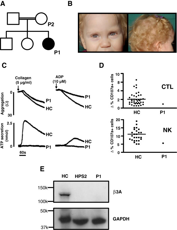
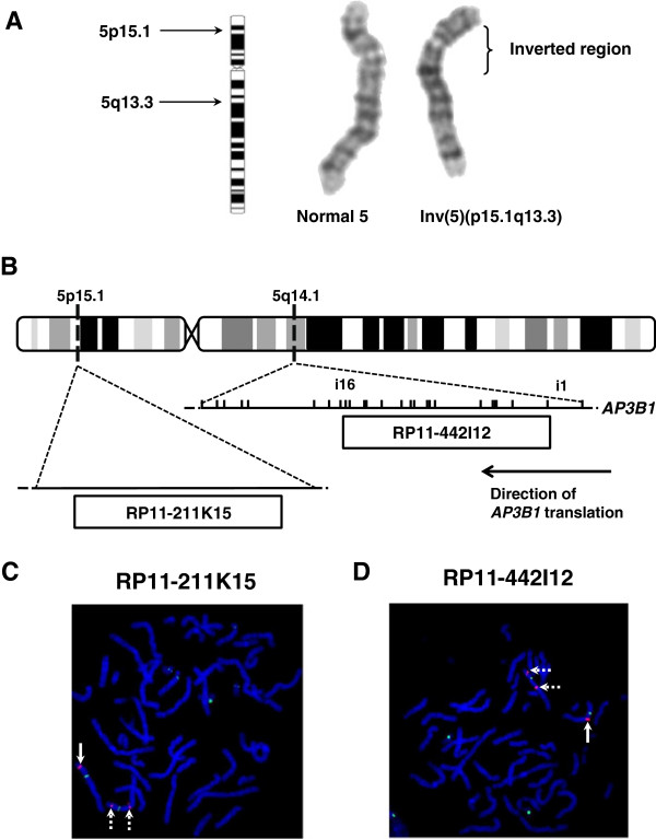
Case presentation: We describe a consanguineous female infant with reduced pigmentation, neutropenia and recurrent infections. Platelets displayed reduced aggregation and absent ATP secretion in response to collagen and ADP, indicating a platelet dense granule defect. There was increased basal surface expression of CD107a (lysosome-associated membrane protein 1(LAMP-1)) on NK cells and CTLs from the study subject and a smaller increase in the percentage of CD107a positive cells after stimulation compared to most healthy controls. Immunoblotting of protein extracts from EBV-transformed lymphoblasts from the index case showed absent expression of full-length AP-3 β3A subunit protein, confirming a phenotypic diagnosis of HPS2.The index case displayed a homozygous pericentric inv(5)(p15.1q14.1), which was also detected as a heterozygous defect in both parents of the index case. No loss of genetic material was demonstrated by microarray comparative genome hybridisation at 60kb resolution. Fluorescence in-situ hybridisation using the 189.6kb probe RP11-422I12, which maps to 5q14.1, demonstrated dual hybridisation to both 5q14.1 and 5p15.1 regions of the inverted Chr5. The RP11-422I12 probe maps from intron 1 to intron 16 of AP3B1, thus localising the 5q inversion breakpoint to within AP3B1. The probe RP11-211K15, which corresponds to an intergenic region on 5p also showed dual hybridisation, enabling localisation of the 5p inversion breakpoint.
Conclusion: This case report extends the phenotypic description of the very rare disorder HPS2. Our demonstration of a homozygous Chr5 inversion predicted to disrupt AP3B1 gene provides a novel pathogenic mechanism for this disorder.
Figures
References
-
- Wei AH, Li W. Hermansky-Pudlak syndrome: pigmentary and non-pigmentary defects and their pathogenesis. Pigment Cell Melanoma Res. 2012;26:176–192. - PubMed
-
- Chiang PW, Oiso N, Gautam R, Suzuki T, Swank RT, Spritz RA. The Hermansky-Pudlak syndrome 1 (HPS1) and HPS4 proteins are components of two complexes, BLOC-3 and BLOC-4, involved in the biogenesis of lysosome-related organelles. J Biol Chem. 2003;278(22):20332–20337. doi: 10.1074/jbc.M300090200. - DOI - PubMed
Publication types
MeSH terms
Substances
Supplementary concepts
LinkOut - more resources
Full Text Sources
Other Literature Sources
Research Materials
Miscellaneous
