. 2013 Apr 4:13:179.
doi: 10.1186/1471-2407-13-179.

Radiation mitigating properties of the lignan component in flaxseed

Affiliations

Radiation mitigating properties of the lignan component in flaxseed

Ralph Pietrofesa et al. BMC Cancer. .

Abstract

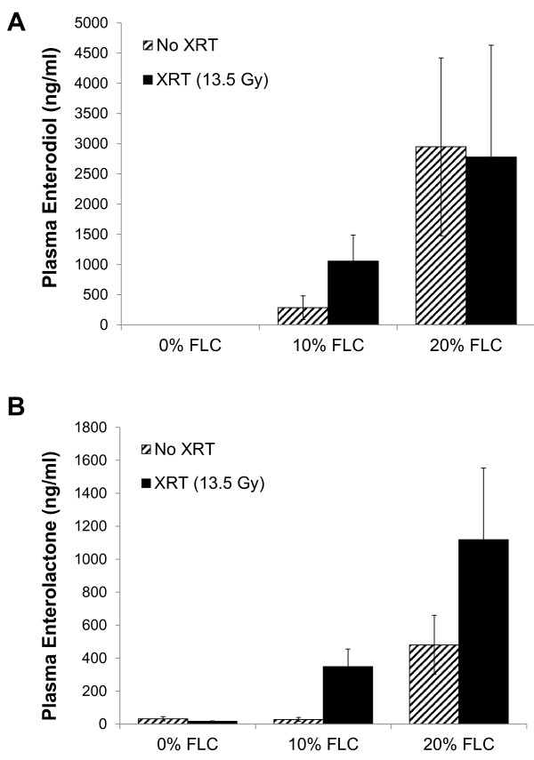
Background: Wholegrain flaxseed (FS), and its lignan component (FLC) consisting mainly of secoisolariciresinol diglucoside (SDG), have potent lung radioprotective properties while not abrogating the efficacy of radiotherapy. However, while the whole grain was recently shown to also have potent mitigating properties in a thoracic radiation pneumonopathy model, the bioactive component in the grain responsible for the mitigation of lung damage was never identified. Lungs may be exposed to radiation therapeutically for thoracic malignancies or incidentally following detonation of a radiological dispersion device. This could potentially lead to pulmonary inflammation, oxidative tissue injury, and fibrosis. This study aimed to evaluate the radiation mitigating effects of FLC in a mouse model of radiation pneumonopathy.

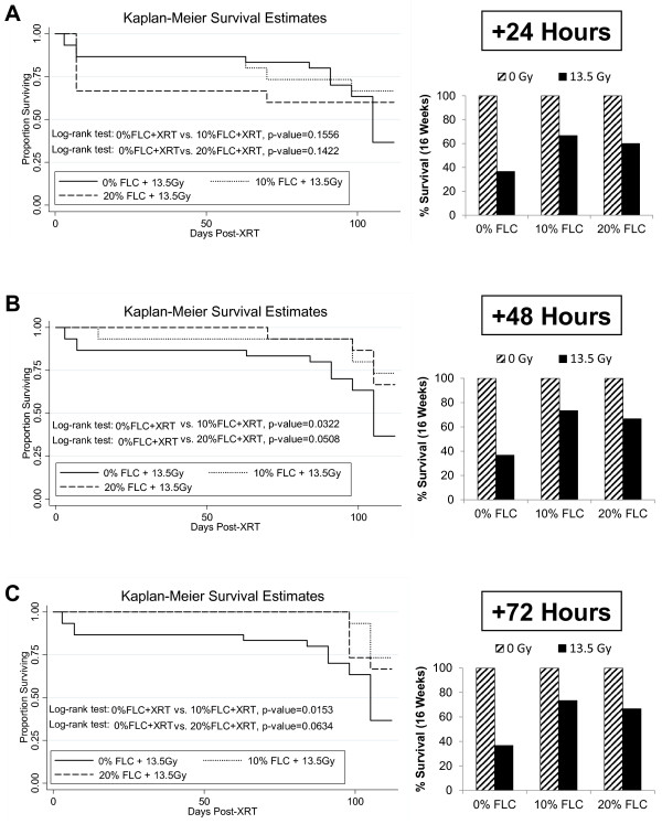
Methods: We evaluated FLC-supplemented diets containing SDG lignan levels comparable to those in 10% and 20% whole grain diets. 10% or 20% FLC diets as compared to an isocaloric control diet (0% FLC) were given to mice (C57/BL6) (n=15-30 mice/group) at 24, 48, or 72-hours after single-dose (13.5 Gy) thoracic x-ray treatment (XRT). Mice were evaluated 4 months post-XRT for blood oxygenation, lung inflammation, fibrosis, cytokine and oxidative damage levels, and survival.

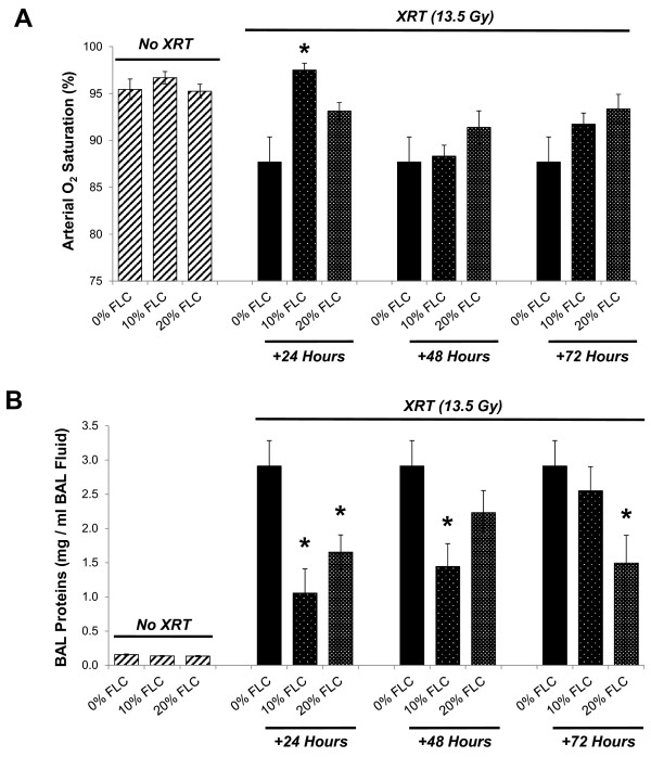
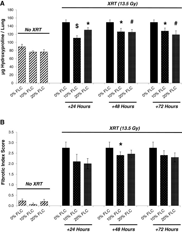
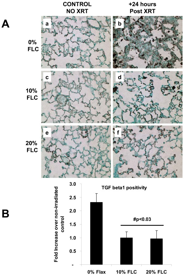
Results: FLC significantly mitigated radiation-related animal death. Specifically, mice fed 0% FLC demonstrated 36.7% survival 4 months post-XRT compared to 60-73.3% survival in mice fed 10%-20% FLC initiated 24-72 hours post-XRT. FLC also mitigated radiation-induced lung fibrosis whereby 10% FLC initiated 24-hours post-XRT significantly decreased fibrosis as compared to mice fed control diet while the corresponding TGF-beta1 levels detected immunohistochemically were also decreased. Additionally, 10-20% FLC initiated at any time point post radiation exposure, mitigated radiation-induced lung injury evidenced by decreased bronchoalveolar lavage (BAL) protein and inflammatory cytokine/chemokine release at 16 weeks post-XRT. Importantly, neutrophilic and overall inflammatory cell infiltrate in airways and levels of nitrotyrosine and malondialdehyde (protein and lipid oxidation, respectively) were also mitigated by the lignan diet.

Conclusions: Dietary FLC given early post-XRT mitigated radiation effects by decreasing inflammation, lung injury and eventual fibrosis while improving survival. FLC may be a useful agent, mitigating adverse effects of radiation in individuals exposed to incidental radiation, inhaled radioisotopes or even after the initiation of radiation therapy to treat malignancy.


Figures

Figure 1
Figure 1
Experimental plan of animal feeding protocol and radiation exposure. Mice were pre-fed 0% FLC for 72 hours prior to single fraction thoracic X-ray radiation therapy (13.5 Gy). Following XRT exposure, mouse cohorts (n=15) were fed 10% FLC or 20% FLC diets initiated 24, 48, or 72 hours post-XRT. Control-fed mouse cohorts remained on 0% FLC diet throughout the course of the study. Mice were sacrificed at 16 weeks post-XRT.
Figure 2
Figure 2
Detection of flaxseed lignan metabolites enterodiol (ED) and enterolactone (EL) in plasma of mice fed FLC diets. Circulating mammalian lignans, Panel A: enterodiol (ED) and Panel B: enterolactone (EL) levels in plasma of mice were determined using GC/MS/MS. Mouse cohorts were initiated on the control 0% FLC diet 3 days prior to XRT exposure. At 24 hours post-XRT 10% FLC and 20% FLC diets were initiated. Control-fed mouse cohorts remained on 0% FLC diet throughout the course of the study. Mouse cohorts were sacrificed at 16 weeks post-XRT and plasma was collected. Data is represented as mean ± SEM (n=5 mice / group). No statistical significance was found between XRT and no XRT.
Figure 3
Figure 3
Irradiation of mouse thorax using the SARRP. Panel A: Whole thorax irradiation jig and beam arrangement. Panel B: Radial arrangement of mice on platform. Panel C: Lead shielding of head area and upper extremities. Panel D: Lateral radiograph of mouse on the SARRP platform using thoracic irradiation jig. Mice were irradiated using the SARRP, to deliver single fraction 13.5 Gy X-ray irradiation to the thorax. Shielding was provided for the head only as the highly collimated field edge already limits dose to the abdomen/pelvis. The red shaded area represents the radiation field.
Figure 4
Figure 4
Effect of FLC diets on the survival of mice through 16 weeks post-XRT. Kaplan-Meier curves for overall survival. Mice were pre-fed 0% FLC for 72 hours prior to single fraction thoracic X-ray radiation therapy (13.5 Gy). Following XRT exposure, mouse cohorts (n=15) were fed 10% FLC or 20% FLC diets initiated Panel A: 24, Panel B: 48 or Panel C: 72 hours post-XRT and survival was observed up to 16 weeks post-XRT. Control-fed mouse cohorts remained on 0% FLC diet throughout the course of the study. No mice were lost in the non-irradiated cohorts (100% survival-not shown). Log-rank p-values (shown in figure) were calculated by log-rank test between irradiated mouse cohorts. Overall survival at 16 weeks post-XRT is depicted in the bar graph.
Figure 5
Figure 5
Evaluation of blood oxygenation levels and lung injury in mice 16 weeks post-XRT. Mice were pre-fed 0% FLC for 72 hours prior to single fraction thoracic X-ray radiation therapy (13.5 Gy). Following XRT exposure, mouse cohorts (n=15) were fed 10% FLC or 20% FLC diets initiated 24, 48, or 72 hours post-XRT. Control-fed mouse cohorts remained on 0% FLC diet throughout the course of the study. Mice were sacrificed at 16 weeks post-XRT. Panel A: Pulse oximetry analysis was performed prior to sacrifice at 16 weeks post-XRT. Data is represented as mean ± SEM. *p< 0.01 for irradiated 0% FLC vs. irradiated 10% FLC (+24 Hours). Panel B: BAL protein levels were determined at 16 weeks post-XRT. Data is represented as mean ± SEM. *p< 0.01 for irradiated 0% FLC vs. irradiated 10% and 20% FLC.
Figure 6
Figure 6
Oxidative and nitrosative stress in lung tissues 16 weeks post radiation exposure. Mice on control diet (0% FLC) were exposed to a single fraction thoracic X-ray radiation therapy (13.5 Gy). Following XRT exposure, mice were switched to 10% FLC or 20% FLC diets that were initiated 24 hours post radiation exposure while control-fed mouse cohorts remained on 0% FLC diet throughout the course of the study. Lungs were harvested at 16 weeks post-XRT, and BAL performed followed by paraffin embedding and immunostaining for nitrotyrosine; brown staining (Panel A). Representative lung sections are shown In Panels a-b: representing 0% FLC, Panels c-d: representing 10% FLC and Panels e-f: representing 20% FLC. Sections were counterstained with methyl green. (Magnification 400X). Levels of thiobarbituric acid reactive substances (TBARS) in BAL samples were determined at 16 weeks post-XRT (Panel B). Data is represented as mean ± SEM. # p< 0.05 for 0% FLC vs. irradiated 0% FLC; *p< 0.01 for irradiated 0% FLC vs. irradiated 10% and 20% FLC.
Figure 7
Figure 7
Evaluation of lung inflammation in mice at 16 weeks post-XRT. Mice were pre-fed 0% FLC for 72 hours prior to single fraction thoracic X-ray radiation therapy (13.5 Gy). Following XRT exposure, mouse cohorts (n=15) were fed 10% FLC or 20% FLC diets initiated 24, 48, or 72 hours post-XRT. Control-fed mouse cohorts remained on 0% FLC diet throughout the course of the study. Mice were sacrificed at 16 weeks post-XRT and bronchoalveolar lavage (BAL) fluid was collected. Panel A: Total WBC counts in BAL fluid. Data is represented as mean ± SEM. *p≤ 0.01 for irradiated 0% FLC vs. Irradiated 10% FLC. Panel B: Total PMN cells in bronchoalveolar lavage (BAL). Data is represented as mean ± SEM. *p< 0.05 for irradiated 0% FLC vs. irradiated 10% and 20% FLC (+48 hours).
Figure 8
Figure 8
Histological evaluation of lung fibrosis at 16 weeks post -XRT. Mice on control diet (0% FLC) were exposed to a single fraction thoracic X-ray radiation therapy (13.5 Gy). Following XRT exposure, mouse cohorts (n=15) were switched to 10% FLC or 20% FLC diets that were initiated 24, 48, or 72 hours post radiation exposure while control-fed mouse cohorts remained on 0% FLC diet throughout the course of the study. Lungs were harvested at 16 weeks post-XRT and processed for Trichrome staining to evaluate collagen deposition and fibrosis. Representative lung sections stained with Trichrome are shown on Panels A-C: representing 0% FLC, Panels D-F: representing 10% FLC and Panels G-I: representing 20% FLC. (Magnification 400X).
Figure 9
Figure 9
Determination of fibrotic changes in murine lungs at 16 weeks post-XRT. Mice were pre-fed 0% FLC for 72 hours prior to single fraction thoracic X-ray radiation therapy (13.5 Gy). Following XRT exposure, mouse cohorts (n=15) were fed 10% FLC or 20% FLC diets initiated 24, 48, or 72 hours post-XRT. Control-fed mouse cohorts remained on 0% FLC diet throughout the course of the study. Lungs were harvested at 16 weeks post-XRT. Panel A: Evaluation of hydroxyproline content in lungs. Data is represented as mean ± SEM. *p< 0.05 for irradiated 0% FLC vs. irradiated 10% FLC and 20% FLC, #p≤ 0.01 for irradiated 0% FLC vs. irradiated 20% FLC (+48 and +72 hours), $p≤ 0.001 for irradiated 0% FLC vs. irradiated 10% FLC (+24 hours). Panel B: Fibrotic Index (range=0-4) scoring. Data is represented as mean ± SEM. *p< 0.05 for irradiated 0% FLC vs. 10% FLC (+48 hours).
Figure 10
Figure 10
Immunohistochemical detection and quantification of TGF-beta1 in lung at 16 weeks post –XRT. Mice on control diet (0% FLC) were exposed to a single fraction thoracic X-ray radiation therapy (13.5 Gy). Following XRT exposure, mice were switched to 10% FLC or 20% FLC diets that were initiated 24 hours post radiation exposure while control-fed mouse cohorts remained on 0% FLC diet throughout the course of the study. Lungs were harvested at 16 weeks post-XRT, paraffin embedded and immunostained for TGF beta1; brown staining (Panel A). Representative lung sections are shown In Panels a-b: representing 0% FLC, Panels c-d: representing 10% FLC and Panels e-f: representing 20% FLC. Sections were counterstained with methyl green. (Magnification 400X). Quantification of TGF-beta1 positivity (Panel B) was made using the Aperio image analysis system. #p<0.03 as compared to irradiated 0% FLC.

References

    1. Brenner DJ, Hall EJ. Computed tomography–an increasing source of radiation exposure. N Engl J Med. 2007;357(22):2277–2284. doi: 10.1056/NEJMra072149. - DOI - PubMed
    1. Vogel H. Rays as weapons. Eur J Radiol. 2007;63(2):167–177. doi: 10.1016/j.ejrad.2007.04.028. - DOI - PubMed
    1. Chin FK. Scenario of a dirty bomb in an urban environment and acute management of radiation poisoning and injuries. Singapore Med J. 2007;48(10):950–957. - PubMed
    1. Dombroski MJ, Fischbeck PS. An integrated physical dispersion and behavioral response model for risk assessment of radiological dispersion device (RDD) events. Risk Anal. 2006;26(2):501–514. doi: 10.1111/j.1539-6924.2006.00742.x. - DOI - PubMed
    1. Moulder JE. Post-irradiation approaches to treatment of radiation injuries in the context of radiological terrorism and radiation accidents: a review. Int J Radiat Biol. 2004;80(1):3–10. doi: 10.1080/09553000310001642920. - DOI - PubMed

Publication types

MeSH terms