. 2013 Apr 4:10:11.
doi: 10.1186/1743-8977-10-11.

Exposure of silver-nanoparticles and silver-ions to lung cells in vitro at the air-liquid interface

Exposure of silver-nanoparticles and silver-ions to lung cells in vitro at the air-liquid interface

Fabian Herzog et al. Part Fibre Toxicol. .

Abstract

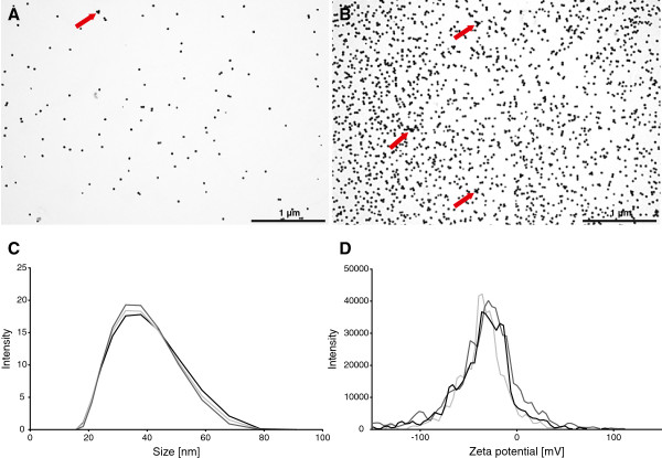
Background: Due to its antibacterial properties, silver (Ag) has been used in more consumer products than any other nanomaterial so far. Despite the promising advantages posed by using Ag-nanoparticles (NPs), their interaction with mammalian systems is currently not fully understood. An exposure route via inhalation is of primary concern for humans in an occupational setting. Aim of this study was therefore to investigate the potential adverse effects of aerosolised Ag-NPs using a human epithelial airway barrier model composed of A549, monocyte derived macrophage and dendritic cells cultured in vitro at the air-liquid interface. Cell cultures were exposed to 20 nm citrate-coated Ag-NPs with a deposition of 30 and 278 ng/cm2 respectively and incubated for 4 h and 24 h. To elucidate whether any effects of Ag-NPs are due to ionic effects, Ag-Nitrate (AgNO3) solutions were aerosolised at the same molecular mass concentrations.

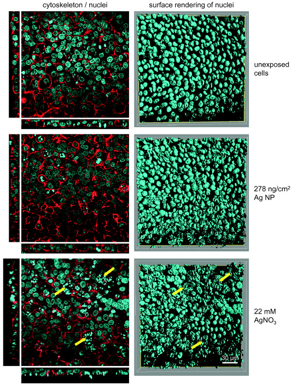
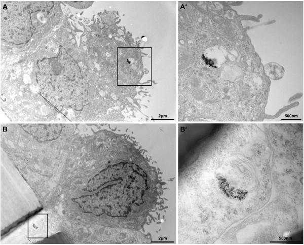
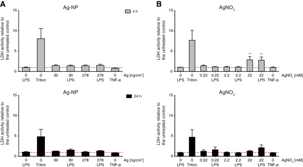
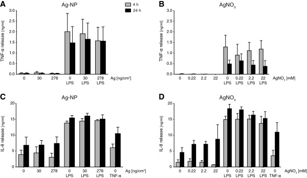
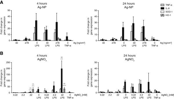
Results: Agglomerates of Ag-NPs were detected at 24 h post exposure in vesicular structures inside cells but the cellular integrity was not impaired upon Ag-NP exposures. Minimal cytotoxicity, by measuring the release of lactate dehydrogenase, could only be detected following a higher concentrated AgNO3-solution. A release of pro-inflammatory markers TNF-α and IL-8 was neither observed upon Ag-NP and AgNO3 exposures as well as was not affected when cells were pre-stimulated with lipopolysaccharide (LPS). Also, an induction of mRNA expression of TNF-α and IL-8, could only be observed for the highest AgNO3 concentration alone or even significantly increased when pre-stimulated with LPS after 4 h. However, this effect disappeared after 24 h. Furthermore, oxidative stress markers (HMOX-1, SOD-1) were expressed after 4 h in a concentration dependent manner following AgNO3 exposures only.

Conclusions: With an experimental setup reflecting physiological exposure conditions in the human lung more realistic, the present study indicates that Ag-NPs do not cause adverse effects and cells were only sensitive to high Ag-ion concentrations. Chronic exposure scenarios however, are needed to reveal further insight into the fate of Ag-NPs after deposition and cell interactions.


Figures

Figure 1
Figure 1
Particle deposition in the ALICE, size characterization and stability. Transmission electron microscopy (TEM) pictures of 1x (A) and 10× (B) concentrated 20 nm Ag NPs deposited onto TEM grids by nebulization with the ALICE system (scale bar = 1 μm). Agglomerates are indicated with red arrows. The size distribution (C) was measured by dynamic light scattering and showed an average hydrodynamic diameter of 33.4 ± 0.23 nm. The average zeta potential (D) was defined at -37.5 ± 0.25 mV.
Figure 2
Figure 2
Cell morphology of exposed cells. Illustrated pictures represent unexposed, 278 ng/cm2 Ag-NP and 22 mM AgNO3 exposed triple cell co-cultures. At 24 h after exposure, the cells were fixed and stained for the actin cytoskeleton (phalloidin rhodamine; red) and DNA (DAPI; blue). Examples of morphological changes in form of augmented DNA condensation and alterations in the cytoskeleton were marked with yellow arrows. Images for the actin cytoskeleton and nuclei on the left side are represented as single optical xy projections with representative side views in xz (bottom) and yz (left) direction. Nuclei on the right side are visualized by surface rendering of xy stacks (scale bar = 30 μm).
Figure 3
Figure 3
Particle uptake in the upper transwell cell layer. Ag-NPs were found in the upper cell layer of the transwell membrane (A) and in cells crossing the transwell insert (B) as aggregates inside vesicles at 24 h post-exposure (scale bar = 2 μm). Overall cell morphology upon Ag-NP exposure was similar to untreated control. A’ and B’ represent a higher magnification of the black marked box of the opposing picture (scale bar = 500 nm). B’ reveals particle agglomerates inside a multilamellar body.
Figure 4
Figure 4
Cytotoxicity upon Ag-NP and AgNO3 exposure. Cell integrity as estimated by quantification of extracellular LDH release relative to the unexposed and untreated control (red dashed line) was measured 4 h (grey bars) and 24 h (black bars) after exposure. Cells were exposed to 30 ng/cm2 and 270 ng/cm2 Ag-NP (A) and equal concentrations of 0.22 mM and 2.2 mM as well as 22 mM AgNO3 (B). As positive control (Triton) cells were treated with Triton X-100 for 4 h and 24 h. LDH release was also measured for cells treated with TNF-α. Error bars represent the standard error of the mean (SEM) for at least 3 independent experiments. A two-way analysis of variance (ANOVA) with a subsequent Bonferroni post-hoc test was performed. Values were considered significantly different compared to the unexposed and untreated control with p < 0.01 (**).
Figure 5
Figure 5
Protein secretion of pro-inflammatory cytokines TNF-α and IL-8. The extracellular release of pro-inflammatory markers (ng/mL) TNF-α (A and B) and IL-8 (C and D) were analysed by ELISA 4 h (grey bars) and 24 h (black bars) after exposure. Error bars represent the SEM for at least 3 independent experiments.
Figure 6
Figure 6
Quantitative gene expression of pro-inflammatory and oxidative stress markers. Exposed cells were harvested 4 h and 24 h after exposure and mRNA levels of pro-inflammatory markers TNF-α (grey) and IL-8 (black) as well as oxidative stress markers SOD-1 (white) and HMOX-1 (striated) were analysed by real-time RT-PCR. Fold changes of gene expression compared to unexposed untreated controls were calculated with 2-ΔΔCt. Error bars represent the SEM for at least 3 independent experiments. A two-way ANOVA with a subsequent Bonferroni post-hoc test was performed. Values were considered significantly different compared to unexposed untreated control (1) and unexposed LPS treated control (2) with p < 0.001 (***) or p < 0.0001 (****).

References

    1. The European Commission. Commission Recommendation of 18 October 2011 on the definition of nanomaterial (2011/696/EU) Brussels, Official Journal of the European Union. 2011;L 275/38
    1. Luxresearch Inc. Nanomaterials State of the Market Q3 2008: stealth success, broad impact. St Market Rep. 2008. https://portal.luxresearchinc.com/research/document_excerpt/3735.
    1. Beer C, Foldbjerg R, Hayashi Y, Sutherland DS, Autrup H. Toxicity of silver nanoparticles - nanoparticle or silver ion? Toxicol Lett. 2012;208:286–292. doi: 10.1016/j.toxlet.2011.11.002. - DOI - PubMed
    1. Edwards-Jones V. The benefits of silver in hygiene, personal care and healthcare. Lett Appl Microbiol. 2009;49:147–152. doi: 10.1111/j.1472-765X.2009.02648.x. - DOI - PubMed
    1. Morones JR, Elechiguerra JL, Camacho A, Holt K, Kouri JB, Ramirez JT, Yacaman MJ. The bactericidal effect of silver nanoparticles. Nanotechnology. 2005;16:2346. doi: 10.1088/0957-4484/16/10/059. - DOI - PubMed

Publication types

MeSH terms