. 2013 Jul-Aug;27(4):307-15.
doi: 10.1016/j.jdiacomp.2013.02.009. Epub 2013 Apr 2.

Arterial insulin resistance in Yucatan micropigs with diet-induced obesity and metabolic syndrome

Affiliations

Arterial insulin resistance in Yucatan micropigs with diet-induced obesity and metabolic syndrome

Cecilia C Low Wang et al. J Diabetes Complications. 2013 Jul-Aug.

Abstract

Aim: Metabolic syndrome affects a large proportion of the population and increases cardiovascular disease risk. Because metabolic syndrome often co-exists clinically with atherosclerosis, it is difficult to distinguish the respective contributions of the components to vascular abnormalities. Accordingly, we utilized a porcine dietary model of metabolic syndrome without atherosclerosis to investigate early abnormalities of vascular function and signaling.

Methods: Thirty-two Yucatan micropigs were fed either a high-fat, high-simple-sugar, high-calorie (HFHS) or standard chow diet (STD) for 6 months. Neither diet contained added cholesterol. Blood pressure and flow-mediated vasodilatation were assessed at baseline and 6 months. Aortas were harvested at 6 months to assess histology, insulin signaling, and endothelial nitric oxide (eNOS) phosphorylation.

Results: HFHS pigs developed characteristics of metabolic syndrome including obesity, dyslipidemia, and insulin resistance, but without histologic evidence of atherosclerosis. Although arterial intima-media thickness did not differ between groups, vascular dysfunction in HFHS was manifest by increased blood pressure and impaired flow-mediated vasodilation of the femoral artery. Compared with STD, aortas from HFHS exhibited increased p85α expression and Ser307 IRS-1 phosphorylation, and blunted insulin-stimulated IRS-1-associated phosphatidylinositol (PI) 3-kinase activity. In the absence of insulin stimulation, aortic Akt Ser473-phosphorylation was greater in HFHS than in STD. With insulin stimulation, Akt phosphorylation increased in STD, but not HFHS. Insulin-induced Ser1177-phosphorylation of eNOS was decreased in HFHS, compared with STD.

Conclusions: Pigs with metabolic syndrome develop early vascular dysfunction and aortic insulin signaling abnormalities, and could be a useful model for early human vascular abnormalities in this condition.


Conflict of interest statement

Conflict of Interest Statement

The authors declare that they have no conflict of interest that could bias the studies described.

Figures

Figure 1
Figure 1
In vivo and histologic vascular studies. A Systolic blood pressure (SBP) was measured in all pigs at baseline and again after 6 months of assigned diet. SBP was similar between groups at baseline and did not change significantly over 6 months in the standard diet (STD) group. However, at 6 months, SBP in the high fat high simple sugar diet (HFHS) group was significantly greater than baseline and significantly greater than STD group at 6 months. Diet x time interaction was also significant (p<0.05). B Femoral artery flow-mediated vasodilatation (FMD) was measured in 8 pigs in each diet group at baseline and again after 6 months of assigned diet. Data are mean ± SE. FMD was similar in both groups at baseline and did not change over time in the STD diet group. However, at 6 months, FMD in the HFHS group was significantly decreased from baseline, and significantly lower than STD at 6 months. Diet x time interaction was also significant (p<0.05). *p<0.05). C Intima-media thickness as measured in the aorta was not different between STD (n=7) and HFHS (n=7) diet groups at 6 months. Horizontal lines and bars indicate mean and SE.
Figure 2
Figure 2
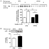
PI 3-kinase and p85alpha expression in aortic lysates. A Insulin-stimulated IRS-1-associated PI 3-kinase activity in the aorta was measured by thin layer chromatography in STD (n=12) and HFHS (n=16) diet groups. Top: representative thin layer chromatography blot. Bottom: Group data, mean ± SE. In the absence of insulin stimulation, PI 3-kinase activity was similar in both diet groups. After exposure to an insulin bolus in vivo, the STD diet group exhibited the expected stimulation of PI 3-kinase activity. In contrast, this response was blunted in the HFHS diet group. The interaction of diet and insulin on PI 3-kinase activity was significant (p<0.001). B Expression of the p85α (regulatory) subunit of PI 3-kinase in aortic wall. Data are mean ± SE with n=15 (HFHS diet group) and n=14 (STD diet group). Total p85α expression was higher in the HFHS diet group compared with STD group. *p<0.05, **p<0.001
Figure 3
Figure 3
Expression of Ser473 phospho-Akt, total Akt and Akt isoforms in aortic wall of STD (n=13) and HFHS (n=16) groups. A Ser473 phospho- and total non-isoform-specific Akt. In the absence of insulin stimulation, Ser473 phospho-Akt was greater in HFHS than STD. With in vivo insulin exposure, Ser473 phospho-Akt increased as expected in STD but did not increase further in HFHS. Total Akt content did not differ between diet groups, with or without in vivo exposure to insulin. Control was set at 100% for STD group in absence of insulin stimulation. Diet x insulin interaction was significant (p<0.001). Data are mean ± SE. **p<0.001. B Ser473 phospho-Akt1 and total Akt1, C Ser473 phospho-Akt2 and total Akt2, D Ser473 phospho-Akt3 and total Akt3. For each isoform, representative immunoblots are shown above group data for STD (n=13) and HFHS (n=16). There was a trend suggesting greater phosphorylation of Ser473 Akt1 in STD than in HFHS. Insulin had no significant effect on Akt1 phosphorylation in either group. There was a modest increase in Akt2 Ser473 phosphorylation in response to insulin, which approached statistical significance in STD (p=0.07). Ser473 phospho-Akt3 increased in response to insulin in STD (p<0.05) but not HFHS. There was a significant interaction of diet and insulin stimulation on phospho-Akt3 (p<0.05). The total content of each Akt isoform did not differ significantly between diet groups or in the absence or presence of insulin.
Figure 3
Figure 3
Expression of Ser473 phospho-Akt, total Akt and Akt isoforms in aortic wall of STD (n=13) and HFHS (n=16) groups. A Ser473 phospho- and total non-isoform-specific Akt. In the absence of insulin stimulation, Ser473 phospho-Akt was greater in HFHS than STD. With in vivo insulin exposure, Ser473 phospho-Akt increased as expected in STD but did not increase further in HFHS. Total Akt content did not differ between diet groups, with or without in vivo exposure to insulin. Control was set at 100% for STD group in absence of insulin stimulation. Diet x insulin interaction was significant (p<0.001). Data are mean ± SE. **p<0.001. B Ser473 phospho-Akt1 and total Akt1, C Ser473 phospho-Akt2 and total Akt2, D Ser473 phospho-Akt3 and total Akt3. For each isoform, representative immunoblots are shown above group data for STD (n=13) and HFHS (n=16). There was a trend suggesting greater phosphorylation of Ser473 Akt1 in STD than in HFHS. Insulin had no significant effect on Akt1 phosphorylation in either group. There was a modest increase in Akt2 Ser473 phosphorylation in response to insulin, which approached statistical significance in STD (p=0.07). Ser473 phospho-Akt3 increased in response to insulin in STD (p<0.05) but not HFHS. There was a significant interaction of diet and insulin stimulation on phospho-Akt3 (p<0.05). The total content of each Akt isoform did not differ significantly between diet groups or in the absence or presence of insulin.
Figure 4
Figure 4
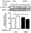
Phosphorylated Ser 1177 and total eNOS protein in aortic lysates from pigs fed STD (n=13) or HFHS (n=16) diet in the absence or presence of insulin treatment in vivo. Data are mean ± SE. Representative immunoblots are shown above group data. There was significant interaction of diet and insulin treatment on Ser1177 phospho-eNOS (p=0.01). The data suggest an effect of insulin to stimulate Ser1177 phospho-eNOS in STD (°p=0.07), but not in HFHS. Ser1177 phospho-eNOS was significantly different between diet groups in the presence of insulin treatment. Total eNOS expression did not differ between groups in the absence or presence of insulin. Insulin-stimulated Ser1177 phospho-eNOS bands in HFHS are in non-contiguous lanes of same blot, demarcated by a space within a double line. All other lanes are contiguous. *p<0.05.

References

    1. Barbour LA, Mizanoor RS, Gurevich I, Leitner JW, Fischer SJ, Roper MD, et al. Increased P85alpha is a potent negative regulator of skeletal muscle insulin signaling and induces in vivo insulin resistance associated with growth hormone excess. J Biol Chem. 2005;280(45):37489–37494. - PubMed
    1. Baron AD. The coupling of glucose metabolism and perfusion in human skeletal muscle. The potential role of endothelium-derived nitric oxide. Diabetes. 1996;45(Suppl 1):S105–S109. - PubMed
    1. Bayturan O, Tuzcu EM, Uno K, Lavoie AJ, Hu T, Shreevatsa A, et al. Comparison of rates of progression of coronary atherosclerosis in patients with diabetes mellitus versus those with the metabolic syndrome. Am J Cardiol. 2010;105(12):1735–1739. - PubMed
    1. Berg AH, Scherer PE. Adipose tissue, inflammation, and cardiovascular disease. Circ Res. 2005;96(9):939–949. - PubMed
    1. Bertacca A, Ciccarone A, Cecchetti P, Vianello B, Laurenza I, Maffei M, et al. Continually high insulin levels impair Akt phosphorylation and glucose transport in human myoblasts. Metabolism. 2005;54(12):1687–1693. - PubMed

Publication types

Substances