. 2013:3:1609.
doi: 10.1038/srep01609.

Bacterial expression strategies for several Sus scrofa diacylglycerol kinase alpha constructs: solubility challenges

Affiliations

Bacterial expression strategies for several Sus scrofa diacylglycerol kinase alpha constructs: solubility challenges

Elizabeth J Petro et al. Sci Rep. 2013.

Abstract

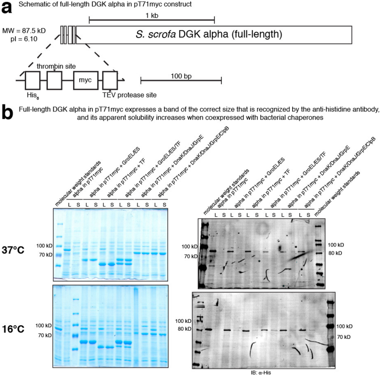
We pursued several strategies for expressing either full-length Sus scrofa diacylglycerol kinase (DGK) alpha or the catalytic domain (alphacat) in Escherichia coli. Alphacat could be extracted, refolded, and purified from inclusion bodies, but when subjected to analytical gel filtration chromatography, it elutes in the void volume, in what we conclude are microscopic aggregates unsuitable for x-ray crystallography. Adding glutathione S-transferase, thioredoxin, or maltose binding protein as N-terminal fusion tags did not improve alphacat's solubility. Coexpressing with bacterial chaperones increased the yield of alphacat in the supernatant after high-speed centrifugation, but the protein still elutes in the void upon analytical gel filtration chromatography. We believe our work will be of interest to those interested in the structure of eukaryotic DGKs, so that they know which expression strategies have already been tried, as well as to those interested in protein folding and those interested in chaperone/target-protein interactions.


Figures

Figure 1
Figure 1. Cloning, expression, refolding, purification, and analytical gel filtration of alphacat in pT71myc.
(a) Top, a schematic of S. scrofa DGK alpha. Middle, a schematic of “alphacat”, which consists of residues 333–733 of DGK alpha, including the LCB5 domain, but missing the C-terminal cysteine. Bottom, a magnification of the epitope tag region of alphacat in pT71myc. pT71myc adds to the N-terminus of the protein a fusion tag consisting of a hexahistidine, a thrombin proteolytic site, a myc tag, and a TEV protease site. The total mass of this protein construct (including the fusion tag) and pI are predicted to be 49.8 kD and 6.99, respectively. (b) SDS-PAGE of 12% acrylamide gels followed by: left, Coomassie staining; right, immunoblot against histidine. IB, immunoblot. (c) SDS-PAGE of a 12% acrylamide gel followed by Coomassie staining. DNAse I was added to the lysate to 10 U per mL. TX-100, Triton X-100. (d) SDS-PAGE of 12% acrylamide gels followed by: top, Coomassie staining; bottom, immunoblot against DGK alpha C-terminus. (e) Elution volumes of molecular weight standards and of purified alphacat from a Sephadex® G-200 column. Left vertical axis, the absorbance at 620 nm (A620) of blue dextran elution and the absorbance at 280 nm (A280) of protein standard elutions. Right vertical axis, protein concentration of eluted alphacat, as measured by the Bio-Rad Protein Assay.
Figure 2
Figure 2. Expression, purification, and analytical gel filtration of alphacat in pT71myc coexpressed with bacterial chaperones.
(a) SDS-PAGE of 12% acrylamide gels followed by: left, Coomassie staining; right, immunoblot against DGK alpha C-terminus; top, induced at 37°C for three hours; bottom, induced at 16°C overnight. U, uninduced; L, lysate; S, supernatant (after removing insoluble from lysate). (b) SDS-PAGE of an 8% acrylamide gel followed by immunoblotting against DGK alpha C-terminus. (c) SDS-PAGE of a 10% acrylamide gel followed by Coomassie staining. (d) Top, elution volumes of molecular weight standards and of purified alphacat from a Sephadex® G-200 column. Left vertical axis, A620 of blue dextran elution and A280 of protein standard elutions. Right vertical axis, quantification of the immunoblot signal from the blot shown at bottom, mean ± standard deviation (SD) (arbitrary units (AU)). Densitometry of the ~50 kD band was measured three times from the same film using ImageJ. Bottom, SDS-PAGE of a 10% acrylamide gel followed by immunoblotting against DGK alpha C-terminus. (e) SDS-PAGE of a 10% acrylamide gel followed by Coomassie staining. The lanes marked “+” were purified in the presence of 1 mM ATP.
Figure 3
Figure 3. Cloning and expression of full-length DGK alpha in pT71myc.
(a) Top, schematic of full-length S. scrofa DGK alpha in pT71myc. Bottom, a magnification of the epitope tag region of alpha in pT71myc. pT71myc adds to the N-terminus of the protein a fusion tag consisting of a hexahistidine, a thrombin proteolytic site, a myc tag, and a TEV protease site. The total mass of this protein construct (including the fusion tag) and pI are predicted to be 87.5 kD and 6.10, respectively. The epitope-tagged region of the construct is magnified for clarity. (b) SDS-PAGE of 8% acrylamide gels followed by: left, Coomassie staining; right, immunoblotting against hexahistidine; top, induced at 37°C for three hours; bottom, induced at 16°C overnight. L, lysate; S, supernatant (after removing insoluble from lysate).
Figure 4
Figure 4. Cloning, expression, purification, and analytical gel filtration of alphacat in pGEX-4T2 coexpressed with bacterial chaperones.
(a) “Alphacat” consists of residues 333–733 of S. scrofa DGK alpha. pGEX-4T2 adds to the N-terminus of the protein a fusion tag consisting of S. japonicum GST followed by a thrombin proteolytic site. The total mass of this protein construct (including the fusion tag) and pI are predicted to be 71.6 kD and 6.97, respectively. (b) SDS-PAGE of 12% acrylamide gels followed by: left, Coomassie staining; right, immunoblotting against against DGK alpha C-terminus. I, induced; U, uninduced; L, lysate; S, supernatant (after removing insoluble from lysate). (c) SDS-PAGE of a 10% acrylamide gel followed by Coomassie staining. This sample was purified after having previously flowed through MagneGST™. The insoluble fraction was removed before loading onto fresh MagneGST™. GSH, glutathione. (d) Elution volumes of molecular weight standards and of purified alphacat from a Sephadex® G-200 column. Left vertical axis, A620 of blue dextran elution and A280 of protein standard elutions. Right vertical axis, quantification of the immunoblot signal from the blot shown at right, mean ± SD (AU). Densitometry of the ~70 kD band was measured three times from the same film using ImageJ. Right, SDS-PAGE of an 8% acrylamide gel followed by immunoblotting against DGK alpha C-terminus.
Figure 5
Figure 5. Cloning and expression of alphacat and full-length DGK alpha in pET32a coexpressed with bacterial chaperones.
(a) Top: schematic of alphacat TEV in pET32a construct. “Alphacat” consists of residues 333–733 of S. scrofa DGK alpha; for this construct, the 734th residue of S. scrofa DGK alpha, cysteine, was added back. pET32a adds to the N-terminus of the protein a fusion tag consisting of E. coli TRX followed by a hexahistidine tag. For alphacat TEV, a TEV protease site site was added between the fusion tag and alphacat. The total mass of this protein construct (including the fusion tag) and pI are predicted to be 64.4 kD and 6.19, respectively. Middle: schematic of alphacat PP in pET32a construct. Instead of a TEV protease site, a PreScission Protease site was added between the fusion tag and alphacat. The total mass of this protein construct (including the fusion tag) and pI are predicted to be 64.4 kD and 6.19, respectively. Bottom: schematic of full-length S. scrofa DGK alpha TEV in pET32a construct. A TEV protease site was added between the fusion tag and DGK alpha, and the initial methionine was removed from DGK alpha. The total mass of this protein construct (including the fusion tag) and pI are predicted to be 102.0 kD and 5.84, respectively. (b) SDS-PAGE of 10% acrylamide gels followed by: top and middle, Coomassie staining; bottom, immunoblot against DGK alpha C-terminus. L, lysate; S, supernatant (after removing insoluble from lysate).
Figure 6
Figure 6. Cloning, expression, partial purification, and analytical gel filtration of alphacat and full-length DGK alpha in pET28-HisMBP-FLAGpp.
(a) Top, schematic of alphacat in HisMBP-FLAGpp construct. “Alphacat” consists of residues 333–733 of S. scrofa DGK alpha; for this construct, the 734th residue of S. scrofa DGK alpha, cysteine, was added back. pET28-HisMBP-FLAGpp adds to the N-terminus of the protein a fusion tag consisting of a hexahistidine tag, followed by P. furiosus maltose-binding protein (MBP) fusion tag, followed by a FLAG tag, followed by a PreScission Protease site. The total mass of the protein construct (including the fusion tag) and pI are predicted to be 92.8 kD and 5.51, respectively. Bottom, schematic of full-length S. scrofa DGK alpha construct. The total mass of the protein construct (including the fusion tag) and pI are predicted to be 130.6 kD and 5.45, respectively. (b) SDS-PAGE of 8% acrylamide gels followed by: top, Coomassie staining; bottom, immunoblotting against DGK alpha C-terminus. U, ininduced; I, induced; L, lysate; S, supernatant (after removing insoluble from lysate). (c) SDS-PAGE followed by: left, Coomassie staining; right, immunoblotting against DGK alpha C-terminus. The soluble fraction after lysis (supernatant) had been stored for two weeks at −80°C (in 20% glycerol); immediately prior to affinity purification, the insoluble fraction was removed by centrifugation at 100,000 × g for one hour at 4°C (supernatant's supernatant). (d) Left, elution volumes of molecular weight standards and of purified alphacat from a Sephadex® G-200 column. Left vertical axis, A620 of blue dextran elution and A280 of protein standard elutions. Right vertical axis, quantification of the immunoblot signal from the blot shown at right, mean ± SD (AU). Densitometry of the ~90 kD band was measured three times on the same film using ImageJ. Right, SDS-PAGE followed by immunoblotting against DGK alpha C-terminus. (e) SDS-PAGE of 8% acrylamide gels followed by: top, Coomassie staining; bottom, immunoblotting against DGK alpha C-terminus. U, uninduced; I, induced; L, lysate; S, supernatant (after removing insoluble from lysate). Cam, chloramphenicol.

Similar articles

Cited by

References

    1. Takeishi Y., Goto K. & Kubota I. Role of diacylglycerol kinase in cellular regulatory processes: a new regulator for cardiomyocyte hypertrophy. Pharmacol. Ther. 115, 352–359 (2007). - PubMed
    1. Ali H. et al. Selective translocation of diacylglycerol kinase zeta in hippocampal neurons under transient forebrain ischemia. Neurosci. Lett. 372, 190–195 (2004). - PubMed
    1. Kanoh H. & Ohno K. Partial purification and properties of diacylglycerol kinase from rat liver cytosol. Arch. Biochem. Biophys. 209, 266–275 (1981). - PubMed
    1. Besterman J. M., Pollenz R. S., Booker E. L. Jr. & Cuatrecasas P. Diacylglycerol-induced translocation of diacylglycerol kinase: use of affinity-purified enzyme in a reconstitution system. Proc. Natl. Acad. Sci. U.S.A. 83, 9378–9382 (1986). - PMC - PubMed
    1. Stathopoulos V. M. et al. Identification of two cytosolic diacylglycerol kinase isoforms in rat brain, and in NIH-3T3 and ras-transformed fibroblasts. Biochem. J. 272, 569–575 (1990). - PMC - PubMed

Publication types

MeSH terms

LinkOut - more resources