. 2013 May 17;288(20):14624-14635.
doi: 10.1074/jbc.M113.459438. Epub 2013 Apr 4.

Structural and biochemical analyses of glycoside hydrolase families 5 and 26 β-(1,4)-mannanases from Podospora anserina reveal differences upon manno-oligosaccharide catalysis

Affiliations

Structural and biochemical analyses of glycoside hydrolase families 5 and 26 β-(1,4)-mannanases from Podospora anserina reveal differences upon manno-oligosaccharide catalysis

Marie Couturier et al. J Biol Chem. .

Abstract

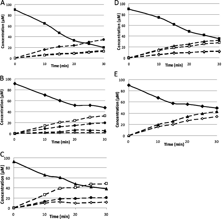
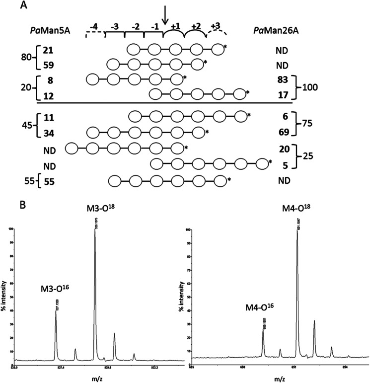
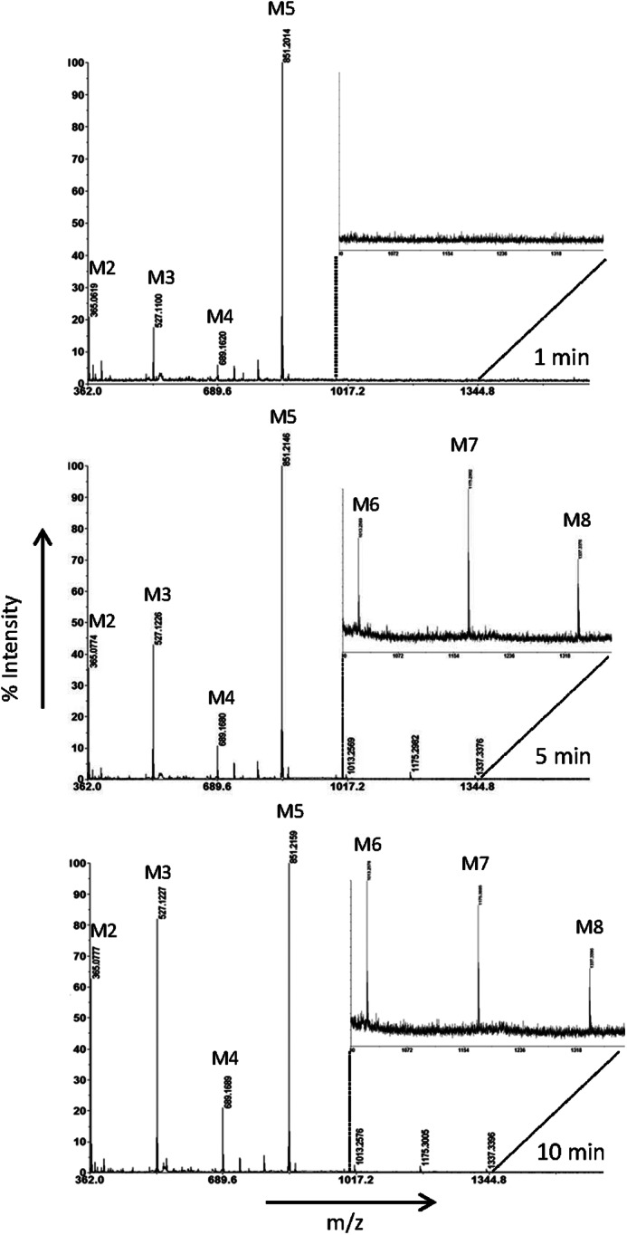
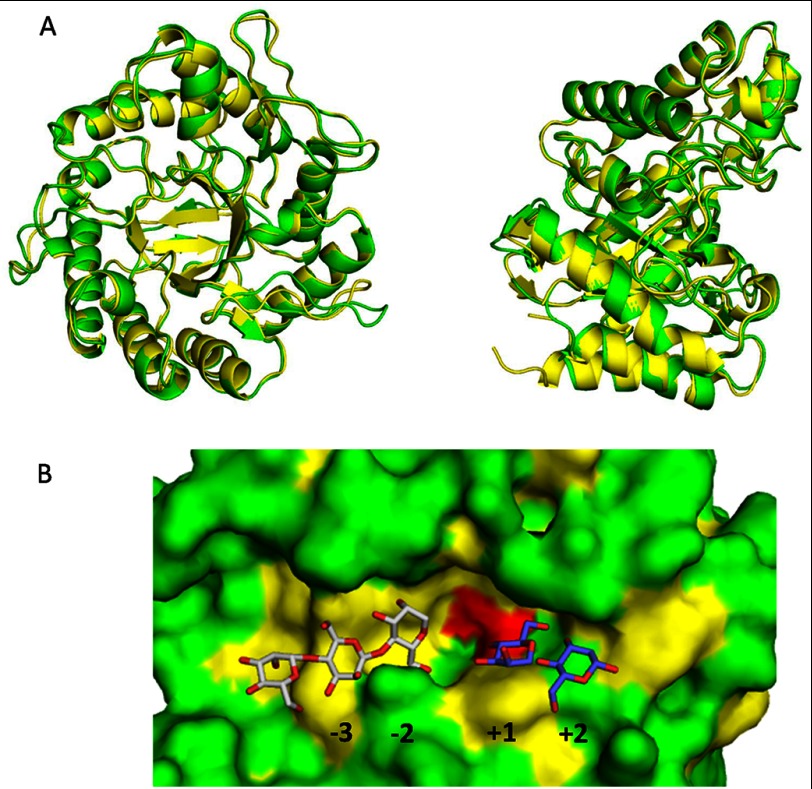
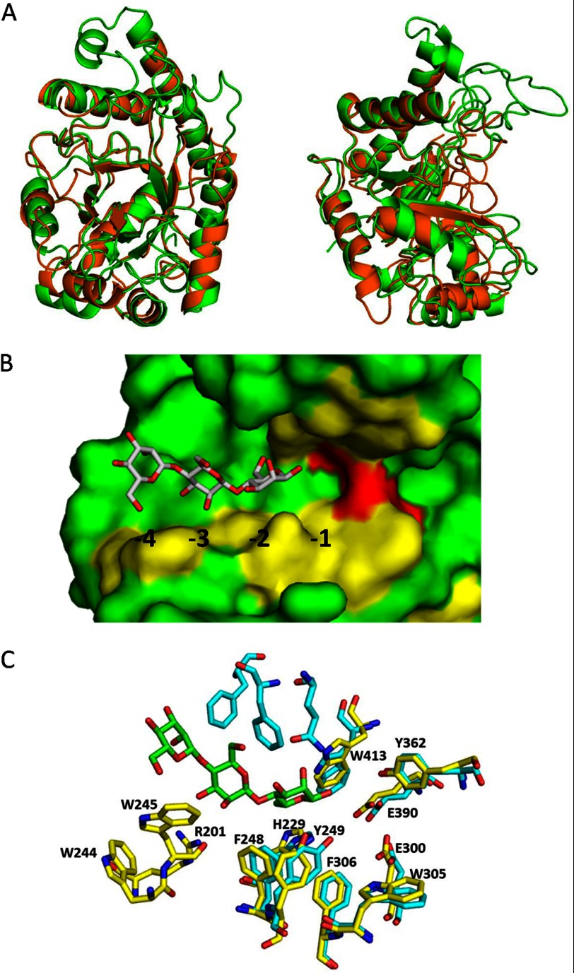
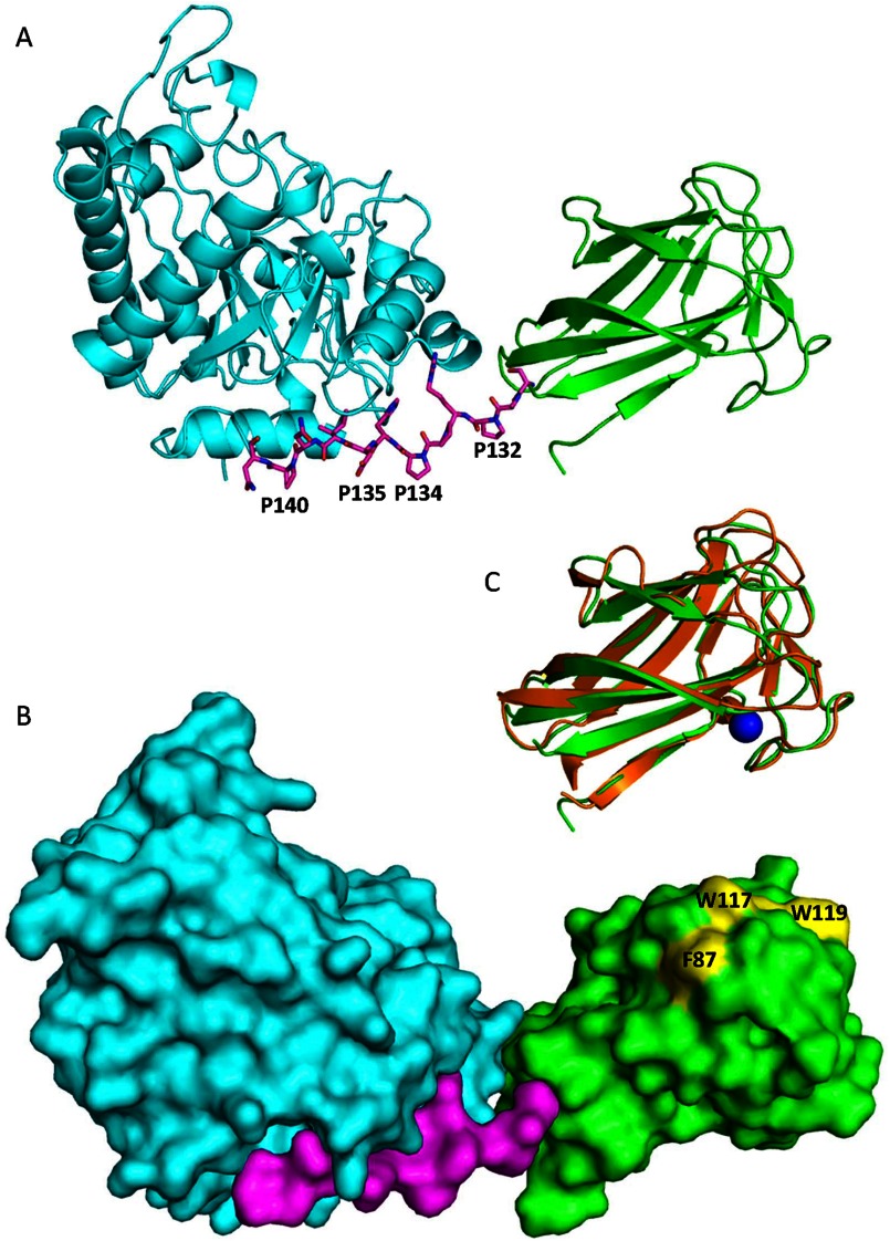
The microbial deconstruction of the plant cell wall is a key biological process that is of increasing importance with the development of a sustainable biofuel industry. The glycoside hydrolase families GH5 (PaMan5A) and GH26 (PaMan26A) endo-β-1,4-mannanases from the coprophilic ascomycete Podospora anserina contribute to the enzymatic degradation of lignocellulosic biomass. In this study, P. anserina mannanases were further subjected to detailed comparative analysis of their substrate specificities, active site organization, and transglycosylation capacity. Although PaMan5A displays a classical mode of action, PaMan26A revealed an atypical hydrolysis pattern with the release of mannotetraose and mannose from mannopentaose resulting from a predominant binding mode involving the -4 subsite. The crystal structures of PaMan5A and PaMan26A were solved at 1.4 and 2.85 Å resolution, respectively. Analysis of the PaMan26A structure supported strong interaction with substrate at the -4 subsite mediated by two aromatic residues Trp-244 and Trp-245. The PaMan26A structure appended to its family 35 carbohydrate binding module revealed a short and proline-rich rigid linker that anchored together the catalytic and the binding modules.

Keywords: CAZymes; Carbohydrate; Enzyme Structure; Fungi; Glycoside Hydrolases; Mannan; Plant Cell Wall; Polysaccharide.


Figures

FIGURE 1.
FIGURE 1.
Progress curve of the manno-oligosaccharides generated by PaMan5A and PaMan26A after the hydrolysis of manno-oligosaccharides. The recombinant enzymes were incubated with 100 μm manno-oligosaccharides in acetate buffer, pH 5.2, at 40 °C. The quantity of mannose (open diamonds), M2 (open squares), M3 (full circles), M4 (full triangles), M5 (full diamonds), and M6 (full squares) produced during the course of the reaction was quantified using HPAEC-PAD. The concentrations of enzymes used were PaMan5A, 18.2 nm with M6 (A), 18.2 nm with M5 (B), and 60 nm with M4 (C), and PaMan26A (D), 15 nm with M6 and 30 nm with M5 (E).
FIGURE 2.
FIGURE 2.
Relative frequency of the productive modes of binding of manno-oligosaccharides to PaMan5A and PaMan26A. A, the numbers represent the percentages of binding in each binding mode. These were calculated from the quantitative product analysis using HPAEC-PAD (numbers to the far left and far right, obtained from Table 4) followed by a detailed analysis of the hydrolytic cleavage patterns of M5 and M6 using MALDI-TOF-MS analysis of 18O-labeled products, allowing to distinguish further between binding modes. The arrow indicates the mannosidic bond to be cleaved. *, reducing end of oligosaccharide. The −4 and +3 dashed subsites are only present in PaMan26A and PaMan5A, respectively. ND, not detected. B, MALDI-TOF-MS spectra of M5 hydrolysis by PaMan5A and PaMan26A show enlarged parts of the spectra with the M3 product formed by PaMan5A at a ratio of 1:2.9 of M3/M3O18 (left) and the M4 product formed by PaMan26A at a ratio of 1:5.0 of M4/M4O18 (right). The peaks in the spectra correspond to the monoisotopic masses of sodium adducts [M + Na]+ of the manno-oligosaccharides.
FIGURE 3.
FIGURE 3.
MALDI-TOF-MS analysis of the transglycosylation product formation from M5 during 1–10 min by PaMan5A. Peaks in the spectra correspond to monoisotopic masses of sodium adducts [M + Na]+ of manno-oligosaccharides: M2, m/z 365.1; M3, m/z 527.1; M4, m/z 689.2; M5, m/z 851.2; M6, m/z 1013.3, M7, m/z 1175.3; M8, m/z 1337.3. The enlarged part in each spectrum corresponds to ∼3% of the relative intensity.
FIGURE 4.
FIGURE 4.
Crystal structure of PaMan5A. A, superposition of PaMan5A (green) and TrMan5A (yellow) structures is shown. The two views are related by a rotation of ∼90° about the vertical axis. B, shown is a surface view of the catalytic cleft of PaMan5A with mannotriose modeled in the −2 and −3 subsites and mannobiose modeled in the +1 and +2 subsites. The structures of GH5 from T. reesei and T. fusca in complex with mannobiose and mannotriose, respectively, were superimposed on the top of the structure of PaMan5A to map the substrate binding subsites.
FIGURE 5.
FIGURE 5.
Crystal structure of PaMan26A. A, superposition of PaMan26A catalytic module (green) and BCMan (orange) structures. The two views are related by a rotation of ∼90° about the vertical axis. B, shown is a surface view of the catalytic cleft of PaMan26A with mannotriose modeled in the −2 to −4 subsites. The structure of GH26 from C. fimi in complex with mannotriose was superimposed on the top of the structure of PaMan26A to map the substrate-binding subsites. C, shown is the organization of the glycone binding subsites in PaMan26A (yellow) compared with C. fimi (cyan).
FIGURE 6.
FIGURE 6.
Views of Modular architecture of PaMan26A. A, shown is a ribbon diagram of PaMan26A catalytic (blue) and CBM (green) domains. The proline-rich linker is shown in stick format. B, shown is a molecular surface representation of PaMan26A structure with the catalytic domain in blue, the PaCBM35 domain in green, and the linker in purple. The three aromatic residues present at the surface of the PaCBM35 domain are shown in yellow. C, shown is superposition of the PaCBM35 domain (green) and C. thermocellum CBM35 (orange). The calcium ion is represented by a blue sphere.

References

    1. Timell T. E. (1967) Recent progress in the chemistry of wood hemicellulose. Wood Sci. Technol. 1, 45–70
    1. Cantarel B. L., Coutinho P. M., Rancurel C., Bernard T., Lombard V., Henrissat B. (2009) The carbohydrate-active EnZymes database (CAZy). An expert resource for glycogenomics. Nucleic Acids Res. 37, 233–238 - PMC - PubMed
    1. Davies G., Henrissat B. (1995) Structures and mechanisms of glycosyl hydrolases. Structure 3, 853–859 - PubMed
    1. Vocadlo D. J., Davies G. J. (2008) Mechanistic insights into glycosidase chemistry. Curr. Opin. Chem. Biol. 12, 539–555 - PubMed
    1. Gilbert H. J., Stålbrand H., Brumer H. (2008) How the walls come crumbling down. Recent structural biochemistry of plant polysaccharide degradation. Curr. Opin. Plant. Biol. 11, 338–348 - PubMed

Publication types