. 2013 May 17;288(20):14276-14286.
doi: 10.1074/jbc.M113.455402. Epub 2013 Apr 4.

Alternative pyrimidine biosynthesis protein ApbE is a flavin transferase catalyzing covalent attachment of FMN to a threonine residue in bacterial flavoproteins

Affiliations

Alternative pyrimidine biosynthesis protein ApbE is a flavin transferase catalyzing covalent attachment of FMN to a threonine residue in bacterial flavoproteins

Yulia V Bertsova et al. J Biol Chem. .

Abstract

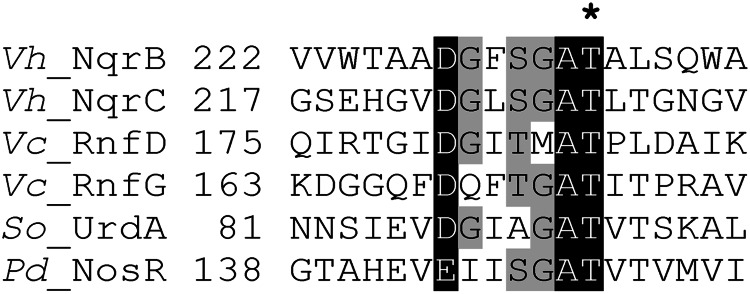
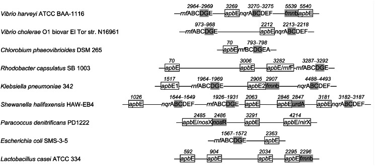
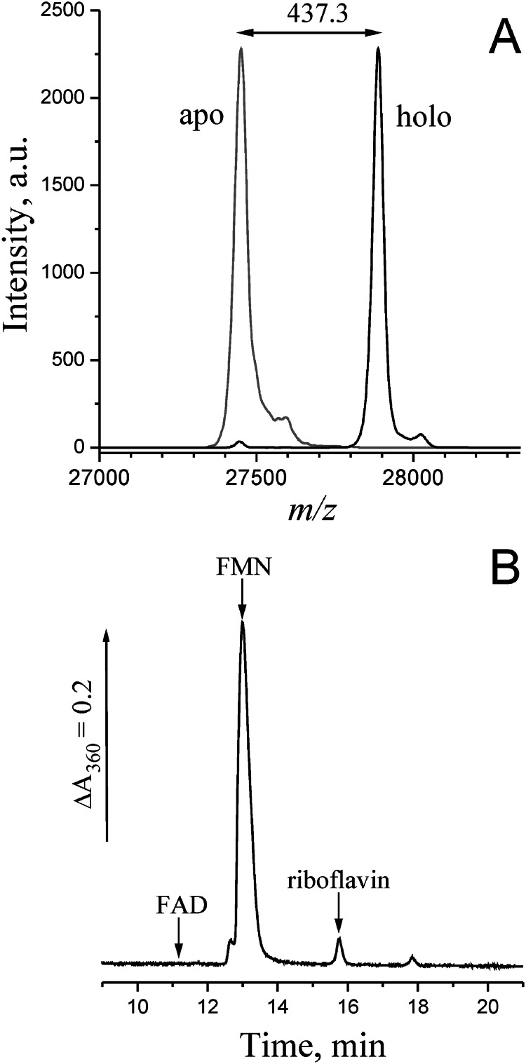
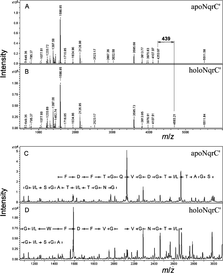
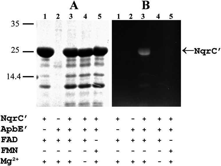
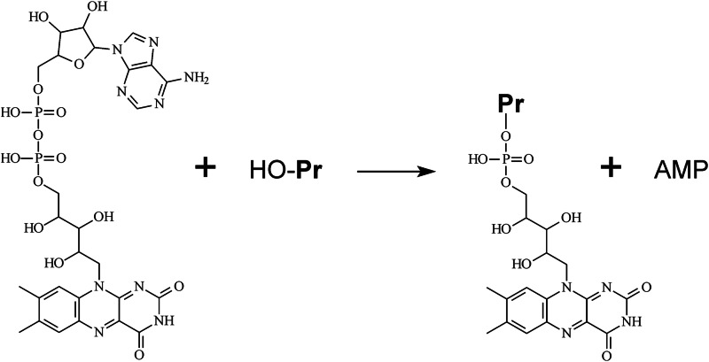
Na(+)-translocating NADH:quinone oxidoreductase (Na(+)-NQR) contains two flavin residues as redox-active prosthetic groups attached by a phosphoester bond to threonine residues in subunits NqrB and NqrC. We demonstrate here that flavinylation of truncated Vibrio harveyi NqrC at Thr-229 in Escherichia coli cells requires the presence of a co-expressed Vibrio apbE gene. The apbE genes cluster with genes for Na(+)-NQR and other FMN-binding flavoproteins in bacterial genomes and encode proteins with previously unknown function. Experiments with isolated NqrC and ApbE proteins confirmed that ApbE is the only protein factor required for NqrC flavinylation and also indicated that the reaction is Mg(2+)-dependent and proceeds with FAD but not FMN. Inactivation of the apbE gene in Klebsiella pneumoniae, wherein the nqr operon and apbE are well separated in the chromosome, resulted in a complete loss of the quinone reductase activity of Na(+)-NQR, consistent with its dependence on covalently bound flavin. Our data thus identify ApbE as a novel modifying enzyme, flavin transferase.

Keywords: Bacterial Metabolism; FMN; Flavoproteins; Na+-translocating NADH:Quinone Oxidoreductase; Post-translational Modification; Respiratory Chain.


Figures

FIGURE 1.
FIGURE 1.
Sequence alignment of FMN-binding motifs in several proteins. Vh_NqrB and Vh_NqrC, V. harveyi Na+-NQR subunits NqrB and NqrC (accession numbers Q9RFW0 and Q9RFV9, respectively); Vc_RnfD and Vc_RnfG, V. cholerae RNF subunits RnfD and RnfG (ACP09041 and ACP09040, respectively); Pd_NosR, P. denitrificans regulator of NO reductase transcription NosR (Pden_4220); So_UrdA, S. oneidensis MR-1 urocanate reductase (NP_720136). The position of the experimentally identified FMN acceptor Thr residue (11, 15) is marked by an asterisk.
FIGURE 2.
FIGURE 2.
Typical arrangements of genes for ApbE, Na+-NQR, RNF, and other FMN-binding proteins in bacterial chromosomes. Genes for Na+-NQR and RNF subunits are indicated by the last letters in their standard designations. apbE genes together with their accepted alternative designations are shown as rectangles, genes for FMN-binding Na+-NQR and RNF subunits, UrdA, NosR, and other FMN-binding proteins (fmnb) are indicated by gray shading. The connecting lines refer to intercalating sequences. The numbers above the genes indicate their numbers in genomes according to the Kyoto Encyclopedia of Genes and Genomes. The genes were detected as hits in a BLAST search of complete prokaryotic genomes using the V. harveyi proteins as the queries. Genes for FMN-binding proteins were identified using search for Pfam motifs PF04205 and PF03116.
FIGURE 3.
FIGURE 3.
Absorption spectra of recombinant NqrC′ protein of V. harveyi. A, red line, NqrC′ isolated from E. coli/pMshC3 strain (nqrC′ only); blue line, NqrC′ isolated from E. coli/pMshC3 + pΔhis3 strain (nqrC′ + apbE′); black line, NqrC′ isolated from E. coli/pMshC3 + pΔhis3 strain and denaturated with 1% (w/v) SDS. Inset, magnified spectra in the 325–600 nm region. B, HoloNqrC′ purified from E. coli/pMshC3 + pΔhis3 strain. Blue line, air oxidized holoNqrC′ (as isolated); red line, holoNqrC′ fully reduced by excess dithionite; green line, partially reduced holoNqrC′ obtained by slow oxidation of the fully reduced protein by air. Peak labels refer to curves of the same color. The assay medium contained 0.81 mg/ml protein, 20 mm Tris-HCl (pH 8.0), and 50 mm NaCl.
FIGURE 4.
FIGURE 4.
SDS-PAGE analysis of recombinant NqrC′ and UrdA′ proteins purified from different E. coli strains. A and C, unstained gels under UV illumination; B and D, Coomassie Blue-stained gels. Lanes 1 and 2 are NqrC′ isolated from E. coli/pMshC3 strain (nqrC′ only) and E. coli/pMshC3 + pΔhis3 strain (nqrC′ + apbE′), respectively. The UrdA′ protein was purified from E. coli/p9DL + pΔhis3 strain (urdA′ + apbE′). Protein load was 10 μg/lane for NqrC′ and 5 μg/lane for UrdA′. The fluorescent bands seen in A and C correspond to flavin-bound NqrC′ and UrdA′, respectively. The bars with numbers on the left side denote the positions and molecular masses of marker proteins.
FIGURE 5.
FIGURE 5.
Mass spectral and chromatographic identification of the flavin prosthetic group in NqrC′. A, MALDI mass spectra of apoNqrC′ (gray line) and holoNqrC′ (black line). The difference in the molecular masses corresponds to the mass of an FMN residue (438.3 Da). B, HPLC separation of flavins extracted from holoNqrC′ by LiOH treatment. The arrows indicate the retention times for FAD, FMN, and riboflavin standards.
FIGURE 6.
FIGURE 6.
MALDI mass spectral identification of the modification site in NqrC′. A and B, tryptic digests of apoNqrC′ and holoNqrC′, respectively. C and D, MS/MS spectra of the selected tryptic peptide of apoNqrC′ (m/z = 4254) and holoNqrC′ (m/z = 4693), respectively. The deduced protein sequences are shown.
FIGURE 7.
FIGURE 7.
In vitro flavinylation of apoNqrC′. The apoNqrC′ protein (4 mg/ml) was incubated in medium containing ApbE′ (0.1 mg/ml), MgSO4 (5 mm), and flavin (1 mm), as indicated under the gel. The reaction products (5 μl) were separated by SDS-PAGE. A, Coomassie-stained gel. B, unstained gel under UV illumination. The bars with numbers on the left side refer to the positions and molecular masses of marker proteins.
FIGURE 8.
FIGURE 8.
The reaction catalyzed by ApbE. HO-Pr, the protein to be modified.
FIGURE 9.
FIGURE 9.
A stereo view of the FAD-binding site in S. enterica ApbE (Protein Data Bank code 3PND) (43). ApbE is shown as a surface model (probe radius, 1.4 Å), and FAD is depicted as a stick model. The pyrophosphate group of FAD is shown in orange. The figure was created with PyMOL (PyMOL Molecular Graphics System, version 1.5.0.4; Schrödinger, LLC).

References

    1. Verkhovsky M. I., Bogachev A. V. (2010) Sodium-translocating NADH:quinone oxidoreductase as a redox-driven ion pump. Biochim. Biophys. Acta 1797, 738–746 - PubMed
    1. Zhou W., Bertsova Y. V., Feng B., Tsatsos P., Verkhovskaya M. L., Gennis R. B., Bogachev A. V., Barquera B. (1999) Sequencing and preliminary characterization of the Na+-translocating NADH:ubiquinone oxidoreductase from Vibrio harveyi. Biochemistry 38, 16246–16252 - PubMed
    1. Häse C. C., Fedorova N. D., Galperin M. Y., Dibrov P. A. (2001) Sodium ion cycle in bacterial pathogens. Evidence from cross-genome comparisons. Microbiol. Mol. Biol. Rev. 65, 353–370 - PMC - PubMed
    1. Nakayama Y., Hayashi M., Unemoto T. (1998) Identification of six subunits constituting Na+-translocating NADH-quinone reductase from the marine Vibrio alginolyticus. FEBS Lett. 422, 240–242 - PubMed
    1. Rich P. R., Meunier B., Ward F. B. (1995) Predicted structure and possible ion-motive mechanism of the sodium-linked NADH-ubiquinone oxidoreductase of Vibrio alginolyticus. FEBS Lett. 375, 5–10 - PubMed

Publication types

MeSH terms